The nervous system constantly receives information from the external and internal environments, allowing us to perceive sensations, maintain balance, and coordinate movement. Somatic sensations, originating from the body’s surface and musculoskeletal structures, are conveyed to the central nervous system (CNS) via specific ascending pathways. These pathways are typically composed of a series of neurons (first-order, second-order, and often third-order) that transmit signals from peripheral receptors through the spinal cord and brainstem to higher brain centers like the thalamus, cerebral cortex, and cerebellum.
Understanding these pathways is fundamental to neurophysiology and clinical neurology, as damage to specific tracts can result in characteristic sensory deficits.
Major Ascending Somatic Sensory Pathways:
- The Dorsal Column-Medial Lemniscus Pathway (Gracile and Cuneate Tracts)
- Sensory Modalities Conveyed: This pathway is responsible for transmitting highly discriminative somatic sensations, including:
- Conscious Proprioception: The sense of the position and movement of the limbs and body in space (kinesthesia). This allows us to know where our body parts are without looking.
- Fine Touch: The ability to perceive light touch with precision, such as identifying textures or recognizing objects by touch (stereognosis).
- Vibration: The perception of rapidly changing pressure stimuli.
- Two-Point Discrimination: The ability to distinguish between two simultaneous points of contact on the skin as separate stimuli.
- Pressure: While other pathways carry crude pressure, the DCML contributes to the perception of sustained, localized pressure.
- Pathway Structure:
- First-Order Neuron: The cell bodies are located in the dorsal root ganglia (DRG) for spinal nerves and cranial nerve ganglia (e.g., Trigeminal) for the head and face (though this description focuses on limbs/trunk). The peripheral axons connect to various receptors in the skin, muscles, tendons, and joints (e.g., Meissner’s corpuscles, Pacinian corpuscles, Merkel discs, Ruffini endings, muscle spindles, Golgi tendon organs). The central axons enter the spinal cord via the dorsal root and ascend ipsilaterally (on the same side) in the dorsal (posterior) funiculus.
- Fibers from the lower body (below spinal cord segment T6) collect in the medial portion of the dorsal column, forming the gracile fasciculus (fasciculus gracilis).
- Fibers from the upper body (above spinal cord segment T6) collect in the lateral portion of the dorsal column, forming the cuneate fasciculus (fasciculus cuneatus).
- These axons ascend the entire length of the spinal cord and terminate in the medulla oblongata. The gracile fasciculus ends in the nucleus gracilis, and the cuneate fasciculus ends in the nucleus cuneatus.
- Second-Order Neuron: The second-order neurons are located in the nucleus gracilis and nucleus cuneatus in the caudal medulla. Their axons, called internal arcuate fibers, cross (decussate) the midline at the level of the medulla. After decussating, they ascend contralaterally (on the opposite side) through the brainstem as a consolidated bundle called the medial lemniscus. The medial lemniscus ascends through the pons and midbrain, terminating in the thalamus.
- Third-Order Neuron: The cell bodies of the third-order neurons are located primarily in the ventral posterior lateral (VPL) nucleus of the thalamus (for input from spinal nerves via the gracile and cuneate nuclei). From the thalamus, these axons project through the internal capsule to the primary somatosensory cortex (Brodmann areas 1, 2, and 3) located in the postcentral gyrus of the parietal lobe. Here, the sensory information is consciously perceived and interpreted, forming a somatotopic map (homunculus) where different body parts are represented in distinct cortical areas.
- First-Order Neuron: The cell bodies are located in the dorsal root ganglia (DRG) for spinal nerves and cranial nerve ganglia (e.g., Trigeminal) for the head and face (though this description focuses on limbs/trunk). The peripheral axons connect to various receptors in the skin, muscles, tendons, and joints (e.g., Meissner’s corpuscles, Pacinian corpuscles, Merkel discs, Ruffini endings, muscle spindles, Golgi tendon organs). The central axons enter the spinal cord via the dorsal root and ascend ipsilaterally (on the same side) in the dorsal (posterior) funiculus.
- Key Characteristics & Function: This pathway is characterized by its speed, high degree of spatial localization, and ability to transmit detailed information about the stimulus (intensity, location, fine discrimination). The ipsilateral ascent in the spinal cord and decussation in the medulla are key anatomical features. It is essential for skilled movements and fine sensory discrimination. Lesions in this pathway lead to deficits in fine touch, vibration, and proprioception on the ipsilateral side below the spinal cord lesion (due to ascending ipsilaterally) or on the contralateral side above the medulla lesion (due to decussation).
- Sensory Modalities Conveyed: This pathway is responsible for transmitting highly discriminative somatic sensations, including:
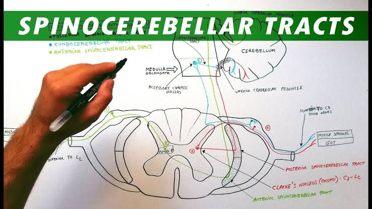
Spinal Cord Pathways for Unconscious Proprioception (Dorsal and Ventral Spinocerebellar Tracts)
-
- Sensory Modalities Conveyed: These pathways transmit information about unconscious proprioception from muscles, tendons, and joints. This information is used by the cerebellum for coordinating movement, maintaining posture, and learning motor skills. It does not reach conscious perception in the traditional sense, although it contributes fundamentally to motor control. Receptors include muscle spindles (sensing muscle length and rate of change), Golgi tendon organs (sensing muscle tension), and joint receptors (sensing joint position and movement).
- Pathway Structure: There are several spinocerebellar tracts, but the primary ones from the limbs and trunk are the dorsal (posterior) and ventral (anterior) spinocerebellar tracts. The cuneocerebellar tract serves a similar function for the upper body and is often considered the upper body equivalent of the dorsal spinocerebellar tract.
- Dorsal Spinocerebellar Tract:
- First-Order Neuron: From receptors (primarily muscle spindles and Golgi tendon organs) in the lower body (below T1), including the leg and trunk. Cell bodies are in the DRG. Peripheral axons connect to receptors. Central axons enter the spinal cord via the dorsal root and synapse on second-order neurons in the nucleus dorsalis (Clark’s nucleus), located in the medial gray matter of the spinal cord, primarily between spinal segments C8 and L2/L3.
- Second-Order Neuron: Cell bodies are in Clark’s nucleus. The axons ascend ipsilaterally in the dorsal part of the lateral funiculus (dorsal spinocerebellar tract). These fibers enter the cerebellum primarily through the inferior cerebellar peduncle, terminating mainly in the cerebellar vermis and intermediate zones.
- (Upper body equivalent: Cuneocerebellar Tract): First-order neurons from the upper body (above C8) synapse on second-order neurons in the Accessory Cuneate Nucleus in the medulla. These second-order axons ascend ipsilaterally and enter the cerebellum via the inferior cerebellar peduncle.
- Ventral Spinocerebellar Tract:
- First-Order Neuron: From receptors (including muscle spindles, Golgi tendon organs, and interneurons involved in spinal reflexes) in the lower body (below L2). Cell bodies are in the DRG. Central axons enter the spinal cord and synapse on second-order neurons in the base of the dorsal horn and intermediate gray matter (primarily laminae V-VII) in the lumbar and sacral spinal cord.
- Second-Order Neuron: Cell bodies are in the lumbar/sacral gray matter. These axons cross the midline twice – first in the ventral gray commissure at the spinal cord level where they originate, and then crossing back in the brainstem before entering the cerebellum. They ascend primarily in the ventral part of the lateral funiculus contralaterally within the spinal cord (ventral spinocerebellar tract). These fibers ascend through the pons and midbrain and enter the cerebellum primarily through the superior cerebellar peduncle.
- (Complexity): While often simplified as crossing twice, some fibers may not cross at all, or cross only once. The key functional outcome is that the information largely ends up on the ipsilateral side of the cerebellum relative to the body part providing the input, after traversing complex paths that include spinal interneuronal populations.
- Third-Order Neuron: While not always described with a strict third-order neuron like the conscious pathways, signals within the cerebellum are processed through complex circuits involving various cerebellar nuclei and cortical layers before influencing descending motor pathways. The cerebellum does not project directly to the cerebral cortex in the same way the DCML or Spinothalamic pathways do.
- Dorsal Spinocerebellar Tract:
- Key Characteristics & Function: These pathways operate below the level of consciousness, providing the cerebellum with continuous, updated information about muscle length, tension, and limb position. This allows the cerebellum to compare intended movements (from the cerebral cortex) with actual movements and make adjustments to motor output for smooth, coordinated, and accurate actions. The complexity of crossing in the ventral tract, often involving spinal interneurons, reflects its role in conveying state information of spinal reflex circuits. Lesions in these tracts lead to ataxia (lack of voluntary coordination of muscle movements) on the ipsilateral side of the body because the cerebellar hemisphere controls the ipsilateral side of the body.
The Anterolateral System: Lateral Spinothalamic Tract
-
- Sensory Modalities Conveyed: This tract is the principal pathway for transmitting:
- Pain: Nociceptive information, including sharp, dull, burning, and aching pain.
- Temperature: Sensations of heat and cold.
- Pathway Structure:
- First-Order Neuron: The cell bodies are located in the DRG. The peripheral axons connect to free nerve endings in the skin and other tissues that function as nociceptors (pain receptors) and thermoreceptors (temperature receptors). These receptors are typically polymodal (responding to mechanical, thermal, and chemical stimuli) for pain or specific for heat/cold. The central axons enter the spinal cord via the dorsal root. Upon entering, they ascend or descend one or two spinal segments within Lissauer’s tract (dorsolateral tract) before synapsing on second-order neurons in the dorsal horn gray matter.
- Second-Order Neuron: The cell bodies are located in the dorsal horn of the spinal cord, primarily in Rexed laminae I (marginal zone), II (substantia gelatinosa), and V (neck of the dorsal horn). The axons of these second-order neurons immediately cross (decussate) the midline in the anterior white commissure of the spinal cord, usually within one or two segments of their origin. After crossing, they ascend contralaterally in the lateral funiculus of the spinal cord as the lateral spinothalamic tract.
- Third-Order Neuron: The lateral spinothalamic tract ascends through the brainstem, running adjacent to and often intermingled with the medial lemniscus and ventral spinothalamic tract (collectively the Anterolateral System). These fibers terminate in the thalamus, primarily in the ventral posterior lateral (VPL) nucleus for discriminative aspects of pain and temperature, but also project to medial and intralaminar nuclei of the thalamus. From the VPL, third-order neurons project to the primary somatosensory cortex (postcentral gyrus) for conscious perception and localization of pain and temperature. Projections to medial/intralaminar nuclei are thought to contribute to the emotional, arousal, and motivational aspects of pain.
- Key Characteristics & Function: This is a vital protective pathway, alerting the brain to potentially harmful stimuli. Compared to the DCML, it is slower and provides less precise localization, especially for pain (though discriminative aspects can be processed by the cortex). The immediate decussation in the spinal cord is a critical feature; a unilateral spinal cord lesion will cause loss of pain and temperature sensation on the contralateral side of the body below the level of the lesion.
- Sensory Modalities Conveyed: This tract is the principal pathway for transmitting:
The Anterolateral System: Ventral Spinothalamic Tract
-
- Sensory Modalities Conveyed: This tract is responsible for transmitting:
- Crude Touch: The ability to perceive light touch without fine localization or discrimination (e.g., feeling that something is touching you, but not exactly what or where).
- Pressure: Contributing to the perception of pressure, distinct from the fine discriminatory pressure carried by the DCML.
- Pathway Structure: The ventral spinothalamic tract runs in the anterolateral system alongside the lateral spinothalamic tract, sharing many similarities in structure.
- First-Order Neuron: The cell bodies are located in the DRG. Peripheral axons connect to various mechanoreceptors (e.g., free nerve endings, some Merkel discs) that respond to light touch or pressure. Central axons enter the spinal cord via the dorsal root and synapse on second-order neurons in the dorsal horn gray matter. They may ascend/descend briefly in Lissauer’s tract.
- Second-Order Neuron: The cell bodies are located in the dorsal horn of the spinal cord, typically in Rexed laminae III and IV. The axons of these second-order neurons also cross (decussate) the midline in the anterior white commissure, but the crossing can occur over several spinal segments. After crossing, they ascend contralaterally in the anterior (ventral) funiculus of the spinal cord as the ventral spinothalamic tract.
- Third-Order Neuron: The ventral spinothalamic tract ascends through the brainstem, terminating in the thalamus, primarily in the ventral posterior lateral (VPL) nucleus, but also potentially in other nuclei like the ventral posterior medial (VPM) nucleus and intralaminar nuclei. From the thalamus, these third-order neurons project to the primary somatosensory cortex (postcentral gyrus), allowing for conscious perception of crude touch and pressure, although with poor localization compared to fine touch via the DCML.
- Key Characteristics & Function: This pathway provides basic, spatially imprecise touch and pressure information. Because fine touch is carried by a separate pathway (DCML), the ventral spinothalamic tract alone is insufficient for tasks requiring fine texture or object discrimination. The decussation occurs in the spinal cord, similar to the lateral spinothalamic tract, meaning a unilateral spinal cord lesion would impair crude touch and pressure sensation on the contralateral side below the lesion, although often less severely or completely than pain/temperature loss due to the multi-segmental crossing and some redundancy or contribution from other pathways.
- Sensory Modalities Conveyed: This tract is responsible for transmitting:
In summary, somatic sensory information from the limbs and trunk ascends to the brain via distinct parallel pathways, each specialized for different types of sensory input. The Dorsal Column-Medial Lemniscus system carries fine, discriminative sensations and proprioception that are consciously perceived and precisely localized in the somatosensory cortex after decussating in the medulla. The Spinocerebellar pathways transmit unconscious proprioceptive information to the cerebellum for motor coordination, with complex crossing patterns but delivering information largely ipsilaterally to the cerebellum. The Anterolateral System, comprising the lateral and ventral spinothalamic tracts, conveys pain, temperature, and crude touch, decussating immediately in the spinal cord and projecting to the thalamus and somatosensory cortex for conscious perception, although with less spatial precision than and often different qualitative aspects compared to the sensations carried by the DCML. The parallel processing and distinct anatomical routes of these pathways underscore the complexity and functional organization of the somatic sensory system.


