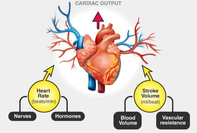
Understanding the dynamics of blood circulation is fundamental to appreciating physiological function and pathology. Central to this is the concept of Cardiac Output (CO), the measure of blood volume pumped by the heart per unit of time.
The Importance of Cardiac Output
The primary function of the cardiovascular system is to deliver oxygen and nutrients to tissues and remove metabolic waste products. Cardiac Output is the key determinant of this delivery rate. It is the volume of blood ejected by the left ventricle into the aorta (or, equivalently in a steady state, by the right ventricle into the pulmonary artery) per minute. Adequate cardiac output is essential for maintaining tissue perfusion and meeting the body’s metabolic demands, which fluctuate significantly between rest and activity.
Defining Cardiac Output (CO) and Cardiac Index (CI)
- Cardiac Output (CO): CO is the product of Stroke Volume (SV) and Heart Rate (HR).
- Equation: CO = SV × HR
- Stroke Volume (SV): The volume of blood ejected by the heart ventricle during each contraction (systole). Typically measured in milliliters (mL).
- Heart Rate (HR): The number of times the heart beats per minute. Typically measured in beats per minute (bpm).
- Units: CO is usually expressed in liters per minute (L/min).
- Typical Resting Value: In an adult at rest, CO is typically around 4.0 to 8.0 L/min.
- Cardiac Index (CI): CI is a refinement of CO that accounts for differences in body size. It is calculated by dividing the CO by the individual’s Body Surface Area (BSA). BSA is calculated using specific formulas based on height and weight.
- Equation: CI = CO / BSA
- Units: CI is usually expressed in liters per minute per square meter (L/min/m²).
- Typical Resting Value: In an adult at rest, CI is typically around 2.5 to 4.0 L/min/m².
- Purpose: Using CI is often more clinically useful than CO alone when comparing values between individuals or assessing a patient’s status, as it normalizes the value for variations in metabolic needs due to size.
The Role of Venous Return (VR) in Determining Cardiac Output
Under steady-state conditions, the amount of blood pumped out by the heart (CO) must equal the amount of blood returning to it (Venous Return, VR). If CO exceeded VR for any sustained period, the systemic circulation would empty, and the heart would stop pumping. Conversely, if VR exceeded CO, blood would accumulate in the systemic veins and heart chambers. Therefore, VR is a crucial determinant of CO.
- Definition of Venous Return: The rate of blood flow back to the heart from the systemic circulation.
- The Frank-Starling Mechanism: This intrinsic property of cardiac muscle explains how increased VR leads to increased CO. When more blood returns to the heart during diastole (filling), the ventricular muscle fibers are stretched to a greater length. This greater stretch leads to a more forceful contraction during systole, ejecting a larger stroke volume. Within physiological limits, the heart pumps out virtually all the blood that returns to it. This mechanism ensures that the CO is matched to the VR.
Cardiac Reserve and its Effect on Cardiac Output
Cardiac Reserve (CR) represents the heart’s ability to increase its output beyond the resting level to meet increased demand, such as during exercise, stress, or illness.
- Definition of Cardiac Reserve: The difference between the maximum cardiac output the heart can achieve and the cardiac output at rest.
- Importance: CR is essential for the body to perform activities requiring increased oxygen delivery (e.g., exercise). A healthy individual might have a resting CO of 5 L/min but can increase this to 20-25 L/min during strenuous exercise, demonstrating a significant cardiac reserve (15-20 L/min).
- Effect on CO: A higher cardiac reserve indicates a healthier, more adaptable cardiovascular system capable of significantly increasing CO when needed. Conditions that impair heart function (e.g., heart failure, myocardial infarction) reduce cardiac reserve, limiting the individual’s ability to increase CO and thus their capacity for physical activity.
The Role of Right Atrial Pressure (RAP) and Mean Circulatory Filling Pressure (MCFP)
Blood flow throughout the circulatory system, including venous return, is driven by pressure gradients. Two key pressures influencing VR are Right Atrial Pressure (RAP) and Mean Circulatory Filling Pressure (MCFP).
- Right Atrial Pressure (RAP): Also known as Central Venous Pressure (CVP), RAP is the pressure in the right atrium. It serves as the downstream pressure for systemic venous return.
- Typical Value: Normally, RAP is very low, close to 0 mmHg, and fluctuates slightly with respiration.
- Role: A low RAP facilitates VR by maintaining a steep pressure gradient between the systemic veins and the right atrium. An increase in RAP (e.g., due to heart failure, volume overload, tricuspid valve stenosis) reduces the pressure gradient for VR, thereby impeding VR and consequently reducing CO (assuming the heart can’t overcome the increased afterload/preload).
- Mean Circulatory Filling Pressure (MCFP): This theoretical pressure is the pressure that would exist everywhere in the systemic circulation if blood flow were instantaneously stopped and pressure equalized throughout the systemic vessels. It represents the average elastic recoil pressure of the circulatory system when filled with blood.
- Influence: MCFP is primarily determined by the volume of blood in the systemic circulation and the tone (contractile state) of the systemic blood vessels, particularly the veins which are highly compliant capacitance vessels.
- Role: MCFP can be considered the upstream pressure driving venous return. The pressure gradient for VR is approximately MCFP – RAP. An increase in MCFP (e.g., due to increased blood volume or widespread venoconstriction) increases this gradient, promoting increased VR and thus CO (via Frank-Starling).
In essence, the interplay between MCFP and RAP sets the primary pressure gradient driving blood from the periphery back to the heart, directly influencing VR and therefore CO.
Effect of Increased Sympathetic Activity on Cardiac Output
The sympathetic nervous system (SNS) is a major regulator of cardiovascular function, particularly in response to stress or increased activity. Increased sympathetic activity significantly impacts CO through its effects on both the heart and the blood vessels.
- Effects on the Heart:
- Increased Heart Rate (Positive Chronotropy): Sympathetic stimulation via norepinephrine released from sympathetic nerve endings and epinephrine from the adrenal medulla increases the rate of firing of the sinoatrial (SA) node, increasing HR.
- Increased Contractility (Positive Inotropy): Sympathetic stimulation increases the force of myocardial contraction, leading to a larger stroke volume at any given end-diastolic volume. This shifts the Frank-Starling curve upwards and to the left, meaning the heart can pump more blood more forcefully.
- Effects on Blood Vessels:
- Vasoconstriction: Sympathetic activity causes constriction of both resistance vessels (arterioles) and capacitance vessels (veins). While arteriolar constriction increases systemic vascular resistance (afterload), which can potentially decrease SV if severe, venoconstriction is particularly important for CO regulation.
- Increased MCFP: Venoconstriction reduces the capacity of the venous system. Blood is displaced from the peripheral veins into the central circulation, increasing the central blood volume and consequently increasing MCFP. This increased MCFP enhances the pressure gradient for VR (MCFP – RAP), thereby increasing VR.
- Overall Effect: Increased sympathetic activity usually leads to a significant increase in CO. The combined effects of increased HR and contractility enhance the heart as a pump, while the venoconstriction and increased MCFP enhance the return of blood to the heart. These effects work in concert to elevate CO rapidly and substantially, meeting the demands of situations like exercise or the “fight-or-flight” response.
The Effect of Increased Blood Volume on Cardiac Output
Blood volume is another critical factor influencing cardiac output, primarily by affecting the filling pressures of the circulation.
- Effect on MCFP: An increase in total blood volume directly leads to an increase in Mean Circulatory Filling Pressure (MCFP). With more fluid in the system, the vessels are stretched more, increasing their elastic recoil pressure when flow is stopped.
- Effect on Venous Return: As discussed in Step 4, an increased MCFP raises the pressure gradient for venous return (MCFP – RAP), promoting a higher rate of blood flow back to the heart.
- Effect on Cardiac Output: The increased venous return delivers a larger volume of blood to the heart during diastole, increasing end-diastolic volume. According to the Frank-Starling mechanism (Step 2), this increased stretch leads to a more forceful contraction and a larger stroke volume. Assuming the heart is healthy and capable of handling the increased volume, the increased stroke volume, often coupled with a reflex increase in heart rate, results in a higher cardiac output. Conversely, decreased blood volume (e.g., due to hemorrhage) leads to decreased MCFP, reduced VR, and typically reduced CO.
Methods for the Measurement of Cardiac Output
Accurate measurement of cardiac output is vital in clinical settings for diagnosing conditions, monitoring patient status (especially in critical care), and guiding therapeutic interventions. Various methods exist, ranging from invasive to non-invasive.
- The Direct Fick Method: Considered the gold standard but complex and rarely used routinely in clinical practice.
- Principle: Based on the Fick principle, which states that the amount of a substance taken up or released by an organ per unit time is equal to the blood flow through the organ multiplied by the difference in concentration of the substance in the arterial and venous blood supplying the organ.
- Application for CO: Measures oxygen uptake by the lungs (VO₂) and the arterial-venous oxygen content difference across the lungs (CaO₂ – CvO₂).
- Equation: CO = VO₂ / (CaO₂ – CvO₂)
- Requirements: Requires measuring VO₂ (e.g., using metabolic cart) and obtaining simultaneous samples of arterial blood and mixed venous blood (from the pulmonary artery via a catheter) to determine oxygen content.
- Thermodilution: A common invasive method, usually performed using a Pulmonary Artery Catheter (PAC).
- Principle: A known volume and temperature (usually cold saline) is injected into the right atrium. The solution mixes with the blood flowing through the right heart and pulmonary artery. A thermistor (temperature sensor) located near the tip of the PAC in the pulmonary artery detects the change in blood temperature over time.
- Calculation: The CO is inversely proportional to the area under the temperature-time curve. A faster return to baseline temperature indicates higher flow (higher CO), while a slower return indicates lower flow (lower CO).
- Advantages: Relatively quick and repeatable measurements.
- Disadvantages: Invasive, carries risks associated with catheter insertion, and can be inaccurate in certain conditions (e.g., tricuspid regurgitation).
- Indicator Dilution: Similar principle to thermodilution but uses an indicator substance (like a dye, e.g., indocyanine green) injected into the circulation, and its concentration is measured downstream over time. Less common now than thermodilution.
- Non-Invasive and Minimally Invasive Methods: A growing number of methods aim to measure or estimate CO less invasively.
- Echocardiography (Doppler Ultrasound): Uses ultrasound to estimate blood flow velocity across heart valves or in the aorta. Calculates stroke volume by multiplying the velocity-time integral by the cross-sectional area of the valve or vessel. CO is then SV x HR.
- Pulse Contour Analysis: Analyzes the shape of the arterial pressure waveform obtained from an arterial line. It estimates stroke volume based on the assumption that the aortic impedance is relatively constant. Requires invasive arterial pressure monitoring.
- Bioreactance/Bioimpedance: Uses electrodes placed on the skin to measure changes in electrical impedance/reactance across the chest, which relate to changes in blood volume during the cardiac cycle. Estimates stroke volume from these changes. Non-invasive but can be less accurate in certain patients or conditions (e.g., obesity, pleural effusion).
- Esophageal Doppler: Uses a Doppler probe inserted into the esophagus to measure blood flow velocity in the descending aorta. Minimally invasive.
The choice of measurement method depends on the clinical setting, the required accuracy, the invasiveness tolerance, and the patient’s condition.
Conclusion
Cardiac output is a vital measure of cardiovascular function, representing the heart’s ability to meet the body’s demands for blood flow. It is directly determined by stroke volume and heart rate. Cardiac index normalizes CO for body size. The regulation of CO is a complex interplay of numerous factors, including the amount of blood returning to the heart (venous return, driven by the gradient between mean circulatory filling pressure and right atrial pressure), the heart’s intrinsic ability to adapt its stroke volume to filling (Frank-Starling mechanism), the heart’s reserve capacity, and the extrinsic control exerted by the nervous system (particularly sympathetic activity) and circulating volume. Accurate measurement of CO is essential for clinical diagnosis and management, utilizing various methods from invasive thermodilution to non-invasive Doppler ultrasound. A thorough understanding of these concepts is crucial for anyone working with cardiovascular physiology and patient care.


