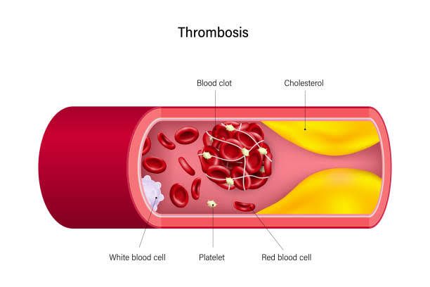
Maintaining the fluidity of blood while being poised to stop bleeding rapidly is a fundamental paradox of the circulatory system. This delicate balance, crucial for life, is orchestrated by a complex interplay of cells and proteins. At the forefront of maintaining blood fluidity in healthy vessels is the endothelium – the single layer of cells lining the inner surface of blood vessels.
The Normal Endothelium – A Dynamic Anticoagulant Surface
The endothelium is far more than a simple barrier; it is a highly active metabolic organ playing a pivotal role in vascular health and preventing thrombosis (unwanted clot formation). In its normal, healthy state, the endothelium actively inhibits coagulation through multiple mechanisms:
- Providing a Physical Barrier: The smooth, continuous surface of the intact endothelium prevents circulating blood cells, particularly platelets, and coagulation factors from coming into contact with the underlying pro-thrombotic subendothelial matrix (collagen, tissue factor). Damage to this layer immediately exposes these pro-thrombotic elements, initiating the coagulation cascade and platelet activation.
- Releasing Vasodilators and Platelet Inhibitors: Healthy endothelial cells produce and release potent molecules that counteract platelet aggregation and cause vasodilation, promoting smooth blood flow and reducing the likelihood of clot formation. Key among these are:
- Nitric Oxide (NO): A powerful vasodilator and inhibitor of platelet adhesion and aggregation.
- Prostacyclin (PGI₂): Another potent vasodilator and inhibitor of platelet activation and aggregation. These molecules work synergistically to keep blood flowing freely.
- Expressing Anticoagulant Molecules on its Surface: The endothelial surface is studded with molecules that actively inhibit the coagulation cascade:
- Heparan Sulfate: A glycosaminoglycan present on the luminal surface that acts as a cofactor for Antithrombin III (ATIII). By binding to both ATIII and certain coagulation factors, heparan sulfate enhances the inhibitory activity of ATIII by several thousandfold, particularly against Thrombin and Factor Xa.
- Thrombomodulin: This transmembrane protein binds Thrombin, fundamentally changing Thrombin’s activity. Instead of cleaving fibrinogen to fibrin (its pro-coagulant role), Thrombin bound to thrombomodulin becomes a potent activator of Protein C. This interaction is a crucial link between the coagulation cascade and the Protein C anticoagulant pathway.
- Producing and Secreting Fibrinolytic Regulators: While not directly anticoagulant, the endothelium contributes to fibrinolysis (the breakdown of clots). Endothelial cells are a primary source of:
- Tissue Plasminogen Activator (t-PA): A key enzyme that converts plasminogen into plasmin, the primary enzyme responsible for degrading fibrin clots.
- Plasminogen Activator Inhibitor-1 (PAI-1): An inhibitor of t-PA (and urokinase-type plasminogen activator, u-PA). The balance between t-PA and PAI-1 influences the rate of fibrinolysis. Under normal conditions, the presence of t-PA contributes to maintaining fluidity by dissolving small fibrin deposits, while PAI-1 acts as a brake, preventing excessive fibrinolysis.
In summary, the normal endothelium actively maintains an antithrombotic state through physical separation, release of inhibitory mediators, expression of anticoagulant cofactors, and modulation of fibrinolysis.
Key Players in the Anticoagulation Cascade – ATIII, Protein C, and Protein S
Beyond the endothelial surface, several proteins circulating in the blood or bound to membranes play critical roles in inhibiting the coagulation cascade. The most important are Antithrombin III, Protein C, and Protein S.
- Antithrombin III (ATIII):
- What it is: ATIII is a small glycoprotein synthesized primarily in the liver, circulating in plasma. It belongs to the serine protease inhibitor (serpin) family.
- How it works: ATIII is a potent inhibitor of key enzymes in the coagulation cascade, primarily Thrombin (Factor IIa) and Factor Xa. It also inhibits Factors IXa, XIa, and XIIa to a lesser extent. ATIII works by forming a stable, irreversible complex with the target protease, effectively neutralizing its enzymatic activity.
- Enhancement by Heparin/Heparan Sulfate: The inhibitory activity of ATIII is dramatically accelerated by binding to heparin or heparan sulfate. These negatively charged molecules act as templates, binding both ATIII and its target protease (especially Thrombin and Factor Xa) and facilitating their interaction. This catalytic effect explains the therapeutic use of heparin as an anticoagulant and highlights the importance of endothelial heparan sulfate in physiological anticoagulation.
- Protein C:
- What it is: Protein C is a vitamin K-dependent protein zymogen (inactive precursor) synthesized in the liver.
- Activation: Protein C is activated on the surface of endothelial cells. Thrombin, upon binding to Thrombomodulin on the endothelial surface, undergoes a conformational change. This Thrombin-Thrombomodulin complex then efficiently activates Protein C to Activated Protein C (APC).
- What it does: APC is a powerful serine protease that, in the presence of its cofactor Protein S and an acidic phospholipid surface (often provided by activated platelets or cell membranes), degrades the activated forms of Factor V (Factor Va) and Factor VIII (Factor VIIIa). Factors Va and VIIIa are crucial cofactors in the coagulation cascade (Factor Va accelerates prothrombin activation by Factor Xa, and Factor VIIIa accelerates Factor X activation by Factor IXa). By inactivating these cofactors, APC effectively dampens down Thrombin generation and clot formation.
- Protein S:
- What it is: Protein S is another vitamin K-dependent protein synthesized in the liver, endothelial cells, and megakaryocytes. It circulates in plasma in two forms: bound to C4b-binding protein and free.
- Role: Only the free form of Protein S functions as a cofactor for Activated Protein C (APC). Protein S enhances the binding of APC to cell membranes and increases the efficiency of APC’s proteolytic degradation of Factors Va and VIIIa. It essentially presents the substrates (Va and VIIIa) to APC in an optimal configuration for inactivation.
These three proteins (ATIII, Protein C, and Protein S) represent major negative regulators of the coagulation cascade, acting at critical points to prevent excessive or unwanted clot formation.
The Pathogenesis of Activated Protein C (APC) Resistance
Activated Protein C (APC) Resistance is a condition where the coagulation system is less responsive to the inhibitory effects of APC. This means that Factors Va and VIIIa are not inactivated efficiently by APC, leading to prolonged activity of these cofactors and an increased tendency for thrombin generation and thrombosis.
- Definition: APC Resistance is functionally defined as a poor anticoagulant response to APC in a plasma-based clotting assay.
- Most Common Cause: Factor V Leiden Mutation: The vast majority (90-95%) of cases of APC Resistance are due to a specific genetic mutation in the gene for Factor V, known as the Factor V Leiden mutation.
- Genetic Basis: This is a single point mutation (Guanine to Adenine substitution) in the Factor V gene at nucleotide position 1691.
- Protein Effect: This mutation results in a single amino acid substitution in the Factor V protein, specifically changing Arginine at position 506 to Glutamine (Arg506Gln).
- Functional Consequence: Arginine at position 506 is one of the primary cleavage sites where Activated Protein C (APC) normally cleaves and inactivates Factor Va. The substitution to Glutamine at this position renders Factor Va resistant to cleavage by APC. While APC can still cleave Factor Va at other sites (like Arg306), the cleavage at 506 is significantly impaired.
- Outcome: This leads to a circulating Factor Va molecule that persists in its activated state for a longer duration than normal Factor Va, promoting extended thrombin generation and increasing the risk of venous thromboembolism (VTE), such as deep vein thrombosis (DVT) and pulmonary embolism (PE).
- Inheritance: Factor V Leiden is inherited in an autosomal dominant pattern. Individuals can be heterozygous (one copy of the mutated gene) or homozygous (two copies). Homozygotes have a higher degree of APC resistance and a significantly increased thrombotic risk compared to heterozygotes.
- Other Causes: While Factor V Leiden is the main culprit, other less common factors can contribute to APC resistance, including certain mutations in Factor V or Factor VIII, or elevated levels of Factor VIII. However, the Factor V Leiden mutation is the prototype and by far the most clinically significant cause.
Understanding APC resistance, particularly the mechanism of Factor V Leiden, is crucial because it is the most common inherited risk factor for venous thrombosis.
Common Conditions Associated with Primary and Acquired Thrombosis
Thrombotic events can occur due to inherited predispositions (primary or genetic thrombophilia) or acquired conditions that increase the likelihood of clotting. Often, a thrombotic event results from an interaction between one or more inherited risk factors and one or more acquired risk factors (the “multiple hit” hypothesis).
Primary (Genetic) Thrombosis Conditions (Inherited Thrombophilias):
These are genetic disorders that increase the risk of thrombosis, typically venous thrombosis.
- Factor V Leiden Mutation: (As discussed in Step 3) – Most common inherited thrombophilia.
- Prothrombin Gene Mutation (G20210A): A mutation in the gene for Prothrombin (Factor II) resulting in higher circulating levels of prothrombin, leading to increased thrombin generation. This is the second most common inherited thrombophilia.
- Antithrombin Deficiency: Genetic mutations leading to reduced levels or impaired function of Antithrombin III. Can be Type I (reduced quantity) or Type II (reduced function).
- Protein C Deficiency: Genetic mutations leading to reduced levels or impaired function of Protein C. Can be Type I (quantitative) or Type II (qualitative).
- Protein S Deficiency: Genetic mutations leading to reduced levels or impaired function of Protein S. Can be Type I (quantitative deficiency of total and free Protein S), Type II (qualitative defect in function), or Type III (quantitative deficiency of free Protein S with normal total).
- Other Rare Inherited Thrombophilias: Less common genetic defects including dysfibrinogenemia (abnormal fibrinogen), elevated levels of Factor VIII, IX, or XI, and defects in fibrinolysis (e.g., plasminogen deficiency, PAI-1 overexpression, t-PA deficiency – though defects in natural inhibitors like ATIII, Protein C, S are generally more potent risk factors).
Acquired Thrombosis Conditions:
These are conditions or circumstances that develop during a person’s lifetime and increase thrombotic risk.
- Immobility/Venous Stasis: Periods of prolonged immobility (e.g., long flights/car rides, bed rest, hospitalization, paralysis) lead to slow blood flow in veins, promoting clot formation.
- Surgery and Trauma: Especially orthopedic surgery (hip/knee replacement), abdominal surgery, and major trauma. Tissue injury releases pro-coagulants, and immobility often follows.
- Malignancy (Cancer): Many cancers are associated with a hypercoagulable state due to tumor-released pro-coagulants, inflammation, immobility, and treatments. This is often referred to as Trousseau’s Syndrome.
- Pregnancy and Puerperium (Postpartum Period): Hormonal changes, venous stasis from uterine compression, and pro-coagulant factor increases contribute to a significantly increased risk.
- Obesity: Associated with chronic low-grade inflammation, impaired fibrinolysis, and sometimes reduced mobility.
- Oral Contraceptives (OCPs) and Estrogen/Hormone Replacement Therapy (HRT): Estrogens can increase the synthesis of certain clotting factors and decrease levels of some anticoagulants. The risk varies depending on the type and dose of hormone.
- Antiphospholipid Syndrome (APS): An autoimmune disorder characterized by the presence of antiphospholipid antibodies (like lupus anticoagulant, anticardiolipin antibodies, anti-beta2-glycoprotein I antibodies) and a history of thrombosis (arterial or venous) and/or recurrent miscarriages. This is a significant cause of acquired thrombophilia.
- Heparin-Induced Thrombocytopenia (HIT): A serious, antibody-mediated reaction to heparin that paradoxically causes platelet activation and a high risk of thrombosis.
- Myeloproliferative Neoplasms (MPNs): Conditions like Polycythemia Vera and Essential Thrombocythemia involve excessive production of blood cells, leading to increased blood viscosity and platelet anomalies, increasing thrombotic risk.
- Inflammatory Conditions: Systemic inflammation (e.g., sepsis, inflammatory bowel disease, autoimmune diseases) can activate the coagulation system.
- Age: Risk of thrombosis increases with age.
- Smoking: Damages endothelium and promotes platelet aggregation.
- Certain Medical Conditions: Heart failure, nephrotic syndrome, paroxysmal nocturnal hemoglobinuria (PNH).
It is important to note that many thrombotic events occur in individuals with multiple risk factors simultaneously (e.g., an individual with Factor V Leiden taking OCPs who undergoes surgery).
Conclusion
The normal endothelium is a cornerstone of physiological anticoagulation, actively preventing thrombosis through a combination of physical, chemical, and enzymatic mechanisms. This intrinsic antithrombotic state is powerfully supplemented by circulating inhibitors like Antithrombin III, the Protein C system (involving Protein C, Protein S, and the endothelial cofactor Thrombomodulin), which collectively act as brakes on the coagulation cascade.
Disruptions to this delicate balance, whether due to inherited defects (like the Factor V Leiden mutation causing APC resistance) or acquired conditions (ranging from immobility and surgery to cancer and autoimmune disorders), can shift the hemostatic system towards a pro-thrombotic state, increasing the risk of potentially life-threatening events like deep vein thrombosis and pulmonary embolism. A comprehensive understanding of these mechanisms is essential for diagnosing, preventing, and managing thrombotic disorders.


