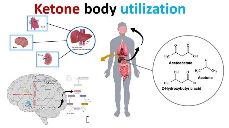
Ketone bodies are water-soluble molecules produced by the liver from fatty acids during periods of low carbohydrate availability. They serve as an important alternative fuel source for various tissues, particularly the brain, when glucose is scarce. While their production is a normal metabolic adaptation, uncontrolled formation can lead to serious clinical conditions.
Types of Ketone Bodies and Conditions During Which They Are Formed
The term “ketone bodies” typically refers to three compounds:
- Acetoacetate (Acetoacetic acid): The primary ketone body formed directly from the breakdown of HMG-CoA in the liver. It is a beta-keto acid.
- Beta-hydroxybutyrate (β-hydroxybutyric acid): While technically not a ketone (it contains a hydroxyl group, not a ketone group), it is metabolically interconverted with acetoacetate and is the most abundant ketone body in the blood during ketosis or ketoacidosis. It is formed reversibly from acetoacetate via the enzyme beta-hydroxybutyrate dehydrogenase, a process consuming NADH.
- Acetone: A volatile compound produced by the spontaneous, non-enzymatic decarboxylation of acetoacetate. It is not metabolized for energy and is excreted via the lungs (giving a characteristic “fruity” odor to the breath) and urine. Its concentration is generally much lower than acetoacetate and beta-hydroxybutyrate.
Ketone bodies are primarily formed in the mitochondria of liver cells under conditions where the body’s primary fuel source, glucose, is either unavailable or cannot be effectively utilized. These conditions are characterized by a low insulin-to-glucagon ratio, which promotes the mobilization of fatty acids from adipose tissue and directs liver metabolism away from glucose synthesis and storage towards fatty acid oxidation and ketogenesis.
Key conditions leading to increased ketone body formation include:
- Prolonged Fasting or Starvation: When energy intake is insufficient and liver glycogen stores are depleted (typically after 12-24 hours of fasting), the body relies heavily on fatty acid oxidation for energy. The large flux of fatty acids into the liver leads to increased acetyl-CoA production, which, coupled with low levels of intermediates of the citric acid cycle (diverted for gluconeogenesis), favors the shunting of acetyl-CoA into the ketogenic pathway.
- Low-Carbohydrate, High-Fat Diets (e.g., Ketogenic Diets): Diets severely restricting carbohydrate intake mimic the metabolic state of prolonged fasting. With minimal glucose available from the diet, insulin levels remain low, while glucagon and other counter-regulatory hormones are relatively elevated. This stimulates fatty acid mobilization and hepatic ketogenesis, leading to a state of nutritional ketosis, where ketone bodies become a significant fuel source.
- Uncontrolled Diabetes Mellitus (especially Type 1): This is the most common pathological cause of severe ketoacidosis (specifically Diabetic Ketoacidosis). In the absence of sufficient insulin, glucose cannot enter many cells, leading to hyperglycemia despite cellular energy starvation. The lack of insulin also fails to suppress hormone-sensitive lipase in adipose tissue, resulting in massive lipolysis and release of free fatty acids. These fatty acids are taken up by the liver and converted into ketone bodies at a high rate. The critical difference from nutritional ketosis is the rate of production and the accompanying metabolic derangements (severe hyperglycemia, dehydration, electrolyte imbalance).
- Prolonged Strenuous Exercise: In prolonged, intense exercise, especially if carbohydrate intake is insufficient, hepatic glycogen can become depleted, and fatty acid oxidation increases, potentially leading to a mild increase in ketone body levels.
- Alcoholic Ketoacidosis: Occurs in chronic excessive alcohol users, often exacerbated by poor nutritional intake, vomiting, and depletion of glycogen stores. Ethanol metabolism alters the NAD+/NADH ratio, favoring the conversion of acetoacetate to beta-hydroxybutyrate and contributing to acidosis.
Important Steps in Synthesis of Ketone Bodies (Ketogenesis) and the Rate-Limiting Step
Ketogenesis occurs exclusively in the mitochondria of liver cells. The process begins with acetyl-CoA, primarily derived from the beta-oxidation of fatty acids.
The key enzymatic steps are:
- Formation of Acetoacetyl-CoA: Two molecules of acetyl-CoA condense to form acetoacetyl-CoA. This reaction is catalyzed by Thiolase (specifically, mitochondrial thiolase, which is also involved in the final step of beta-oxidation).
- 2 Acetyl-CoA → Acetoacetyl-CoA + CoA
- Formation of HMG-CoA: Acetoacetyl-CoA reacts with a third molecule of acetyl-CoA to form beta-hydroxy-beta-methylglutaryl-CoA (HMG-CoA). This reaction is catalyzed by HMG-CoA Synthase (specifically, mitochondrial HMG-CoA Synthase).
- Acetoacetyl-CoA + Acetyl-CoA + H₂O → HMG-CoA + CoA
- Cleavage of HMG-CoA to Acetoacetate: HMG-CoA is cleaved to form acetoacetate and acetyl-CoA. This reaction is catalyzed by HMG-CoA Lyase.
- HMG-CoA → Acetoacetate + Acetyl-CoA
Acetoacetate is the first true ketone body produced. Once formed, acetoacetate can undergo further transformations:
- Reduction to Beta-hydroxybutyrate: Acetoacetate is reversibly reduced to beta-hydroxybutyrate by Beta-hydroxybutyrate Dehydrogenase, utilizing NADH as a cofactor. The ratio of beta-hydroxybutyrate to acetoacetate in the blood is determined by the mitochondrial NAD+/NADH ratio, which is high during active fatty acid oxidation.
- Acetoacetate + NADH + H⁺ ⇌ Beta-hydroxybutyrate + NAD⁺
- Spontaneous Decarboxylation to Acetone: Acetoacetate can non-enzymatically lose a carboxyl group to form acetone and carbon dioxide, particularly at lower pH values.
- Acetoacetate → Acetone + CO₂
The Rate-Limiting Step of Ketone Body Synthesis:
The rate-limiting step of ketogenesis is the reaction catalyzed by mitochondrial HMG-CoA Synthase. This enzyme’s activity is highly regulated. Its expression and activity are increased under conditions favoring ketogenesis (e.g., fasting, diabetes) due to hormonal signals (low insulin, high glucagon) that promote transcription factors involved in fatty acid oxidation and ketogenesis. The availability of its substrates, acetoacetyl-CoA and acetyl-CoA, is also crucial, as these are increased by high rates of fatty acid oxidation. The activity of HMG-CoA Synthase ultimately determines the flux through the ketogenic pathway.
Steps in Utilization of Ketone Bodies (Ketolysis) and the Organs Which Use It
Ketone bodies serve as an alternative fuel source for many extrahepatic tissues, including the brain, heart, skeletal muscle, and renal cortex. The liver produces ketone bodies but cannot utilize them for energy because it lacks a key enzyme required for ketolysis.
The process of ketone body utilization, or ketolysis, occurs in the mitochondria of these peripheral tissues. The steps are essentially the reverse of synthesis, converting ketone bodies back into acetyl-CoA for entry into the citric acid cycle:
- Conversion of Beta-hydroxybutyrate to Acetoacetate: Beta-hydroxybutyrate is first oxidized back to acetoacetate by Beta-hydroxybutyrate Dehydrogenase, using NAD⁺ as the electron acceptor. This is the reverse of the reaction in the liver.
- Beta-hydroxybutyrate + NAD⁺ ⇌ Acetoacetate + NADH + H⁺ (Note: If starting with acetoacetate, this step is skipped).
- Activation of Acetoacetate: Acetoacetate is activated by transferring a CoA group from succinyl-CoA to form acetoacetyl-CoA. This reaction is catalyzed by Succinyl-CoA:3-ketoacid CoA transferase, also known as Thiophorase.
- Acetoacetate + Succinyl-CoA → Acetoacetyl-CoA + Succinate
- Cleavage of Acetoacetyl-CoA: Acetoacetyl-CoA is cleaved into two molecules of acetyl-CoA by Thiolase.
- Acetoacetyl-CoA + CoA → 2 Acetyl-CoA
The two molecules of acetyl-CoA produced can then enter the citric acid cycle (TCA cycle) to be completely oxidized, generating ATP via oxidative phosphorylation.
Organs That Use Ketone Bodies:
- Brain: The brain is a major consumer of glucose, but during prolonged fasting or starvation, it can adapt to utilize ketone bodies (primarily beta-hydroxybutyrate and acetoacetate) as a significant fuel source, sparing glucose for other tissues like red blood cells. Ketones can meet up to 60-70% of the brain’s energy needs during extended periods of carbohydrate deprivation.
- Heart Muscle: Cardiac muscle is a highly aerobic tissue and readily utilizes fatty acids and ketone bodies for energy, often preferring them over glucose when available.
- Skeletal Muscle: Skeletal muscle can utilize ketone bodies for energy, particularly during prolonged exercise or fasting.
- Renal Cortex: The kidneys can also utilize ketone bodies as fuel.
- Adrenal Cortex, Pancreas, Adipose Tissue: These tissues can also utilize ketone bodies to varying degrees.
Organs That Do NOT Use Ketone Bodies:
- Liver: The liver produces ketone bodies but lacks the enzyme Thiophorase (Succinyl-CoA:3-ketoacid CoA transferase) required for step 2 of ketolysis. This enzyme’s absence ensures that the liver acts solely as a producer and exporter of ketone bodies, preventing a futile cycle where it would consume the fuel it is producing for other tissues.
- Red Blood Cells: Red blood cells lack mitochondria and therefore cannot perform the metabolic reactions required for ketolysis (or fatty acid oxidation or TCA cycle activity). They rely solely on glucose for energy via anaerobic glycolysis.
The rate-limiting step of ketone body utilization is the thiophorase reaction (catalyzed by Succinyl-CoA:3-ketoacid CoA transferase), as this enzyme is required to activate acetoacetate for subsequent cleavage.
Pathogenesis, Clinical Presentation, and Management of Diabetic Ketoacidosis (DKA)
Diabetic Ketoacidosis (DKA) is a severe, life-threatening complication most commonly seen in patients with Type 1 Diabetes Mellitus, but it can also occur in Type 2 Diabetes under conditions of extreme stress (e.g., severe infection, trauma).
Pathogenesis:
DKA is fundamentally caused by an absolute or relative deficiency of insulin coupled with an excess of counter-regulatory hormones (glucagon, catecholamines, cortisol, growth hormone). This hormonal imbalance leads to a cascade of metabolic derangements:
- Reduced Glucose Utilization & Increased Glucose Production: Insufficient insulin severely impairs glucose uptake by insulin-sensitive tissues (muscle, adipose tissue). Simultaneously, the high glucagon-to-insulin ratio promotes hepatic glycogenolysis and gluconeogenesis. The net result is severe hyperglycemia.
- Increased Lipolysis: Insulin is a potent inhibitor of hormone-sensitive lipase in adipose tissue. Its deficiency leads to uncontrolled breakdown of triglycerides into free fatty acids and glycerol. Large quantities of free fatty acids are released into the circulation and transported to the liver.
- Accelerated Ketogenesis: In the liver, the high flux of fatty acids entering the mitochondria undergoes beta-oxidation, producing massive amounts of acetyl-CoA. The hormonal milieu (low insulin, high glucagon) inhibits key enzymes of the TCA cycle and fatty acid synthesis while activating enzymes of the ketogenic pathway (like mitochondrial HMG-CoA Synthase and CPT-1, which facilitates fatty acid entry into mitochondria). This shunts the excess acetyl-CoA into ketone body synthesis, leading to a rapid and excessive production of acetoacetate and beta-hydroxybutyrate.
- Ketoacidosis: Acetoacetate and beta-hydroxybutyrate are organic acids. Their rapid accumulation in the blood overwhelms the body’s buffering capacity, leading to a significant drop in blood pH (metabolic acidosis). Beta-hydroxybutyrate accounts for the majority of the excess acid due to the higher NADH/NAD+ ratio in the liver during active ketogenesis.
- Osmotic Diuresis and Dehydration: The severe hyperglycemia results in an osmotic effect, drawing water from cells into the extracellular space. When the blood glucose level exceeds the renal threshold (typically ~180 mg/dL), glucose spills into the urine, creating an osmotic gradient that pulls large amounts of water and electrolytes (sodium, potassium, phosphate, magnesium) with it. This leads to polyuria, profound dehydration, and significant electrolyte imbalances.
- Electrolyte Imbalance (especially Potassium): Despite total body potassium depletion due to diuresis and vomiting, serum potassium levels may initially appear normal or even elevated due to the shift of potassium out of cells caused by acidosis and insulin deficiency. However, with insulin therapy and correction of acidosis, potassium rapidly shifts back into cells, leading to potentially dangerous hypokalemia if not preemptively replaced.
Clinical Presentation:
The symptoms of DKA typically develop over hours to a couple of days and are often preceded by a trigger (e.g., infection, missed insulin doses, new diabetes diagnosis, stress). Key clinical manifestations include:
- Symptoms of Hyperglycemia: Polyuria (frequent urination), polydipsia (excessive thirst), polyphagia (increased hunger).
- Gastrointestinal Symptoms: Nausea, vomiting, and severe abdominal pain (often mimicking acute surgical abdomen).
- Acidosis Symptoms:
- Kussmaul Respirations: Deep, rapid breathing representing the body’s attempt to blow off CO₂ to compensate for the metabolic acidosis.
- Fruity Odor on Breath: Due to the exhalation of acetone.
- Dehydration & Hypovolemia: Dry mucous membranes, decreased skin turgor, sunken eyes, tachycardia, hypotension, reduced urine output (later stage).
- Neurological Symptoms: Fatigue, lethargy, confusion, stupor, coma (in severe cases).
- Other: Generalized weakness, malaise, hypothermia (rare, poor prognostic sign).
Diagnosis: Diagnosis is based on the presence of the following triad:
- Hyperglycemia (blood glucose typically > 250 mg/dL, though lower in rare “euglycemic DKA”).
- Metabolic acidosis (arterial pH < 7.3, serum bicarbonate < 18 mEq/L).
- Presence of Ketones (moderate to large ketones in urine or blood).
Management:
Management of DKA is a medical emergency requiring prompt and aggressive treatment in a hospital setting. The cornerstones of therapy are:
- Fluid Resuscitation: This is the immediate priority to correct dehydration and restore circulating volume. Intravenous fluids (usually 0.9% saline initially, switching to 0.45% saline once hemodynamically stable and sodium levels permit) are administered rapidly. Fluid resuscitation improves tissue perfusion and helps lower glucose and ketone levels by improving glomerular filtration and renal excretion.
- Insulin Therapy: Intravenous regular insulin infusion is started at a continuous low dose. Insulin works by:
- Inhibiting further lipolysis and ketogenesis (most crucial effect in reversing acidosis).
- Promoting glucose uptake by cells, lowering blood glucose.
- Driving potassium back into cells. (Insulin is typically continued until the acidosis resolves, even if glucose normalizes, at which point dextrose must be added to the IV fluids to prevent hypoglycemia).
- Electrolyte Replacement: Potassium is the most critical electrolyte to monitor and replace. Despite initial normal or high serum levels, patients are total-body potassium depleted. As insulin is given, potassium rapidly shifts back into cells, potentially causing lethal hypokalemia. Potassium replacement is usually initiated once serum potassium levels are normal or low and adequate urine output is established. Phosphate and magnesium may also require replacement.
- Bicarbonate Administration: Generally not recommended for mild to moderate DKA due to potential adverse effects (worsening hypokalemia, paradoxical central nervous system acidosis, prolonged lactate acidosis). It is typically reserved for severe acidosis (pH < 6.9 or 7.0) that is refractory to initial fluid and insulin therapy, or in cases of severe hyperkalemia or hemodynamic instability unresponsive to other measures.
- Identification and Treatment of Precipitating Factors: It is essential to search for and treat the underlying cause of DKA (e.g., infection with antibiotics, myocardial infarction, pancreatitis).
Monitoring of metabolic status (blood glucose, electrolytes, beta-hydroxybutyrate, arterial or venous blood gases) is crucial to guide therapy and assess response. DKA is a serious condition requiring meticulous monitoring and adjustment of treatment based on serial laboratory values and the patient’s clinical status.
In summary, ketone bodies are vital alternative fuels during periods of low carbohydrate availability, produced by the liver. While physiological ketosis is adaptive, excessive, uncontrolled production, as seen in conditions like Diabetic Ketoacidosis, leads to dangerous metabolic acidosis requiring urgent medical intervention. Understanding the pathways of ketogenesis and ketolysis is fundamental to appreciating their role in normal metabolism and their critical involvement in disease states.


