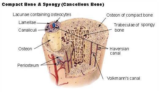
Bone tissue is a dynamic, living connective tissue that serves multiple critical functions, including structural support, protection of vital organs, facilitation of movement, storage of minerals (primarily calcium and phosphate), and housing the bone marrow for hematopoiesis. Its remarkable strength and resilience are attributed to its unique biochemical composition and highly organized structure. Unlike purely organic tissues, bone is a composite material, combining an organic matrix with an inorganic mineral phase. Understanding the biochemical intricacies of bone is essential for comprehending its normal physiology, disease states (like osteoporosis or rickets), and the mechanisms of repair and regeneration.
Here is a breakdown of the key biochemical aspects of bone tissue:
Biochemical Structure of Bone Tissue: The Collagen Matrix and the Hydroxyapatite Cement
Bone tissue is composed of two primary components: an organic matrix and an inorganic mineral phase. The synergy between these two components provides bone with both flexibility (from the organic matrix) and rigidity and compressive strength (from the inorganic mineral).
-
- Organic Matrix: This constitutes approximately 20-30% of the bone’s dry weight. The predominant component (>90%) of the organic matrix is Type I Collagen.
- Type I Collagen Structure: Collagen is a fibrous protein characterized by its unique triple-helical structure. Three polypeptide chains, known as alpha chains, wrap around each other to form a rigid, rope-like structure. In Type I collagen, there are two alpha1(I) chains and one alpha2(I) chain. These triple helices then assemble into larger structures:
- Fibrils: Collagen triple helices self-assemble into microfibrils, which then aggregate into larger collagen fibrils. The fibrils exhibit a characteristic staggered arrangement with “gap” and “overlap” zones, visible under electron microscopy as a 67 nm repeat banding pattern. These gaps are crucial as primary sites for initial mineral deposition.
- Fibers: Bundles of collagen fibrils form collagen fibers, which are macroscopic structures providing the framework for bone tissue.
- Function of Collagen: The collagen matrix provides bone with its tensile strength and flexibility, preventing it from becoming brittle and easily fractured. It acts as a scaffold upon which the mineral phase is deposited. The specific arrangement and cross-linking of collagen fibers are vital for bone’s mechanical properties.
- Type I Collagen Structure: Collagen is a fibrous protein characterized by its unique triple-helical structure. Three polypeptide chains, known as alpha chains, wrap around each other to form a rigid, rope-like structure. In Type I collagen, there are two alpha1(I) chains and one alpha2(I) chain. These triple helices then assemble into larger structures:
- Inorganic Mineral Phase: This constitutes approximately 60-70% of the bone’s dry weight and provides bone with its hardness and rigidity. The primary mineral component is a form of calcium phosphate known as Hydroxyapatite (HA).
- Hydroxyapatite Composition: The ideal chemical formula for hydroxyapatite is Ca10(PO4)6(OH)2. However, in biological bone, the composition is non-stoichiometric and impure. It contains substitutions, such as carbonate (CO3) and fluoride (F), which can replace phosphate and hydroxyl groups, respectively, and other ions like magnesium (Mg) and sodium (Na) substituting for calcium. This biological apatite is often referred to as bone apatite.
- HA Crystal Structure: HA forms tiny, plate-like or needle-like crystals, typically around 20-80 nm long, 10-30 nm wide, and 3-10 nm thick. These crystals are primarily deposited within and on the surface of the collagen fibrils, particularly within the “gap” zones of the staggered collagen arrangement.
- Function of Hydroxyapatite: The HA crystals provide bone with its compressive strength and rigidity, allowing it to bear weight and resist deformation. The intimate association between the mineral crystals and the collagen matrix is critical; the organic framework reinforces the brittle mineral, while the mineral stiffens the flexible collagen. This composite structure is analogous to reinforced concrete, where steel bars (collagen) are embedded in concrete (hydroxyapatite).
- Organic Matrix: This constitutes approximately 20-30% of the bone’s dry weight. The predominant component (>90%) of the organic matrix is Type I Collagen.
Bone Matrix Proteins (Non-Collagenous Proteins) and Their Functions
While Type I collagen forms the bulk of the organic matrix, the remaining 10% consists of a diverse array of non-collagenous proteins (NCPs). These NCPs, synthesized primarily by osteoblasts, play crucial roles in regulating bone formation, mineralization, cell adhesion, and communication within the bone microenvironment. Key bone matrix proteins include:
- Osteocalcin (Bone Gla Protein): One of the most abundant NCPs. It contains gamma-carboxyglutamic acid (Gla) residues, which are formed by vitamin K-dependent carboxylation and enable binding to calcium ions.
- Function: Heavily involved in regulating bone mineralization, specifically by binding to hydroxyapatite crystals. It is a marker of osteoblast activity. Emerging evidence suggests it may function as a hormone, influencing glucose metabolism and male fertility.
- Osteopontin (Secreted Phosphoprotein 1 – SPP1): A highly phosphorylated acidic glycoprotein containing Arg-Gly-Asp (RGD) sequences.
- Function: Plays multiple roles. The RGD motif allows it to bind to integrin receptors on cells (like osteoclasts and osteoblasts), mediating cell adhesion and signaling. It binds strongly to hydroxyapatite and can act as both a negative regulator (inhibitor) and, in specific contexts, a positive nucleator of mineralization. Involved in bone modeling and remodeling, particularly at cement lines and resorption pits. Also has roles in the immune response.
- Bone Sialoprotein (BSP): Another highly acidic, phosphorylated glycoprotein, also containing RGD sequences. More restricted expression than osteopontin.
- Function: Considered a potent nucleator of hydroxyapatite formation, potentially initiating mineralization in specific locations within the matrix. Like osteopontin, it mediates cell attachment via integrins.
- Osteonectin (SPARC – Secreted Protein Acidic and Rich in Cysteine): A glycoprotein that binds to both collagen and hydroxyapatite.
- Function: Proposed roles include regulating collagen assembly, modulating cell proliferation, and influencing mineralization by potentially inhibiting excessive crystal growth or promoting nucleation depending on concentration and context. It acts as a bridge between the organic and inorganic phases.
- Proteoglycans (e.g., Decorin, Biglycan, Lumican): Glycoproteins with heavily glycosylated side chains (glycosaminoglycans – GAGs) that carry a high negative charge.
- Function: Bind large amounts of water, contributing to tissue hydration and resilience. They interact with collagen fibrils, influencing their diameter and organization. They also bind to various growth factors and cytokines, regulating their activity and availability. Some proteoglycans can interact with mineral and influence mineralization.
- Growth Factors and Cytokines: Bone matrix serves as a reservoir for numerous growth factors (e.g., Insulin-like Growth Factors – IGF-I, IGF-II; Bone Morphogenetic Proteins – BMPs; Transforming Growth Factor-beta – TGF-beta) and cytokines.
- Function: Released during bone resorption, these molecules regulate the activity of osteoblasts, osteoclasts, and other local cells, playing critical roles in bone remodeling, repair, and development. BMPs, in particular, can induce differentiation of mesenchymal stem cells into osteoblasts.
- Other Proteins: Including fibronectin, thrombospondins, and enzymes. These proteins also play roles in cell adhesion, matrix organization, and local regulation of matrix turnover.
Collectively, these NCPs fine-tune the mechanical properties of the bone matrix, precisely control the timing and location of mineralization, recruit and regulate bone cells (osteoblasts, osteocytes, osteoclasts), and store and release signaling molecules.
Composition of Calcified Tissues, Calcification in Bones and Teeth, and Formation of Hydroxyapatite
Calcified tissues are biological tissues that have been hardened by the deposition of mineral crystals, primarily forms of calcium phosphate. The main calcified tissues in vertebrates are bone, dentin, enamel, and cementum (all part of the skeletal and dental systems). While they share the presence of mineral, their composition and the process of mineralization vary.
-
- Composition Differences:
- Bone: ~60-70% mineral (biological apatite), ~20-30% organic matrix (mainly Type I collagen and NCPs), ~5-10% water.
- Dentin: Similar to bone in mineral and organic content (~70% mineral, ~20% organic – mainly Type I collagen), but formed by odontoblasts and has a tubular structure.
- Cementum: Similar to bone and dentin (~50% mineral, ~50% organic – mainly Type I collagen), covering the tooth root.
- Enamel: Unique in its high mineral content (~97% mineral, mainly biological apatite), making it the hardest substance in the human body. Its organic matrix (<1%) is non-collagenous, consisting of specialized proteins like amelogenins and enamelins, secreted by ameloblasts.
- Calcification (Mineralization) Process in Bones and Dentin: This is a highly regulated biological process, not simply passive precipitation of mineral. It primarily occurs within the extracellular matrix laid down by osteoblasts (for bone) and odontoblasts (for dentin). The process can be broadly divided into stages:
- Matrix Vesicle Formation: Osteoblasts/odontoblasts bud off small membrane-bound vesicles called matrix vesicles into the extracellular matrix. These vesicles are crucial initiation sites for calcification.
- Mineral Nucleation: Matrix vesicles accumulate high concentrations of calcium and phosphate ions. They contain proteins (like annexins, alkaline phosphatase) and lipids that facilitate the transport and concentration of these ions. Initial crystals of calcium phosphate (possibly amorphous calcium phosphate or octacalcium phosphate) form within the protected environment of the vesicle. Proteins like BSP are thought to reside around or within these vesicles and act as nucleators.
- Crystal Growth and Propagation: As crystals grow within the matrix vesicles, they eventually rupture the vesicle membrane. The newly formed crystals then propagate outwards, depositing along the collagen fibrils, primarily within the “gap” zones, and on the surface of the fibrils. This organized deposition along the collagen scaffold results in the characteristic structure of bone and dentin.
- Regulation: The process is tightly controlled by local factors. Molecules that inhibit mineralization (like pyrophosphate, PPi) are present and must be degraded or overcome for mineralization to proceed. NCPs promoting nucleation (BSP) or modulating crystal growth (osteocalcin, osteopontin, osteonectin) play critical regulatory roles.
- Calcification in Enamel: This is a distinct process. Ameloblasts secrete a protein matrix (amelogenins, enamelins) that guides the formation of large, highly oriented hydroxyapatite crystals. Unlike bone/dentin, the matrix is largely removed as mineralization matures, resulting in the high mineral content. Matrix vesicles are not involved in enamel mineralization initiation.
- Formation of Hydroxyapatite: Biological hydroxyapatite forms from calcium (Ca2+) and phosphate (Pi) ions in the extracellular fluid. For mineralization to occur, the local concentration of Ca2+ and Pi must reach a state of supersaturation relative to hydroxyapatite. However, extracellular fluid is generally supersaturated with respect to HA, yet spontaneous, widespread precipitation doesn’t occur thanks to inhibitors like pyrophosphate (PPi). Therefore, controlled mineralization requires:
- Sufficient local concentrations of Ca2+ and Pi.
- Presence of nucleation sites (e.g., matrix vesicles, collagen gaps, specific NCPs like BSP).
- Removal or inactivation of mineralization inhibitors in the specific location where mineralization is to occur.
- Composition Differences:
Role of Alkaline Phosphatase, Calcium, and Phosphate.
These three components are fundamental players in the process of bone mineralization.
- Alkaline Phosphatase (ALP):
- Enzyme Activity: ALP is a membrane-bound enzyme found on the surface of osteoblasts and within matrix vesicles. Its primary biochemical function in bone is the hydrolysis of phosphate monoesters.
- Key Substrate: One crucial substrate is pyrophosphate (PPi). PPi is a potent inhibitor of hydroxyapatite crystal nucleation and growth. It binds to potential mineralization sites and prevents Ca/Pi deposition.
- Role in Mineralization: By hydrolyzing PPi into two molecules of inorganic phosphate (Pi), ALP achieves two critical things:
- It removes the local inhibition of mineralization caused by PPi.
- It increases the local concentration of inorganic phosphate, contributing to the supersaturation required for HA formation.
- Clinical Significance: ALP activity is high in actively mineralizing tissue. Serum ALP levels are often used as a marker of bone formation activity. Genetic defects leading to low ALP activity cause hypophosphatasia, a disorder characterized by severe impairment of bone and tooth mineralization.
- Calcium (Ca2+):
- Essential Component: Calcium ions are the major cationic component of hydroxyapatite (Ca10(PO4)6(OH)2).
- Concentration: The concentration of free calcium ions in the extracellular fluid is tightly regulated (normally around 2.2-2.6 mM or 8.8-10.4 mg/dL) because of its critical roles in nerve conduction, muscle contraction, blood clotting, and enzyme activity.
- Role in Mineralization: A sufficient local concentration of Ca2+ is necessary for HA formation. Calcium ions bind to the organic matrix (collagen, NCPs with Gla or phosphate residues), helping organize the mineralization front.
- Phosphate (Pi):
- Essential Component: Phosphate ions (PO43-, existing as HPO42- at physiological pH) are the major anionic component of hydroxyapatite.
- Concentration: Inorganic phosphate concentration in extracellular fluid is also regulated, though with wider fluctuations than calcium (normally around 0.8-1.5 mM or 2.5-4.5 mg/dL in adults, higher in children). Pi is essential for energy metabolism (ATP), nucleic acids, phospholipids, etc.
- Role in Mineralization: A sufficient local concentration of Pi is necessary for HA formation. As mentioned, ALP increases local Pi by hydrolyzing PPi, facilitating supersaturation.
The interplay of these factors is critical: sufficient systemic Ca and Pi levels are needed to provide the building blocks, while local factors like ALP activity and the presence of nucleation sites (matrix vesicles, NCPs) ensure that mineralization occurs specifically within the bone matrix and not in soft tissues.
Role of Vitamin D and 1,25-Dihydroxyvitamin D in Bone Formation and Remodeling
Vitamin D is a steroid hormone precursor vital for calcium and phosphate homeostasis and bone health. It is obtained from the diet (as cholecalciferol, D3, or ergocalciferol, D2) or synthesized in the skin upon exposure to ultraviolet B (UVB) radiation (producing D3).
To become biologically active, vitamin D undergoes two hydroxylation steps:
- In the liver, it is hydroxylated at the 25-position, forming 25-hydroxyvitamin D [25(OH)D], also known as calcidiol.
- In the kidneys, under the influence of Parathyroid Hormone (PTH), 25(OH)D is further hydroxylated at the 1-alpha position by the enzyme 1-alpha-hydroxylase, producing 1,25-dihydroxyvitamin D [1,25(OH)₂D], also known as calcitriol. This is the most biologically active form of vitamin D.
Role in Bone Formation and Remodeling:
- Indirect Effects (Major): The primary role of 1,25(OH)₂D is to increase the availability of calcium and phosphate for mineralization. It achieves this mainly by:
- Increasing Intestinal Absorption: It significantly enhances the absorption of calcium and phosphate from the small intestine, the primary source of these minerals in the body. This ensures that sufficient building blocks are available in the bloodstream to support bone mineralization.
- Direct Effects (Less prominent but significant): 1,25(OH)₂D has receptors (Vitamin D Receptor – VDR) on bone cells (osteoblasts and osteoclasts/their precursors).
- On Osteoblasts: It influences osteoblast differentiation and activity, and stimulates the synthesis of key matrix proteins like osteocalcin.
- On Osteoclasts: While often associated with bone resorption (breaking down bone), 1,25(OH)₂D doesn’t directly stimulate mature osteoclast activity. Instead, it acts on osteoblast lineage cells, stimulating them to produce factors (like RANKL) that promote the differentiation and activity of osteoclast precursors, thereby linking bone formation and resorption during remodeling. This role is crucial for maintaining bone structure and releasing calcium if needed by the body.
Vitamin D deficiency leads to inadequate calcium and phosphate absorption, resulting in impaired mineralization of osteoid (rickets in children, osteomalacia in adults), causing soft and deformed bones.
Calcium and Phosphate Homeostasis
Maintaining stable concentrations of calcium and phosphate in the blood and extracellular fluid is vital for numerous physiological processes, not just bone mineralization. This delicate balance is achieved through a complex hormonal regulatory system primarily involving three hormones:
- Parathyroid Hormone (PTH): Produced by the parathyroid glands. PTH is the primary sensor and regulator of calcium levels. It is released when blood calcium levels fall. Its main actions are:
- On Bone: Stimulates osteoclast activity (via osteoblasts) to increase bone resorption, releasing calcium and phosphate into the blood. This is a relatively rapid way to raise blood calcium.
- On Kidneys: Increases calcium reabsorption from the glomerular filtrate back into the blood, reducing calcium loss in urine. It also inhibits phosphate reabsorption, promoting phosphate excretion in urine. This helps prevent the formation of calcium-phosphate precipitates when both levels are high and ensures a higher calcium-to-phosphate ratio in the blood, facilitating calcium delivery to tissues without premature precipitation. Crucially, PTH stimulates the kidney’s 1-alpha-hydroxylase enzyme, thus increasing the production of active 1,25(OH)₂D.
- 1,25-Dihydroxyvitamin D (Calcitriol): As discussed, its main action is to increase the absorption of both calcium and phosphate from the intestine. It synergizes with PTH in bone resorption and renal calcium reabsorption.
- Calcitonin: Produced by the parafollicular (C) cells of the thyroid gland. Calcitonin is released when blood calcium levels are high. Its main action is to inhibit osteoclast activity, thereby reducing bone resorption and lowering blood calcium. Its physiological role in normal human calcium homeostasis is generally considered less significant than that of PTH and Vitamin D.
Regulation Mechanism: When blood calcium is low, PTH is secreted. PTH acts on bone, kidney, and stimulates 1,25(OH)₂D production. This leads to increased bone resorption, increased calcium reabsorption in the kidney, decreased phosphate reabsorption in the kidney, and increased intestinal absorption of both calcium and phosphate (via 1,25(OH)₂D). The net effect is a rise in blood calcium and a slight decrease or stability in blood phosphate (due to increased renal excretion offsetting increased intestinal absorption and bone release). When blood calcium is high, PTH secretion is suppressed, leading to decreased bone resorption, decreased renal calcium reabsorption, and decreased 1,25(OH)₂D production (reducing intestinal absorption). Calcitonin may be released, providing an additional inhibitory signal to osteoclasts. The net effect is a decrease in blood calcium.
This coordinated interplay between bone metabolism (formation and resorption) and renal and intestinal handling of calcium and phosphate ensures that serum mineral concentrations are maintained within narrow limits, providing the necessary building blocks for bone mineralization while protecting overall physiological function.


