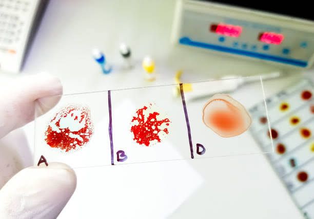
Human blood and tissue types are defined by specific proteins, glycoproteins, or glycolipids (antigens) present on the surface of cells. These antigens play crucial roles in the body’s immune response. Understanding the principles governing these antigen systems is fundamental in various medical fields, including transfusion medicine, transplantation, immunology, and the study of disease associations.
Principles of the ABH Blood Group System
Discovered by Karl Landsteiner in 1900, the ABH system is the most well-known and clinically significant blood group system for transfusion due to the presence of naturally occurring antibodies.
- Antigens:
- The primary ABH antigens are A, B, and H.
- These antigens are complex carbohydrate structures attached to lipids (glycolipids) or proteins (glycoproteins) on the surface of red blood cells (RBCs) and many other tissues and bodily secretions.
- The H antigen is the precursor structure upon which the A and B antigens are built.
- Individuals inherit genes that determine which enzymes are produced to modify the H antigen.
- The A gene produces an enzyme that adds N-acetylgalactosamine to the H structure, creating the A antigen.
- The B gene produces an enzyme that adds D-galactose to the H structure, creating the B antigen.
- The O gene is essentially non-functional and does not add a sugar, leaving the H structure unmodified (resulting in the H antigen remaining as the primary structure).
- The expression of the H antigen itself is controlled by the H gene (also known as FUT1). Almost all individuals have at least one copy of the dominant H allele, ensuring H antigen production. Very rare individuals (<1 in a million) are homozygous for the recessive h allele (hh genotype), resulting in the absence of the H antigen. These individuals have the “Bombay” phenotype and cannot form A or B antigens.
- The expression of ABH antigens in bodily secretions (like saliva, tears, urine) is controlled by the Secretor gene (Se or FUT2). Individuals with at least one dominant Se allele (Secretors) express ABH antigens in their secretions. Those homozygous for the recessive se allele (sese genotype, Non-secretors) do not.
- Antibodies:
- A unique principle of the ABH system is the presence of naturally occurring antibodies in the plasma of individuals.
- These antibodies (Anti-A and Anti-B) develop early in life (usually by 3-6 months of age) and are thought to be stimulated by exposure to similar carbohydrate structures present on common bacteria, viruses, or environmental substances.
- Individuals normally produce antibodies against the ABH antigens they lack on their own red blood cells.
- These antibodies are primarily Immunoglobulin M (IgM) class, which are large pentameric molecules that can efficiently activate the complement system and cause rapid, severe agglutination (clumping) and hemolysis (destruction) of incompatible RBCs.
- Blood Groups and Corresponding Antigens/Antibodies: Based on the combination of inherited A and B genes, individuals fall into one of four major ABH phenotypes:
- Group A: Possesses the A antigen on RBCs; has Anti-B antibody in plasma.
- Group B: Possesses the B antigen on RBCs; has Anti-A antibody in plasma.
- Group AB: Possesses both A and B antigens on RBCs; has neither Anti-A nor Anti-B antibody in plasma.
- Group O: Possesses neither A nor B antigens on RBCs (only H antigen is readily available); has both Anti-A and Anti-B antibodies in plasma.
- Genetics:
- The ABH genes (A, B, O) are located on chromosome 9 at the ABO locus.
- The A and B alleles are co-dominant (if both are present, both antigens are expressed, as in Group AB).
- The O allele is recessive to both A and B (an individual is Group O only if homozygous for the O allele, OO genotype).
- Possible genotypes and their corresponding phenotypes: AA or AO -> Group A; BB or BO -> Group B; AB -> Group AB; OO -> Group O.
- Clinical Significance:
- Blood Transfusion: This is the most critical application. Transfusing blood from a donor with antigens that the recipient has antibodies against (e.g., giving Group A blood to a Group B recipient) causes a severe and potentially fatal acute hemolytic transfusion reaction due to rapid antibody binding, complement activation, and RBC destruction. Therefore, strict ABH compatibility is essential.
- Hemolytic Disease of the Newborn (HDN): ABO incompatibility can cause HDN, usually when a Group O mother carries a Group A or B baby. The mother’s Anti-A and Anti-B antibodies (which can be IgG, unlike the typical IgM) can cross the placenta and attack fetal RBCs. ABO HDN is generally less severe than Rh HDN.
- Disease Associations: Certain ABH blood groups have been associated with susceptibility or resistance to specific diseases (e.g., Group O with increased risk of duodenal ulcers, Group A with increased risk of gastric cancer), although the mechanisms are not fully understood.
Principles of the Rh Blood Group System
The Rh system is the second most important blood group system in transfusion medicine, primarily known for the D antigen, which determines “Rh-positive” or “Rh-negative” status. It was also discovered by Landsteiner and Wiener, initially based on reactions with antibodies raised against Rhesus monkey RBCs.
- Antigens:
- There are over 50 recognized Rh antigens, but the five most significant are D, C, c, E, and e.
- The D antigen is the most immunogenic; its presence or absence defines an individual as Rh-positive or Rh-negative, respectively. About 85% of Caucasians are Rh-positive, while about 15% are Rh-negative. Frequencies vary among different populations.
- Rh antigens are non-glycosylated proteins embedded in the RBC membrane, not found on other cells or in secretions like ABH antigens.
- Rh antigen expression is controlled by two closely linked genes on chromosome 1: RHD and RHCE.
- The RHD gene encodes the RhD protein (carrying the D antigen). Most Rh-negative individuals have a complete deletion of the RHD gene, although other mechanisms (like mutations) can also result in a lack of functional D antigen.
- The RHCE gene encodes the RhCE protein, which carries the C, c, E, and e antigens in various combinations (e.g., RhCe, RhcE, Rhce, RhCE).
- Rh antigens are structural components of the RBC membrane and play a role in maintaining membrane integrity and potentially transporting ammonium or carbon dioxide.
- Antibodies:
- A key principle of the Rh system is that Rh antibodies (Anti-D, Anti-C, Anti-c, Anti-E, Anti-e) are not naturally occurring.
- They are almost exclusively produced as a result of immune stimulation — exposure to foreign Rh antigens through transfusion of incompatible blood or feto-maternal hemorrhage during pregnancy.
- Rh antibodies are primarily Immunoglobulin G (IgG) class. IgG antibodies are monomers that can cross the placenta and are highly effective sensitizers of RBCs, leading to extravascular hemolysis mediated by macrophages in the spleen and liver.
- Sensitization (initial exposure leading to antibody production) can occur following a single transfusion of Rh-positive blood into an Rh-negative individual, or during pregnancy when an Rh-negative mother is exposed to Rh-positive fetal blood. Subsequent exposure (transfusion or pregnancy) can lead to a rapid and strong secondary immune response with high levels of IgG antibodies.
- Rh Status and Antibody Formation:
- Rh-Positive Individuals (D antigen present): Do not produce Anti-D. Can produce antibodies against other Rh antigens (C, c, E, e) if exposed to RBCs carrying those antigens.
- Rh-Negative Individuals (D antigen absent): Will produce Anti-D if exposed to D antigen (via transfusion of Rh-positive blood or carrying an Rh-positive fetus). Can also produce antibodies against C, c, E, or e if exposed.
- Genetics:
- Rh inheritance is complex, involving alleles of the RHD and RHCE genes on chromosome 1.
- Individuals inherit a haplotype (a specific combination of alleles on the same chromosome) from each parent. For example, a common Rh-positive haplotype is DCe, and a common Rh-negative haplotype is dce (where ‘d’ represents the absence of the RHD gene product).
- Understanding Rh genetics helps predict inheritance patterns and assess the risk of Rh HDN.
- Clinical Significance:
- Blood Transfusion: Transfusion of Rh-positive blood to an Rh-negative individual is contraindicated, particularly in females of childbearing potential who have not yet completed their families, due to the risk of sensitizing them and causing Rh HDN in future pregnancies. While a first exposure might not cause a severe acute reaction (as antibodies haven’t formed yet), subsequent exposures can lead to delayed hemolytic reactions.
- Hemolytic Disease of the Newborn (HDN): Rh incompatibility is the most common and often most severe cause of HDN. This occurs when an Rh-negative mother becomes sensitized to the D antigen from an Rh-positive fetus. Maternal Anti-D antibodies cross the placenta and destroy fetal RBCs. Prevention strategies, primarily involving the administration of Rh immunoglobulin (e.g., RhoGAM) to non-sensitized Rh-negative mothers during pregnancy and after delivery of an Rh-positive baby, have dramatically reduced the incidence of severe Rh HDN.
Principles of the Human Leukocyte Antigen (HLA) System
The HLA system is the human version of the Major Histocompatibility Complex (MHC), a complex group of genes encoding proteins crucial for immune recognition. Unlike ABH and Rh, which are primarily known as blood group antigens on RBCs, HLA proteins are found on the surface of most nucleated cells.
- Antigens (HLA Molecules):
- HLA molecules are cell surface glycoproteins that play a central role in presenting peptide fragments to T lymphocytes, enabling the immune system to distinguish “self” from “non-self.”
- There are two main classes of HLA molecules involved in adaptive immunity:
- Class I MHC (HLA-A, HLA-B, HLA-C):
- Present on the surface of nearly all nucleated cells.
- Bind and present small peptides derived from proteins synthesized within the cell (e.g., viral proteins, tumor proteins, or normal cellular proteins).
- These peptide-HLA Class I complexes are recognized by cytotoxic CD8+ T lymphocytes. If the presented peptide is foreign, the CD8+ T cell can kill the target cell.
- Class II MHC (HLA-DR, HLA-DQ, HLA-DP):
- Primarily found on the surface of antigen-presenting cells (APCs), such as dendritic cells, macrophages, and B lymphocytes.
- Bind and present larger peptides derived from proteins taken up by the cell from the extracellular environment (e.g., bacterial components).
- These peptide-HLA Class II complexes are recognized by helper CD4+ T lymphocytes, which then help orchestrate the immune response (e.g., activating B cells to produce antibodies, activating macrophages).
- Class I MHC (HLA-A, HLA-B, HLA-C):
- Genetics:
- The genes encoding HLA molecules are located in a cluster on the short arm of chromosome 6 (the MHC region).
- The HLA system is characterized by extreme polymorphism. This means there are a vast number of different alleles (gene variants) at each HLA locus (e.g., HLA-A, HLA-B, HLA-DRB1). For instance, there are thousands of known alleles for HLA-B. This high variability is a key principle and contributes to the uniqueness of an individual’s immune response repertoire.
- Individuals inherit one chromosome 6 (and thus one set of HLA alleles, called a haplotype) from each parent. Because HLA alleles are co-dominantly expressed, both the maternal and paternal HLA haplotypes are expressed on the individual’s cells.
- Sibling inheritance of HLA haplotypes follows Mendelian genetics: there is a 25% chance of being HLA-identical, a 50% chance of sharing one haplotype (haploidentical), and a 25% chance of sharing no haplotypes.
- Clinical Significance:
- Organ Transplantation: HLA differences between donor and recipient are the major barrier to successful solid organ transplantation. The recipient’s T lymphocytes recognize the donor’s foreign HLA molecules (or peptides presented by them) as non-self, triggering an immune response that attacks and rejects the transplanted organ. HLA matching between donor and recipient is critical, especially for matching HLA-A, HLA-B, and HLA-DR, to minimize the risk of rejection.
- Hematopoietic Stem Cell Transplantation (HSCT, Bone Marrow Transplant): For HSCT, the requirements for HLA matching are even more stringent. Not only is rejection a concern, but the donor’s immune cells (within the graft) can also attack the recipient’s tissues (Graft-versus-Host Disease – GvHD). Very close HLA matching (often at multiple loci including HLA-A, B, C, DRB1, DQB1) significantly improves outcomes.
- Disease Associations: Certain HLA alleles are statistically associated with an increased or decreased risk of developing specific diseases, particularly autoimmune diseases (e.g., HLA-B27 and ankylosing spondylitis, HLA-DRB1*0401 and rheumatoid arthritis, HLA-DQ2/DQ8 and celiac disease). These associations highlight the role of HLA in presenting self-peptides that may trigger autoimmune responses in susceptible individuals.
- Platelet Transfusion Refractoriness: Patients receiving multiple platelet transfusions can develop antibodies against donor HLA antigens on the platelets, leading to rapid destruction of transfused platelets and a failure to achieve a therapeutic increase in platelet count. HLA-matched platelet transfusions may be required in such cases.
- Forensic Science: Due to its high polymorphism, HLA typing can be used in paternity testing and individual identification (though DNA fingerprinting methods are now more common).
Conclusion
The ABH, Rh, and HLA systems represent fundamental pillars of human antigen systems with profound implications for health and medicine. While ABH and Rh are primarily known for their importance in red blood cell compatibility and transfusion safety, the HLA system is paramount in immune recognition, particularly in the context of transplantation and autoimmune disease. Understanding the principles of their antigen structures, antibody formation mechanisms (naturally occurring vs. immune induced), genetic control, and clinical consequences provides a robust framework for diagnosis, treatment, and preventative strategies in transfusion medicine, transplantation biology, and immunology. Their study continues to reveal insights into human genetic diversity and the complex interplay between our genes and the environment in shaping immune responses and disease susceptibility.


