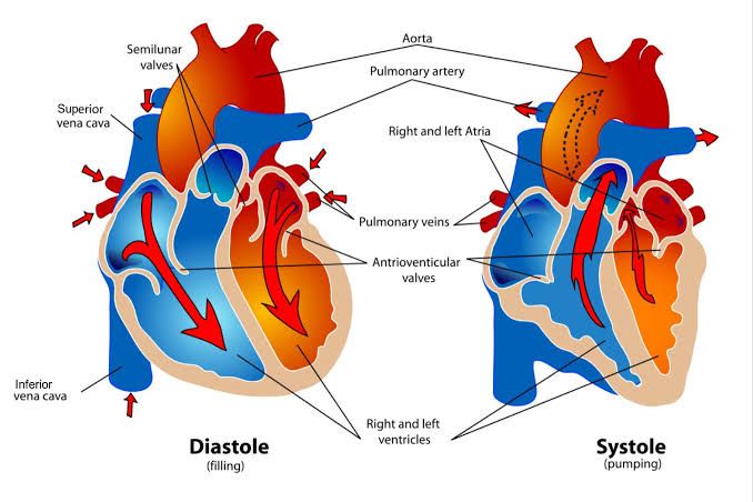
The cardiac cycle represents the complete sequence of events that occur within the heart during a single heartbeat. This complex, rhythmic process is fundamental to the circulatory system, ensuring the efficient pumping of blood throughout the body. Understanding its phases, the interplay of pressure and volume within cardiac chambers, and the corresponding valve dynamics is crucial for comprehending cardiovascular function.
Identification of Systolic and Diastolic Periods
The cardiac cycle is broadly divided into two main phases:
- Systole: This is the contraction phase of the heart chambers. During ventricular systole, the ventricles contract to eject blood into the arteries (aorta and pulmonary artery). Atrial systole, which precedes ventricular systole, involves the contraction of the atria to pump blood into the ventricles. While often used interchangeably with ventricular systole, the term “systole” typically refers specifically to ventricular contraction when discussing the overall cycle phases in relation to blood ejection into the systemic or pulmonary circulation.
- Diastole: This is the relaxation phase of the heart chambers. During ventricular diastole, the ventricles relax and fill with blood from the atria. Atrial diastole occurs concurrently with much of ventricular systole and continues through ventricular diastole as the atria fill with blood returning from the body and lungs. Like systole, “diastole” commonly refers to ventricular diastole when discussing the heart’s filling phase.
The cardiac cycle proceeds sequentially, with periods of systole and diastole alternating in a continuous flow. A healthy heart typically completes one cycle in approximately 0.8 seconds at rest (corresponding to a heart rate of 75 beats/minute), with diastole generally lasting longer than systole.
Pressure and Volume Changes in the Left Ventricle, Left Atrium, and the Aorta During the Cardiac Cycle
To understand the cardiac cycle in detail, it’s best to follow the sequence of events, observing how pressure gradients drive blood flow and valve movements on the left side of the heart:
- Phase 1: Ventricular Filling (Diastole)
- Early Diastole (Isovolumic Relaxation End to Rapid Filling): At the very beginning of diastole, ventricular pressure has dropped significantly following ejection. When the Left Ventricular (LV) pressure falls below the Left Atrial (LA) pressure, the Mitral valve (AtrioVentricular valve between LA and LV) opens. Blood that has accumulated in the LA during ventricular systole rapidly flows into the LV down a steep pressure gradient (LA pressure > LV pressure).
- LA Pressure: Initially rising as it fills, then drops sharply when the mitral valve opens.
- LV Pressure: Low and falling, then remains low or rises slightly as it fills.
- Aortic Pressure: Falling steadily (diastolic runoff) after the closure of the aortic valve (see Isovolumic Relaxation).
- LV Volume: Increases rapidly.
- Mid-Diastole (Diastasis): As the LV fills, the pressure gradient between the LA and LV diminishes. Blood flow into the LV slows down.
- LA Pressure: Slowly rising as it continues to receive blood from the pulmonary veins.
- LV Pressure: Slowly rising as volume increases.
- Aortic Pressure: Continues to fall slowly.
- LV Volume: Increases slowly.
- Late Diastole (Atrial Systole or “Atrial Kick”): The SA node fires, leading to atrial contraction. This contraction increases LA pressure, pushing an additional volume of blood into the LV. This “kick” contributes significantly to LV filling, especially at higher heart rates.
- LA Pressure: Increases significantly during contraction, then drops sharply.
- LV Pressure: Increases slightly due to the influx of additional blood.
- Aortic Pressure: Continues to fall.
- LV Volume: Increases due to atrial contraction, reaching its maximum volume for the cycle, known as the End-Diastolic Volume (EDV).
- Early Diastole (Isovolumic Relaxation End to Rapid Filling): At the very beginning of diastole, ventricular pressure has dropped significantly following ejection. When the Left Ventricular (LV) pressure falls below the Left Atrial (LA) pressure, the Mitral valve (AtrioVentricular valve between LA and LV) opens. Blood that has accumulated in the LA during ventricular systole rapidly flows into the LV down a steep pressure gradient (LA pressure > LV pressure).
- Phase 2: Isovolumic Contraction (Systole Starts)
- Ventricular excitation and contraction begin. As LV pressure rises, it quickly exceeds LA pressure. This pressure reversal causes the Mitral valve to snap shut. For a brief period, the LV is a closed chamber – the Mitral valve is closed (LV > LA pressure), and the Aortic valve is still closed (LV pressure < Aortic diastolic pressure). The ventricle is contracting, but the volume inside is not changing.
- LA Pressure: Shows a small, transient increase (the ‘c’ wave on a pressure trace) due to the Mitral valve bulging back into the atrium. Then starts rising again steadily as it fills.
- LV Pressure: Rises very rapidly and steeply.
- Aortic Pressure: Continues to fall slowly as blood flows into the systemic circulation (diastolic decay).
- LV Volume: Remains constant at EDV.
- Ventricular excitation and contraction begin. As LV pressure rises, it quickly exceeds LA pressure. This pressure reversal causes the Mitral valve to snap shut. For a brief period, the LV is a closed chamber – the Mitral valve is closed (LV > LA pressure), and the Aortic valve is still closed (LV pressure < Aortic diastolic pressure). The ventricle is contracting, but the volume inside is not changing.
- Phase 3: Ventricular Ejection (Systole Continues)
- Once LV pressure exceeds the pressure in the aorta (which is at its diastolic level), the Aortic valve is forced open. Blood is then rapidly ejected from the LV into the aorta. Initially, ejection is rapid, then slows down.
- LA Pressure: Steadily increases as the atrium continues to receive blood from the pulmonary veins while the Mitral valve is closed.
- LV Pressure: Rises to a peak during early ejection, then starts to fall during reduced ejection.
- Aortic Pressure: Rises sharply initially as blood is ejected, reaching a peak pressure (systolic pressure). Then falls slightly during reduced ejection.
- LV Volume: Decreases significantly as blood is ejected into the aorta, reaching its minimum volume for the cycle, known as the End-Systolic Volume (ESV). The volume of blood ejected is the Stroke Volume (SV = EDV – ESV).
- Once LV pressure exceeds the pressure in the aorta (which is at its diastolic level), the Aortic valve is forced open. Blood is then rapidly ejected from the LV into the aorta. Initially, ejection is rapid, then slows down.
- Phase 4: Isovolumic Relaxation (Diastole Starts)
- Ventricular contraction ends, and the ventricle begins to relax. As LV pressure falls rapidly, it drops below aortic pressure. This causes the Aortic valve to close. Like isovolumic contraction, the LV becomes a closed chamber again – the Aortic valve is closed (LV < Aortic pressure), and the Mitral valve is still closed (LV pressure > LA pressure). The ventricle is relaxing without a change in volume.
- LA Pressure: Continues to rise as it fills with blood from the pulmonary veins.
- LV Pressure: Falls very rapidly and steeply.
- Aortic Pressure: Shows a brief, small increase (the dicrotic notch or incisura) due to the elastic recoil of the aorta after valve closure, then falls steadily as blood flows into the periphery.
- LV Volume: Remains constant at ESV. This phase transitions into the ventricular filling phase when LV pressure finally drops below LA pressure and the Mitral valve opens.
- Ventricular contraction ends, and the ventricle begins to relax. As LV pressure falls rapidly, it drops below aortic pressure. This causes the Aortic valve to close. Like isovolumic contraction, the LV becomes a closed chamber again – the Aortic valve is closed (LV < Aortic pressure), and the Mitral valve is still closed (LV pressure > LA pressure). The ventricle is relaxing without a change in volume.
Meaning of Isovolumic Contraction, Period of Ejection, and Isovolumic Relaxation
These terms describe specific, critical phases within the cardiac cycle:
- Isovolumic Contraction: This is the very beginning of ventricular systole. It is defined by the ventricle contracting without a change in volume. This occurs because all heart valves are closed during this brief period. The mitral and tricuspid valves close at the very start (as ventricular pressure exceeds atrial pressure), and the aortic and pulmonary valves remain closed because ventricular pressure has not yet exceeded the pressure in the great arteries. This phase is purely about building sufficient pressure within the ventricle to open the semilunar valves and eject blood.
- Period of Ejection: This is the main part of ventricular systole where blood is actively pumped out of the ventricles. It begins when ventricular pressure exceeds the pressure in the corresponding great artery (aorta or pulmonary artery), forcing the semilunar valves open. Blood flows out of the ventricle, and thus the ventricular volume decreases significantly. This phase continues until ventricular pressure falls below arterial pressure and the semilunar valves close.
- Isovolumic Relaxation: This is the very beginning of ventricular diastole. It is defined by the ventricle relaxing without a change in volume. This occurs because all heart valves are closed during this brief period. The aortic and pulmonary valves close at the start (as arterial pressure exceeds ventricular pressure), and the mitral and tricuspid valves remain closed because ventricular pressure has not yet fallen below atrial pressure. This phase allows the ventricular pressure to drop sufficiently low to eventually fall below atrial pressure, enabling the mitral and tricuspid valves to open for ventricular filling.
Volume-Pressure Relationship in the Left Ventricle (The Pressure-Volume Loop)
The relationship between pressure and volume in the left ventricle over a single cardiac cycle can be graphically represented as a closed loop, known as the Pressure-Volume (PV) Loop. This loop is a powerful tool for visualizing and analyzing ventricular performance. The X-axis represents Left Ventricular Volume, and the Y-axis represents Left Ventricular Pressure. Starting from the bottom right and moving counter-clockwise, the loop traces the four key phases:
- Point A to Point B: Isovolumic Contraction: Starting at the End-Diastolic Volume (EDV) at point A (end of filling, mitral valve just closed), the pressure rises steeply from A to B while volume remains constant. This vertical line represents the isovolumic contraction phase, building pressure until it reaches the aortic diastolic pressure.
- Point B to Point C: Ejection: At point B, LV pressure exceeds aortic pressure, the aortic valve opens. Volume decreases from B to C (ejection), while pressure initially rises to a peak and then falls. Point C represents the End-Systolic Volume (ESV) at the end of ejection, just before the aortic valve closes. The horizontal distance between A (EDV) and C (ESV) represents the Stroke Volume (SV).
- Point C to Point D: Isovolumic Relaxation: At point C (end of ejection, aortic valve just closed), the ventricle begins to relax. Pressure falls steeply from C to D while volume remains constant at ESV. This vertical line represents the isovolumic relaxation phase, with pressure dropping until it falls below atrial pressure.
- Point D to Point A: Ventricular Filling: At point D, LV pressure falls below LA pressure, the mitral valve opens. Volume increases from D back to A (filling) while pressure remains low. This phase encompasses the rapid filling, diastasis, and atrial systole (bringing the volume back to EDV at point A).
The area enclosed by the LV PV loop represents the Stroke Work, which is the mechanical energy transferred by the ventricle during a single beat. Changes in EDV, ESV, and the slopes of the pressure-volume relationships affect the size and shape of the loop, providing insights into ventricular contractility, preload (related to EDV), and afterload (related to aortic pressure).
Development of First and Second Heart Sounds
The familiar “lub-dub” sounds of the heartbeat are produced by the vibrations caused by the sudden closure of the heart valves and the subsequent oscillation of the blood and chamber walls. These sounds are typically identified as S1 (the first sound) and S2 (the second sound).
- First Heart Sound (S1): This sound, often described as “lub,” marks the beginning of ventricular systole. It is primarily caused by the simultaneous closure of the Atrioventricular (AV) valves – the Mitral valve on the left side and the Tricuspid valve on the right side.
- Development: As ventricular contraction begins, ventricular pressure rapidly rises and quickly exceeds the pressure in the atria. This pressure gradient forces the AV valves shut, preventing backflow of blood into the atria. The sudden tensioning of the valve leaflets, the attached chordae tendineae, and the papillary muscles, along with the vibration of the blood and chamber walls, produces the S1 sound. The Mitral valve closure typically contributes more significantly to S1 than the tricuspid closure due to the higher pressures on the left side.
- Timing: S1 occurs at the beginning of the isovolumic contraction phase, immediately after atrial contraction and ventricular filling are complete.
- Second Heart Sound (S2): This sound, often described as “dub,” marks the beginning of ventricular diastole. It is primarily caused by the simultaneous closure of the Semilunar valves – the Aortic valve on the left side and the Pulmonary valve on the right side.
- Development: As ventricular ejection ends and the ventricles begin to relax, ventricular pressure falls rapidly. When the pressure in the ventricles drops below the pressure in the great arteries (aorta and pulmonary artery), the pressure gradient reverses, and the columns of blood in the arteries push back, forcing the semilunar valves shut. The sudden snapping shut of these valves and the subsequent vibrations produce the S2 sound. The Aortic valve closure typically occurs slightly before the Pulmonary valve closure, leading to a physiological split of S2, which can become more pronounced during inspiration.
- Timing: S2 occurs at the beginning of the isovolumic relaxation phase, immediately after ventricular ejection is complete.
Understanding the timing and cause of S1 and S2 allows clinicians to auscultate the heart sounds and interpret the physiological state of the valves and cardiac cycle phases. Abnormal sounds (murmurs) can indicate turbulent blood flow often due to valvular stenosis (narrowing) or regurgitation (leakage).
In summary, the cardiac cycle is a finely tuned sequence of electrical and mechanical events driven by pressure gradients and regulated by the opening and closing of heart valves. The intricate interplay of pressure and volume changes within the left ventricle, left atrium, and aorta during systole and diastole, marked by specific phases like isovolumic contraction, ejection, and isovolumic relaxation, culminates in the efficient pumping of blood. The pressure-volume loop provides a valuable graphical summary of this cycle, and the clear sounds of S1 and S2 serve as audible markers of crucial valve closures that delineate the major phases of the cardiac cycle. A comprehensive grasp of these processes is fundamental to diagnosing and managing cardiovascular conditions.


