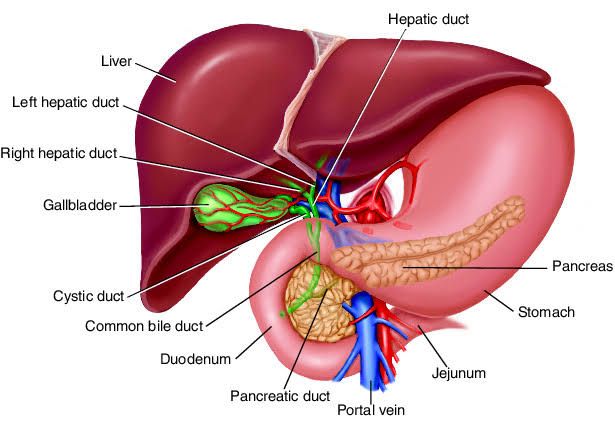
Inherited metabolic disorders represent a diverse group of genetic conditions that affect the body’s ability to produce enzymes or other proteins necessary for various biochemical processes. When these processes involve substances handled or processed by the liver, the liver often becomes a primary site of pathology.
Exploring Glycogen Storage Diseases (GSDs) Affecting the Liver
Glycogen storage diseases (GSDs) are a group of inherited metabolic disorders characterized by defects in the synthesis or breakdown of glycogen, the primary storage form of glucose in animals. While glycogen is stored in various tissues, particularly muscle and liver, defects affecting hepatic glycogen metabolism are the most common and can lead to significant liver dysfunction. These disorders are typically autosomal recessive.
Normally, glycogen serves as a readily accessible glucose reserve, crucial for maintaining blood glucose homeostasis, especially between meals. In hepatic GSDs, enzyme deficiencies disrupt this process, leading to either excessive glycogen accumulation in liver cells (hepatocytes) or an inability to release glucose from stored glycogen.
Several types of GSDs primarily affect the liver:
- GSD Type I (von Gierke’s disease): Caused by a deficiency of glucose-6-phosphatase or the glucose-6-phosphate transporter. This enzyme is critical for the final step of both glycogenolysis (glycogen breakdown) and gluconeogenesis (glucose synthesis from non-carbohydrate sources). The deficiency prevents the liver from releasing glucose into the bloodstream, leading to severe fasting hypoglycemia, buildup of glycogen and fat in the liver (hepatomegaly), elevated lactate, uric acid, and lipids in the blood.
- GSD Type III (Forbes-Cori disease): Due to a deficiency of the debranching enzyme (amylo-1,6-glucosidase), which is needed to break down branched glycogen molecules completely. This results in the accumulation of abnormal, short-branched glycogen structures in the liver and muscle. Manifestations include hepatomegaly, hypoglycemia (varying severity), and muscle weakness. Liver disease can progress to fibrosis or cirrhosis in some cases.
- GSD Type IV (Andersen’s disease): Caused by a deficiency of the branching enzyme (amylo-(1,4 to 1,6)-transglucosidase), which synthesizes the branches in glycogen. This leads to the accumulation of abnormal, long unbranched glycogen chains. These abnormal chains are poorly soluble and trigger immune responses, causing progressive liver fibrosis and cirrhosis, often leading to liver failure in infancy or early childhood.
- GSD Type VI (Hers’ disease): Results from a deficiency of hepatic glycogen phosphorylase, an enzyme initiating glycogen breakdown. This causes excessive glycogen storage in the liver and mild to moderate fasting hypoglycemia. It is generally less severe than Type I or III.
Diagnosis of GSDs involves evaluating blood glucose levels, liver size, measuring related metabolites (lactate, uric acid, lipids), imaging studies, enzyme assays in liver tissue or blood cells, and increasingly, genetic testing. Management focuses on maintaining normoglycemia through frequent feeding, continuous nocturnal feeding (e.g., with cornstarch), and dietary modifications. Liver transplantation may be considered for severe progressive liver disease (especially Type IV and severe Type III) or metabolic complications uncontrollable by medical therapy (severe Type I).
Inherited Disorders of Bilirubin Metabolism
Bilirubin is a yellow pigment produced from the breakdown of heme, a component of hemoglobin in red blood cells. It undergoes a complex metabolic pathway primarily processed by the liver before being excreted from the body. Inherited disorders of bilirubin metabolism affect specific steps in this pathway, leading to elevated levels of bilirubin in the blood (hyperbilirubinemia) and causing jaundice (yellowing of the skin and eyes).
The normal bilirubin pathway involves:
- Production of unconjugated (indirect) bilirubin.
- Transport of unconjugated bilirubin bound to albumin to the liver.
- Uptake of unconjugated bilirubin by hepatocytes.
- Conjugation of bilirubin with glucuronic acid by the enzyme UDP-glucuronosyltransferase (UGT1A1) within hepatocytes, forming conjugated (direct) bilirubin.
- Excretion of conjugated bilirubin into bile canaliculi, then into bile and the intestines.
- Further metabolism of bilirubin in the intestine and excretion in stool and urine.
Inherited disorders can disrupt these steps:
- Disorders of Bilirubin Conjugation:
- Gilbert’s Syndrome: An autosomal recessive condition characterized by a mild deficiency in the UGT1A1 enzyme activity (typically 10-30% of normal). This impairs bilirubin conjugation, leading to unconjugated hyperbilirubinemia. Jaundice is usually mild and intermittent, often triggered by stress, fasting, or illness. It is generally benign and requires no treatment.
- Crigler-Najjar Syndrome: A more severe deficiency or complete absence of UGT1A1 activity.
- Type I: Autosomal recessive, near-total absence of UGT1A1 activity. Results in severe unconjugated hyperbilirubinemia from birth, posing a risk of kernicterus (brain damage due to bilirubin toxicity). Requires aggressive treatment like phototherapy for life or liver transplantation.
- Type II (Arias Syndrome): Autosomal recessive or dominant, severe reduction (but not absence) of UGT1A1 activity. Causes significant unconjugated hyperbilirubinemia, usually less severe than Type I, with lower risk of kernicterus. May respond to phenobarbital, which induces UGT1A1 activity.
- Disorders of Bilirubin Excretion: These affect the transport of conjugated bilirubin from hepatocytes into the bile.
- Dubin-Johnson Syndrome: An autosomal recessive disorder caused by a defect in the multidrug resistance-associated protein 2 (MRP2/ABCC2), a transporter protein on the canalicular membrane of hepatocytes. This prevents conjugated bilirubin from being secreted into bile. Characterized by chronic or intermittent conjugated hyperbilirubinemia and deposition of a dark pigment in hepatocytes, giving the liver a black appearance. Generally benign, requires no specific treatment.
- Rotor Syndrome: Another rare autosomal recessive disorder causing conjugated hyperbilirubinemia. The defect is less well-defined than Dubin-Johnson and involves impaired uptake and storage of conjugated bilirubin by hepatocytes, potentially due to defects in organic anion transporters (OATP1B1 and OATP1B3). The liver is not pigmented. Also generally benign, requires no specific treatment.
Diagnosis involves evaluating bilirubin levels (total, conjugated, unconjugated), liver function tests, and sometimes genetic testing or liver biopsy (e.g., for pigment in Dubin-Johnson). Management depends on the specific disorder and its severity, ranging from observation for benign conditions to intensive phototherapy or liver transplantation for severe Crigler-Najjar Type I.
Alpha-1 Antitrypsin Deficiency (A1ATD) and Liver Disorders
Alpha-1 antitrypsin (AAT) is a protein primarily synthesized in the liver. Its main function is to protect tissues, especially the lungs, from damage by neutrophil elastase and other proteases. Alpha-1 antitrypsin deficiency (A1ATD) is an autosomal recessive disorder characterized by low levels of functional AAT in the blood. The most common severe deficiency allele is the Z mutation (PiZ), leading to the PiZZ phenotype, where a single amino acid substitution causes the AAT protein to misfold and polymerize within the endoplasmic reticulum of hepatocytes.
While A1ATD is widely recognized for causing early-onset emphysema due to lack of protection in the lungs, it is also a significant cause of liver disease. The liver pathology in A1ATD is not caused by the deficiency of AAT in the blood but by the accumulation of the misfolded Z variant protein polymers within the hepatocytes themselves. This intracellular accumulation triggers cellular stress, inflammation, and ultimately can lead to fibrosis and cirrhosis.
Liver disease in A1ATD has a highly variable presentation:
- Neonatal Hepatitis: Up to 10-20% of infants with severe deficiency (PiZZ) develop cholestatic jaundice within the first few months of life. This can range from transient mild hepatitis to severe liver failure requiring transplantation.
- Childhood/Adolescent Liver Disease: Some children present later with elevated liver enzymes, hepatomegaly, or signs of chronic liver disease.
- Adult Liver Disease: Many individuals with A1ATD develop liver disease in adulthood, often insidiously. This can manifest as chronic hepatitis, cirrhosis, and an increased risk (up to 20-30% lifetime risk in PiZZ) of developing hepatocellular carcinoma (liver cancer), even in the absence of significant cirrhosis. Conversely, some individuals with severe deficiency never develop clinically significant liver disease.
Diagnosis involves measuring serum AAT levels (low levels suggest deficiency), followed by AAT phenotyping or genotyping to identify specific alleles (e.g., PiMM is normal, PiMZ is heterozygote, PiZZ is severe deficiency). Liver biopsy may show characteristic PAS-diastase resistant globules in hepatocytes, representing accumulated abnormal AAT protein.
There is no specific treatment to remove the accumulated protein from hepatocytes. Management is supportive and focuses on managing complications of liver disease. Liver transplantation is a curative option for severe liver failure or hepatocellular carcinoma, as a healthy transplanted liver will produce normal AAT protein. Augmentation therapy (infusing purified AAT) is used for lung disease but does not prevent or treat liver disease because it does not address the accumulation issue within hepatocytes.
Lysosomal Storage Diseases (LSDs) with Liver Involvement
Lysosomal storage diseases (LSDs) are a group of over 70 distinct genetic disorders, most inherited in an autosomal recessive manner, caused by deficiencies in specific lysosomal enzymes or proteins. Lysosomes are cellular organelles containing enzymes that break down various macromolecules (lipids, carbohydrates, proteins) into smaller components for recycling or excretion. In LSDs, deficient enzyme activity leads to the accumulation of undegraded substrates within lysosomes in various cell types, including hepatocytes and Kupffer cells in the liver.
Liver involvement is common in many LSDs due to the liver’s role in metabolism and the presence of abundant hepatocytes and macrophages (Kupffer cells) which are prone to accumulating these substances. The accumulation in hepatocytes and/or Kupffer cells can lead to cellular dysfunction, inflammation, and ultimately fibrosis and cirrhosis.
Examples of LSDs with significant liver involvement include:
- Gaucher Disease (Type 1): Deficiency of glucocerebrosidase leads to accumulation of glucocerebroside, primarily in macrophages (Gaucher cells). Liver (and spleen) are typically enlarged (hepatosplenomegaly), and liver dysfunction and fibrosis can occur.
- Niemann-Pick Disease (Types A, B, C):
- Types A and B: Deficiency of acid sphingomyelinase leads to sphingomyelin accumulation. Type A is severe neurological form with hepatosplenomegaly and early death. Type B is non-neuropathic with significant hepatosplenomegaly and potential liver fibrosis/cirrhosis.
- Type C: Defect in cholesterol transport proteins (NPC1 or NPC2) leading to accumulation of unesterified cholesterol and other lipids. Progressive neurological disease is prominent, but hepatosplenomegaly and liver disease are also common findings, sometimes progressing to cirrhosis.
- Mucopolysaccharidoses (MPS): Deficiencies in enzymes needed to break down glycosaminoglycans (GAGs). Several types (e.g., MPS I Hurler, MPS II Hunter, MPS VI Maroteaux-Lamy, MPS VII Sly) involve hepatomegaly due to GAG accumulation in hepatocytes and Kupffer cells, with potential for liver fibrosis.
- Glycogen Storage Disease Type II (Pompe Disease): Although categorized as a GSD, it’s also a LSD caused by deficiency of acid alpha-glucosidase (acid maltase), an enzyme in the lysosome that breaks down glycogen. While muscle and heart are primarily affected, liver glycogen accumulation and hepatomegaly can occur.
- Wolman Disease / Cholesterol Ester Storage Disease (CESD): Deficiency of lysosomal acid lipase (LAL), leading to accumulation of triglycerides and cholesterol esters. Wolman disease is a severe infant form with massive hepatosplenomegaly, malabsorption, and early death. CESD is a later-onset, less severe form with hepatomegaly, elevated lipids, and premature atherosclerosis; liver fibrosis and cirrhosis can develop.
Diagnosis involves clinical suspicion, biochemical tests (measuring enzyme activity in blood or tissue), and genetic testing. Liver biopsy may show characteristic storage material within cells. Management options include enzyme replacement therapy (ERT) for certain conditions (e.g., Gaucher Type 1, several MPS types, Pompe, LAL deficiency), substrate reduction therapy (SRT), chaperone therapy, and hematopoietic stem cell transplantation (HSCT) for some types. Liver transplantation may be needed for end-stage liver disease but does not correct the systemic nature of most LSDs.
Hepatolenticular Degeneration (Wilson’s Disease)
Wilson’s disease is an autosomal recessive genetic disorder of copper metabolism characterized by excessive copper accumulation in various organs, most notably the liver, brain, eyes, and kidneys. It is caused by mutations in the ATP7B gene, which encodes a copper-transporting ATPase protein primarily located in hepatocytes.
The ATP7B protein is crucial for two main functions in copper homeostasis within the liver:
- Transporting copper into the trans-Golgi network for incorporation into ceruloplasmin, a protein that carries copper in the blood.
- Transporting excess cellular copper into bile for excretion, which is the primary route for eliminating copper from the body.
In Wilson’s disease, defective ATP7B function leads to impaired incorporation of copper into ceruloplasmin (resulting in low serum ceruloplasmin levels, although ceruloplasmin itself is not the primary defect) and, more critically, impaired excretion of copper into bile. This causes copper to accumulate first in hepatocytes. When the liver’s storage capacity is overwhelmed, copper spills into the bloodstream and deposits in other tissues.
Liver disease is often the initial presentation of Wilson’s disease, particularly in childhood and adolescence. The manifestations are highly variable:
- Asymptomatic: Elevated liver enzymes may be the only sign.
- Acute Hepatitis: Can mimic viral hepatitis.
- Chronic Hepatitis: Progressive inflammation and fibrosis.
- Cirrhosis: The most common presentation in adults, potentially leading to complications like portal hypertension.
- Fulminant Hepatic Failure: Although less common, it can be life-threatening, often associated with hemolysis (destruction of red blood cells).
Neurological or psychiatric symptoms (tremors, dystonia, parkinsonism, depression, psychosis) typically manifest later than liver disease, as copper accumulates in the basal ganglia of the brain. Kayser-Fleischer rings (copper deposits in the cornea) are characteristic eye findings, present in most individuals with neurological symptoms but may be absent in those with purely hepatic presentation, especially children.
Diagnosis is based on a combination of clinical findings, biochemical tests (low serum ceruloplasmin, elevated 24-hour urinary copper excretion, elevated liver copper content on biopsy), presence of Kayser-Fleischer rings, and genetic testing for ATP7B mutations.
Wilson’s disease is treatable, but early diagnosis is critical before irreversible organ damage occurs. Management involves lifelong therapy focused on removing accumulated copper and preventing reaccumulation:
- Chelation Therapy: Medications like penicillamine or trientine bind to copper, forming complexes that are excreted in urine. Used to remove excess copper.
- Zinc Therapy: Zinc stimulates intestinal cells to produce a protein that binds copper, blocking its absorption from the diet and inducing fecal excretion. Used for maintenance therapy after chelation or as initial therapy in asymptomatic/mildly symptomatic individuals.
- Dietary Restriction: Avoiding high-copper foods (e.g., shellfish, liver, nuts, chocolate).
Liver transplantation is indicated for individuals who present with fulminant hepatic failure or who develop end-stage liver disease despite medical therapy. Transplantation cures the metabolic defect.
Conclusion
Inherited metabolic diseases affecting the liver represent a significant area within hepatology and medical genetics. As detailed in these steps, conditions ranging from glycogen storage defects and bilirubin pathway disorders to protein misfolding diseases like A1ATD, lysosomal storage disorders, and metal metabolism defects like Wilson’s disease, can all lead to substantial hepatic pathology.
Understanding the specific biochemical defect underpinning each disorder is crucial for accurate diagnosis and appropriate management. While some conditions are relatively benign, others are progressive and potentially life-threatening. Early identification, often requiring a high index of suspicion in patients presenting with unexplained liver dysfunction, hepatomegaly, or jaundice, is paramount. Timely intervention, whether through dietary management, enzyme replacement, chelation therapy, or ultimately liver transplantation, can profoundly impact outcomes, preventing or mitigating severe liver damage and systemic complications. Ongoing research continues to improve diagnostic tools and therapeutic strategies for these complex genetic conditions.


