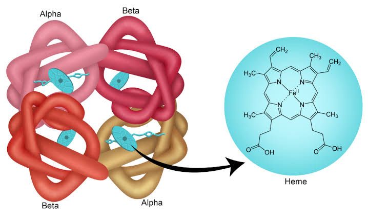
Heme is a vital biological molecule, integral to the function of numerous proteins involved in oxygen transport, electron transfer, and metabolism. Its structure is based on a complex organic ring system known as porphyrin. Understanding the structure, synthesis, and breakdown of heme and porphyrin is fundamental to comprehending various physiological processes and pathological conditions.
The Structure of Heme and Porphyrin
The foundational component is the porphyrin ring. Porphyrins are cyclic compounds featuring a central ring structure derived from four pyrrole rings linked by methine bridges (=CH-).
- Porphyrin Structure:
- Consists of four modified pyrrole subunits.
- These pyrrole rings are connected by alpha-carbon bridges (methine bridges).
- The structure contains a network of conjugated double bonds, which is responsible for the characteristic strong absorption of light (Soret band around 400 nm) and the often vibrant colors of porphyrins.
- Various side chains can be attached to the pyrrole rings, leading to different types of porphyrins (e.g., protoporphyrin, uroporphyrin, coproporphyrin). The arrangement and type of these side chains determine the specific porphyrin species.
- The central cavity formed by the four nitrogen atoms of the pyrrole rings is capable of chelating (binding) a metal ion.
- Heme Structure:
- Heme is specifically an iron-containing porphyrin.
- In most biologically important hemes, the porphyrin is Protoporphyrin IX.
- Iron (usually in the ferrous, Fe²⁺, state in functional proteins like hemoglobin and myoglobin, but can be ferric, Fe³⁺, in others like cytochromes and catalase) is chelated within the central cavity of the Protoporphyrin IX ring.
- The iron atom is coordinated to the four nitrogen atoms of the pyrrole rings. In many heme-containing proteins (hemoproteins), the iron atom also forms coordinate bonds with amino acid residues from the protein and/or ligands like oxygen (O₂) or carbon monoxide (CO).
- This iron-porphyrin complex is the functional unit responsible for binding oxygen (in hemoglobin/myoglobin), transporting electrons (in cytochromes), or acting as a catalytic center (in enzymes like catalase and peroxidases).
In summary, a porphyrin is the complex organic ring system, and heme is this ring system (specifically Protoporphyrin IX) with an iron atom chelated in its center.
The Biosynthesis of Heme and Porphyrin
Heme biosynthesis is a complex, multi-step pathway involving enzymes located in both the mitochondria and the cytosol of cells. While almost all mammalian cells synthesize heme, the most active sites are the bone marrow (for hemoglobin synthesis in red blood cell precursors) and the liver (for synthesis of heme-containing enzymes like cytochromes).
- Starting Materials: The synthesis begins with two simple precursors:
- Glycine (an amino acid)
- Succinyl CoA (an intermediate from the citric acid cycle)
- Key Steps: The pathway involves approximately eight enzymatic reactions, each adding or modifying components to build the porphyrin ring and insert the iron. The key steps are:
- Formation of δ-aminolevulinate (ALA): In the mitochondrial matrix, Glycine and Succinyl CoA condense to form α-amino-β-ketoadipate, which is rapidly decarboxylated to produce δ-aminolevulinate (ALA). This reaction is catalyzed by the enzyme ALA Synthase (ALAS).
- Regulation: This step is the rate-limiting step in heme biosynthesis. ALAS activity is tightly regulated, primarily by negative feedback from free heme. When heme levels are high, it inhibits ALAS synthesis and activity. There are two main isoforms: ALAS1 (ubiquitous, regulated by free heme) and ALAS2 (specific to erythroid cells, regulated by iron availability and ALAS protein synthesis).
- Formation of Porphobilinogen (PBG): Two molecules of ALA are condensed in the cytosol to form porphobilinogen. This reaction is catalyzed by ALA Dehydratase (also known as PBG Synthase). This enzyme is sensitive to inhibition by heavy metals like lead.
- Formation of Hydroxymethylbilane: Four molecules of PBG are linked together in the cytosol to form the linear tetrapyrrole hydroxymethylbilane. This step is catalyzed by Porphobilinogen Deaminase (also known as Hydroxymethylbilane Synthase or “Erythropoietic Uroporphyrinogen I Synthase”).
- Formation of Uroporphyrinogen III: Hydroxymethylbilane is rapidly converted to the cyclic molecule uroporphyrinogen III. This step requires the enzyme Uroporphyrinogen III Synthase. (Note: In the absence of this enzyme, hydroxymethylbilane can non-enzymatically cyclize to form uroporphyrinogen I, an abnormal isomer).
- Modification of Side Chains: Uroporphyrinogen III is converted through a series of reactions involving decarboxylations and modifications carried out by enzymes like Uroporphyrinogen Decarboxylase and Coproporphyrinogen Oxidase (located in the mitochondria) to form Protoporphyrinogen IX.
- Formation of Protoporphyrin IX: Protoporphyrinogen IX is oxidized by Protoporphyrinogen Oxidase (in the mitochondria) to form Protoporphyrin IX.
- Insertion of Iron: Finally, in the mitochondrial matrix, an iron atom (Fe²⁺) is inserted into the center of Protoporphyrin IX to form Heme. This step is catalyzed by the enzyme Ferrochelatase. This enzyme is also sensitive to inhibition by lead.
- Formation of δ-aminolevulinate (ALA): In the mitochondrial matrix, Glycine and Succinyl CoA condense to form α-amino-β-ketoadipate, which is rapidly decarboxylated to produce δ-aminolevulinate (ALA). This reaction is catalyzed by the enzyme ALA Synthase (ALAS).
The synthesized heme is then incorporated into nascent apoproteins (precursors of hemoproteins) or used to regulate ALAS activity.
Exploring the Degradation Process of Heme and Porphyrin
Unlike many biological molecules that are completely broken down and excreted, the porphyrin ring of heme is dismantled primarily in the reticuloendothelial system, particularly in the spleen, liver, and bone marrow. This process is crucial for the breakdown of heme derived from senescent (aged) red blood cells, which are the main source of heme requiring degradation.
- Location: Primarily in macrophages of the reticuloendothelial system. The liver also plays a significant role in the uptake and processing of degradation products.
- Initiation: The degradation process begins with the oxidation of the heme ring.
- Key Enzyme: The enzyme Heme Oxygenase (HO) is the principal enzyme responsible for the initial breakdown of heme. HO is found in the endoplasmic reticulum of cells.
- The Reaction: Heme Oxygenase cleaves the α-methine bridge of the protoporphyrin ring. This reaction requires oxygen (O₂) and NADPH.
- Products: The catalytic action of Heme Oxygenase on heme yields three main products:
- Biliverdin: A linear tetrapyrrole molecule, which is a green pigment. The porphyrin ring structure is opened.
- Carbon Monoxide (CO): Released during the cleavage of the methine bridge.
- Ferric Iron (Fe³⁺): The iron atom is released from the porphyrin ring.
- Subsequent Step: Biliverdin is then rapidly reduced to Bilirubin by the cytosolic enzyme Biliverdin Reductase. This reaction requires NADPH. Bilirubin is a yellow pigment.
This initial process converts the cyclic heme structure into the linear biliverdin, and then into the yellow bilirubin, releasing iron and carbon monoxide.
Substances Produced by Heme Destruction and Their Fate in the Body
Following the initial breakdown catalyzed by Heme Oxygenase and Biliverdin Reductase, the products undergo further processing and excretion.
- Substances Produced:
- Biliverdin (transient, rapidly converted)
- Carbon Monoxide (CO)
- Ferric Iron (Fe³⁺)
- Bilirubin (the main product that is further metabolized and excreted)
- Fate in the Body:
- Iron (Fe³⁺): The released iron is a valuable resource. It is typically bound to transport proteins (like transferrin) and transported to sites of utilization (e.g., bone marrow for new heme synthesis) or stored in the liver and spleen bound to storage proteins like ferritin and hemosiderin. This prevents cellular damage from free iron and ensures its availability for future use.
- Carbon Monoxide (CO): A small amount of the produced CO can bind to hemoglobin (forming carboxyhemoglobin) but is primarily transported in the blood and ultimately exhaled via the lungs. Endogenously produced CO may also have signaling functions.
- Biliverdin: As noted, biliverdin is rapidly converted to bilirubin.
- Bilirubin: The fate of bilirubin is the most complex and clinically significant:
- Transport: The bilirubin produced in the reticuloendothelial system (called unconjugated bilirubin or indirect bilirubin) is lipophilic and insoluble in water. It is transported in the blood tightly bound to albumin to prevent it from depositing in tissues, particularly the brain (where it can be toxic, especially in newborns).
- Hepatic Uptake: Unconjugated bilirubin is taken up by hepatocytes (liver cells) from the blood via specific membrane transporters.
- Conjugation: Inside the hepatocytes, unconjugated bilirubin is made water-soluble by conjugating it with one or two molecules of glucuronic acid. This reaction is catalyzed by the enzyme UDP-glucuronosyltransferase (UGT) in the endoplasmic reticulum. The product is conjugated bilirubin or direct bilirubin.
- Biliary Excretion: Conjugated bilirubin is water-soluble and is actively transported out of the hepatocytes into the bile canaliculi and then into the bile. This transport step is a major rate-limiting step in bilirubin excretion.
- Intestinal Fate: Bile containing conjugated bilirubin is released into the small intestine. In the large intestine, gut bacteria deconjugate the bilirubin and further metabolize it through a series of reactions to form a group of colorless compounds called urobilinogens.
- Urobilinogen Recirculation and Excretion: Most of the urobilinogen remains in the large intestine, where it is oxidized by bacteria and air to form stercobilin. Stercobilin is a brown pigment and is primarily excreted in the feces, giving stool its characteristic color. A small fraction of urobilinogen (about 10-15%) is reabsorbed from the intestine into the portal blood. Most of this reabsorbed urobilinogen is taken up by the liver and re-excreted into the bile (enterohepatic circulation of urobilinogen). A very small amount (about 1-2%) escapes hepatic uptake, enters the systemic circulation, and is excreted by the kidneys into the urine after being oxidized to urobilin (a yellow pigment, contributing to urine color).
Basic Abnormalities That May Result in Heme Degradation
Disruptions at various points in the heme degradation and bilirubin processing pathway can lead to an accumulation of bilirubin in the blood (hyperbilirubinemia), which manifests clinically as jaundice (icterus) – a yellowing of the skin, eyes (sclerae), and mucous membranes. Abnormalities can be broadly categorized based on where the problem occurs relative to the liver’s handling of bilirubin:
- Pre-Hepatic Abnormalities (Increased Bilirubin Production):
- These occur before bilirubin reaches the liver in significant amounts.
- Cause: Primarily increased destruction of red blood cells (hemolysis), leading to an overload of heme/bilirubin for the liver to process. Conditions include hemolytic anemias (e.g., sickle cell anemia, thalassemias, autoimmune hemolytic anemia), large hematomas (blood clots), or ineffective erythropoiesis (premature destruction of red blood cell precursors in the bone marrow).
- Result: The liver’s conjugation and excretion capacity are overwhelmed, although the liver itself is often functioning normally.
- Clinical/Lab Findings: Elevated levels of unconjugated bilirubin in the blood. Urine urobilinogen may be increased (due to increased bilirubin load leading to increased intestinal urobilinogen formation and subsequent renal excretion), but conjugated bilirubin is not typically found in urine. Stool color is usually normal or dark (due to increased stercobilin).
- Hepatic Abnormalities (Impaired Hepatic Uptake, Conjugation, or Excretion):
- These involve dysfunction of the liver itself.
- Causes: Liver diseases such as hepatitis (viral, alcoholic, drug-induced), cirrhosis, liver cancer, or inherited genetic disorders affecting bilirubin metabolism enzymes or transporters.
- Specific examples:
- Gilbert’s Syndrome: A common, mild genetic disorder with reduced activity of UDP-glucuronosyltransferase (mild conjugation deficit). Results in slightly elevated unconjugated bilirubin, often apparent during stress or fasting.
- Crigler-Najjar Syndrome (Type I and II): More severe genetic disorders with severely reduced or absent UGT activity (severe conjugation deficit). Type I can be life-threatening, especially in infants, due to uncontrolled unconjugated hyperbilirubinemia.
- Dubin-Johnson Syndrome & Rotor Syndrome: Genetic disorders affecting the transport of conjugated bilirubin out of hepatocytes into the bile. Result in elevated conjugated bilirubin.
- Specific examples:
- Result: Impaired processing of bilirubin by liver cells.
- Clinical/Lab Findings: Can show elevated unconjugated bilirubin (if uptake or conjugation is affected) or elevated conjugated bilirubin (if excretion is affected), or a mix of both (common in hepatocellular damage like hepatitis). Conjugated bilirubin can appear in urine (“bilirubinuria”) because it is water-soluble and filtered by the kidneys (unlike unconjugated bilirubin). Urine urobilinogen levels can vary depending on the cause, sometimes decreased if bile flow is partially reduced. Stool color may be normal or pale if bile flow is significantly impaired.
- Post-Hepatic Abnormalities (Impaired Biliary Excretion/Obstruction):
- These occur after bilirubin has been conjugated by the liver, involving a blockage in the bile ducts that prevent bile (containing conjugated bilirubin) from reaching the intestine.
- Causes: Obstruction of the bile ducts by gallstones, tumors (e.g., pancreatic cancer, bile duct tumors), strictures, or inflammation (e.g., cholangitis).
- Result: Backflow of conjugated bilirubin into the bloodstream.
- Clinical/Lab Findings: Characterized by significantly elevated levels of conjugated bilirubin in the blood. Bilirubinuria is typically present. Urine urobilinogen is significantly decreased or absent because conjugated bilirubin does not reach the intestine to be converted to urobilinogen. Stool color is typically pale or clay-colored because stercobilin formation is reduced or absent. Patients may also experience pruritus (itching) due to the accumulation of bile salts.
Understanding which type of bilirubin (unconjugated or conjugated) is elevated is a critical step in diagnosing the underlying cause of jaundice and assessing the site of the abnormality in the heme degradation/bilirubin processing pathway.


