Mastitis is an inflammatory condition of the breast tissue that can be a significant source of pain and distress, particularly for lactating individuals. While commonly associated with breastfeeding, it can occur in any woman and, rarely, in men. A thorough understanding of its causes, presentation, diagnosis, and treatment is essential for healthcare providers and crucial for patient education and well-being.
Understanding Mastitis: The Inflammatory Spectrum
Mastitis is fundamentally an inflammation of the mammary gland. It is best understood not as a single event but as a spectrum of conditions that begins with ductal narrowing and can progress to more severe states if not properly managed. The primary cause in lactating individuals is milk stasis—the incomplete removal of milk from the breast. This stasis can occur due to various factors, including an improper latch, missed feedings, abrupt weaning, or external pressure on the breast from tight clothing or a poorly fitting bra.
When milk remains static in the ducts, it can cause an inflammatory response within the breast tissue, leading to what is termed inflammatory mastitis. The stagnant milk can also create a favorable environment for bacterial growth, which can subsequently lead to infectious mastitis. The most common pathogen implicated in infectious mastitis is Staphylococcus aureus, which often colonizes the skin and can enter the breast tissue through small cracks or fissures in the nipple. Other bacteria, such as Streptococcus species and, increasingly, methicillin-resistant Staphylococcus aureus (MRSA), can also be causative agents. Risk factors that predispose an individual to mastitis include a previous history of the condition, nipple trauma, maternal stress, and fatigue, all of which can compromise both milk flow and the immune system. Non-lactational mastitis is less common and includes conditions like periductal mastitis, often associated with smoking, and idiopathic granulomatous mastitis, a rare chronic inflammatory condition.
Symptoms and Signs: Recognizing the Clinical Presentation
The diagnosis of mastitis is primarily clinical, based on a characteristic constellation of symptoms and signs. Differentiating mastitis from a simple plugged duct is key; while a plugged duct presents with a localized, tender lump, mastitis involves a more widespread inflammatory response and often includes systemic symptoms.
Local Signs in the Breast:
- Pain and Tenderness: The affected breast will be tender to the touch, often with a constant, throbbing, or burning pain.
- Erythema (Redness): A characteristic wedge-shaped or diffuse area of redness is often visible on the skin overlying the affected tissue.
- Swelling and Firmness: The area will feel swollen, firm, and indurated compared to the rest of the breast tissue.
- Warmth: The affected skin will be noticeably warm or hot to the touch.
- Palpable Mass: A distinct lump may be felt, corresponding to the area of inflammation and milk stasis.
Systemic Signs and Symptoms: The presence of systemic symptoms is a hallmark of mastitis and distinguishes it from less severe breast issues. These flu-like symptoms are a result of the body’s systemic inflammatory response.
- Fever: A temperature of 38.3°C (101°F) or higher is common.
- Chills and Body Aches: Individuals often report feeling achy, shivery, and generally unwell (malaise).
- Fatigue and Headache: Profound fatigue and general malaise are frequently reported.
If left untreated, mastitis can progress to a more serious complication known as a breast abscess, which presents as a localized, fluctuant (fluid-filled), and exquisitely tender mass that does not resolve with initial management.
Diagnosis: Clinical Assessment and Targeted Investigations
As mentioned, the diagnosis of mastitis is typically made based on the clinical history and physical examination. A healthcare provider will assess the breast for the classic signs of inflammation and inquire about systemic symptoms. However, in certain situations, further investigations may be warranted to confirm the diagnosis, identify the causative organism, or rule out complications.
- Clinical Evaluation: The cornerstone of diagnosis is a careful physical breast examination confirming the presence of a tender, erythematous, indurated area, often accompanied by a history of fever and malaise in a lactating individual.
- Breast Milk Culture: A culture of the breast milk is not routinely necessary for an initial, uncomplicated case of mastitis. However, it is strongly recommended in the following scenarios:
- Severe or persistent symptoms that do not respond to initial antibiotic therapy within 48-72 hours.
- Recurrent episodes of mastitis.
- Mastitis acquired in a hospital setting.
- If the patient has a known allergy to standard first-line antibiotics. A mid-stream milk sample should be collected after cleaning the nipple to minimize skin contamination.
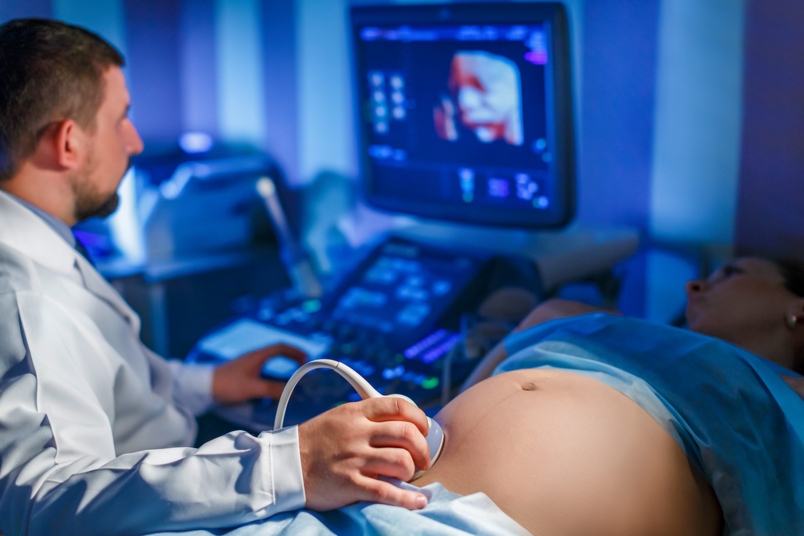
- Imaging (Ultrasound): A breast ultrasound is the imaging modality of choice if a complication, such as a breast abscess, is suspected. It is highly effective at differentiating between cellulitis or a phlegmon (a region of non-liquefied inflammation) and a drainable, fluid-filled abscess. Ultrasound is crucial for guiding needle aspiration or surgical drainage if an abscess is confirmed. For non-lactating mastitis or cases with an atypical presentation, mammography and/or biopsy may be considered to rule out underlying pathologies, including the rare but critical diagnosis of inflammatory breast cancer.
Treatment: A Multifaceted Management Approach
The management of mastitis is centered on two primary goals: relieving the milk stasis and treating the inflammation and/or infection. A comprehensive treatment plan includes effective milk removal, supportive care, and, when indicated, antibiotic therapy.
1. Effective and Frequent Milk Removal: This is the most critical component of treatment. Continuing to breastfeed or pump from the affected breast is essential to clear the milk stasis and relieve ductal pressure.
- Continue Breastfeeding: It is safe and recommended to continue breastfeeding from both breasts. The milk is not harmful to the infant.
- Frequency: Nurse or pump frequently, at least every 2-3 hours, including throughout the night.
- Positioning: Begin feedings on the affected breast, as the infant’s suck is strongest at the start. Ensure a proper latch and try different feeding positions to facilitate drainage of all quadrants of the breast.
- Massage: Gently massage the firm area of the breast towards the nipple during feedings or pumping sessions to assist milk flow.
2. Supportive Care: These measures aim to alleviate pain and reduce inflammation.
- Rest: Adequate rest is vital to support the body’s immune response.
- Hydration and Nutrition: Maintaining good fluid intake and nutrition is important for recovery.
- Pain Relief: Non-steroidal anti-inflammatory drugs (NSAIDs) like ibuprofen are preferred as they target both pain and inflammation. Acetaminophen can also be used for pain control.
- Temperature Therapy: Applying cold packs or cool compresses to the breast after feedings can help reduce swelling and alleviate pain. The traditional recommendation of warm compresses before feeding has become more nuanced, with some guidelines now favoring cold to reduce inflammation-induced swelling that can further compress ducts.
3. Antibiotic Therapy: Antibiotics are indicated for infectious mastitis, typically when symptoms are moderate to severe, have not improved within 12-24 hours of conservative management, or if a nipple fissure is present.
- Choice of Antibiotic: The antibiotic chosen must provide coverage against S. aureus. First-line options include dicloxacillin or cephalexin. For patients with a penicillin allergy, clindamycin is a suitable alternative.
- MRSA Coverage: If MRSA is suspected based on local resistance patterns, a history of MRSA infection, or failure to respond to first-line therapy, trimethoprim-sulfamethoxazole or clindamycin should be considered.
- Duration: A full course of antibiotics, typically lasting 10-14 days, is necessary to fully eradicate the infection and prevent recurrence.
In conclusion, mastitis is a common and treatable condition when approached with a clear, systematic strategy. By focusing on the core principles of effective milk removal, supportive care for inflammation and pain, and the judicious use of antibiotics, healthcare providers can effectively manage the condition, prevent serious complications like abscesses, and support the continuation of the breastfeeding journey.
References
- Mitchell, K. B., Johnson, H. M., Rodríguez, J. M., Eglash, A., Scherzinger, C., Zakarija-Grkovic, I., … & Academy of Breastfeeding Medicine. (2022). Academy of Breastfeeding Medicine Clinical Protocol #36: The Mastitis Spectrum, Revised 2022. Breastfeeding Medicine, 17(5), 360-376.
- Amir, L. H., & The Academy of Breastfeeding Medicine Protocol Committee. (2014). ABM Clinical Protocol #4: Mastitis, Revised March 2014. Breastfeeding Medicine, 9(5), 239-243. [Note: While superseded by the 2022 protocol, this remains a foundational reference].
- Boakes, E., Woods, A., Johnson, N., & Kadoglou, N. (2018). Breast infection: a review of diagnosis and management practices. European Journal of Breast Health, 14(3), 136–143.
- Centers for Disease Control and Prevention (CDC). (2022). Mastitis. Division of Nutrition, Physical Activity, and Obesity. Retrieved from https://www.cdc.gov/breastfeeding/breastfeeding-special-circumstances/maternal-or-infant-illnesses/mastitis.html
- Kataria, K., Srivastava, A., & Dhar, A. (2013). Management of lactational mastitis and breast abscesses. The Indian Journal of Surgery, 75(6), 430–435.


