Of all facial injuries, fractures of the nose are the most common, resulting from the prominent and relatively delicate structure of the nasal pyramid. Caused by direct trauma from sources such as altercations, sports injuries, falls, and motor vehicle accidents, these fractures can range from simple, non-displaced breaks of the nasal bones to complex injuries involving the nasal septum and surrounding facial structures. Effective management requires a systematic approach, beginning with the recognition of clinical signs, followed by a thorough assessment, timely intervention to prevent complications, and comprehensive medical and nursing care to restore both function and aesthetics.
Clinical Manifestations of Nasal Fractures
The clinical presentation of a nasal fracture is often immediate and apparent following the traumatic event. The most common and immediate sign is epistaxis (nosebleed), which can range from minor to significant, originating from the rich vascular supply of the nasal mucosa that is inevitably torn during the fracture. This is typically accompanied by significant pain and tenderness over the nasal bridge.
Within minutes to hours, swelling (edema) and bruising (ecchymosis) develop. The swelling can be extensive, obscuring the underlying bony deformity. Bruising often extends to the periorbital areas, resulting in bilateral periorbital ecchymosis, colloquially known as “raccoon eyes.” This finding suggests a significant impact and should raise suspicion for associated facial or basilar skull fractures.
One of the most defining characteristics is a visible deformity of the nose. The nose may appear deviated, crooked, or depressed (caved in), a condition referred to as a “saddle nose deformity.” On gentle palpation, a healthcare provider may feel crepitus, a crackling or grating sensation caused by fractured bone fragments rubbing against each other. The patient will almost invariably complain of nasal obstruction or difficulty breathing through one or both nostrils, caused by a combination of internal swelling, clotted blood, and displacement of the nasal septum. In some cases, clear, watery drainage from the nose (rhinorrhea) may be present. This is a critical red flag, as it may indicate a cerebrospinal fluid (CSF) leak from a concurrent skull base fracture, most often involving the cribriform plate.
Assessment and Diagnostic Findings
A thorough assessment is paramount and relies more heavily on clinical examination than on imaging. The assessment begins with a detailed history of the injury, including the mechanism, force, and direction of the impact. This information can help predict the pattern of fracture and the likelihood of associated injuries.
The physical examination is the cornerstone of diagnosis.
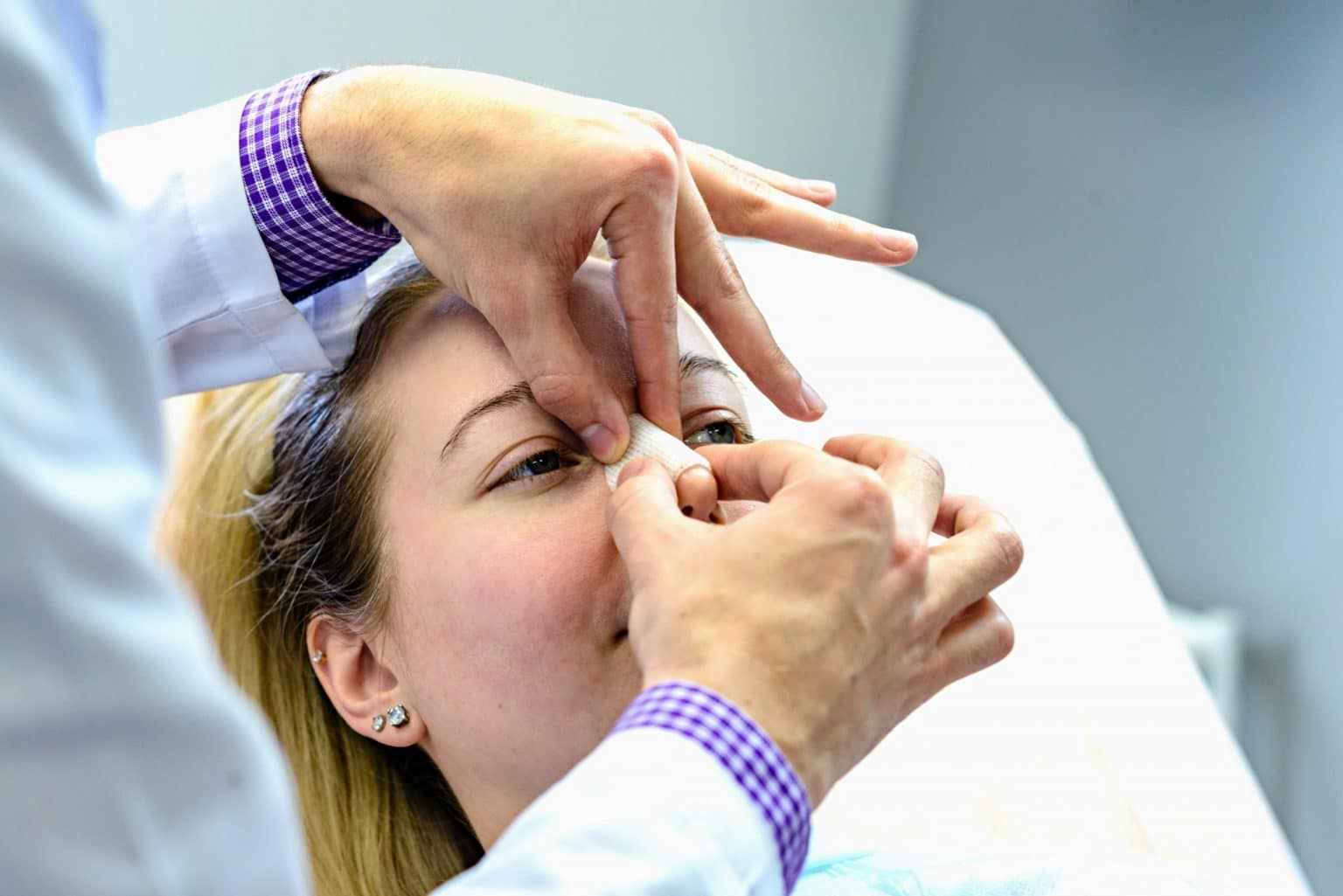
- External Inspection and Palpation: The provider carefully inspects the nose from the front, sides, and from a “bird’s-eye view” (looking down from the patient’s forehead) to best appreciate any deviation or depression. Gentle palpation along the nasal dorsum and sidewalls can identify points of maximal tenderness, instability, and crepitus.
- Internal Nasal Examination: This is the most crucial part of the assessment. After applying a topical anesthetic and decongestant to improve visualization and patient comfort, the provider uses an otoscope or a nasal speculum and a headlight to inspect the inside of the nose. The primary goal is to identify a septal hematoma, which appears as a fluctuant, bluish, or purplish swelling along the nasal septum. This is a clinical emergency. The examination also assesses for mucosal lacerations and the position of the septum.
Diagnostic imaging plays a secondary role in isolated nasal fractures. Plain facial X-rays are generally not recommended as they have low sensitivity and specificity and rarely alter the management plan for a simple, uncomplicated fracture. The diagnosis is clinical. However, a computed tomography (CT) scan is the gold standard and is indicated when there is suspicion of more extensive injuries, such as:
- Associated facial fractures (e.g., orbital blowout, Le Fort, zygomatic fractures).
- Suspected CSF leak (confirmed by testing the fluid for beta-2 transferrin).
- Failure of a closed reduction, requiring more complex surgical planning.
Complications of Nasal Fractures
Failure to promptly and correctly identify and manage nasal fractures can lead to several significant complications, which can be categorized as early or late.
Early Complications:
- Septal Hematoma: This is the most urgent complication. It is a collection of blood between the septal cartilage and its overlying perichondrium, which supplies its nutrients. If left untreated, the cartilage is deprived of its blood supply and can become necrotic within 24-48 hours. This leads to the collapse of the nasal dorsum, resulting in a permanent “saddle nose deformity,” and can also become infected, forming a septal abscess. It requires immediate incision and drainage.
- Cerebrospinal Fluid (CSF) Rhinorrhea: A leak of CSF signifies a fracture of the skull base and creates a direct pathway for bacteria to enter the central nervous system, posing a high risk of meningitis. This is a neurosurgical emergency.
- Severe or Persistent Epistaxis: While some bleeding is expected, uncontrolled bleeding may require nasal packing or, in rare cases, surgical intervention.
Late Complications:
- Cosmetic Deformity: If the fracture is not reduced or heals in a displaced position, a permanent crooked or collapsed nose can result, leading to significant patient dissatisfaction.
- Functional Nasal Obstruction: Chronic difficulty breathing can result from an uncorrected septal deviation, collapse of the nasal valve, or internal scarring.
- Septal Perforation or Deviation: A hole in the septum or a persistent deviation can cause chronic crusting, whistling during breathing, and recurrent nosebleeds.
- Anosmia: Damage to the olfactory nerves at the cribriform plate can lead to a partial or complete loss of smell.
Medical Management
The management strategy for a nasal fracture depends on the severity of the fracture and the presence of complications.
Initial Management: The immediate goals are to control pain, bleeding, and swelling. This is typically achieved with:
- Applying ice packs to the nose and surrounding area for 15-20 minutes every 1-2 hours.
- Keeping the head elevated to minimize edema.
- Administering analgesics such as acetaminophen. NSAIDs are often avoided initially due to their potential to increase bleeding.
- For epistaxis, patients are instructed to pinch the soft part of their nose and lean forward. If this fails, topical vasoconstrictors or nasal packing may be required.
Reduction of the Fracture: If the fracture has resulted in a cosmetic deformity or airway obstruction, a reduction (setting the bones) is necessary.
- Timing: The ideal time for a closed reduction is after the initial swelling has subsided but before the bones begin to set, typically between 5 and 10 days post-injury.
- Closed Reduction: This is the most common procedure and is often performed in an outpatient or emergency department setting under local or general anesthesia. Using specialized instruments (e.g., Boies elevator, Walsham forceps), the surgeon manipulates the depressed or deviated nasal bones back into their proper alignment. Following reduction, the nose is typically supported with internal packing and an external thermoplastic splint, which remains in place for 7-10 days.
- Open Reduction (Rhinoplasty/Septorhinoplasty): This surgical procedure is reserved for complex fractures, fractures involving significant septal damage, or cases where a closed reduction was unsuccessful. It is often delayed for several months to allow for complete resolution of swelling and a more accurate assessment of the deformity.
Nursing Management
Nursing care is critical throughout the patient’s journey, from initial presentation to post-procedure recovery. The focus is on assessment, intervention, and patient education.
Assessment:
- The primary nursing assessment is to ensure a patent airway, monitoring for any signs of respiratory distress.
- Assess for and quantify bleeding.
- Assess pain levels using a standardized scale.
- Check for clear nasal drainage and perform the “halo sign” test on gauze if a CSF leak is suspected.
- Monitor for changes in vision or extraocular movements, which could indicate an orbital fracture.
Interventions:
- Administer prescribed analgesics and apply ice packs.
- Position the patient with the head of the bed elevated at least 30 degrees.
- Provide an emesis basin and tissues and instruct the patient to avoid swallowing blood to prevent nausea.
- Assist the physician during the internal examination or closed reduction procedure, ensuring patient comfort and safety.
Patient Education and Discharge Planning: This is a cornerstone of nursing management. The nurse must provide clear instructions on:
- Care of the nasal splint and packing.
- Pain and swelling management at home (continued ice, head elevation).
- Avoiding activities that could reinjure the nose or increase pressure, such as strenuous exercise, contact sports, and blowing the nose for several weeks.
- Instructing patients to sneeze with their mouth open.
- Warning signs of complications to report immediately, including fever, persistent headache, increased pain or swelling, or purulent drainage.
- The importance of attending follow-up appointments for splint removal and final evaluation.
References
- Higuera, S., & Lee, E. I. (2019). Nasal Trauma and Fractures. In J. J. Johnson & C. A. Rosen (Eds.), Bailey’s Head & Neck Surgery – Otolaryngology (6th ed.). Wolters Kluwer.
- Mondin, V., Rinaldo, A., & Ferlito, A. (2005). Management of nasal fractures. American Journal of Otolaryngology, 26(3), 181–185.
- Das, S. K., & Al-Juhaiman, A. K. (2021). Nasal Bone Fracture. In StatPearls. StatPearls Publishing. Available from: https://www.ncbi.nlm.nih.gov/books/NBK538294/
- Duffy, M. F., & Lantry, J. (2018). Management of Nasal Fractures. UpToDate. Retrieved from https://www.uptodate.com/contents/management-of-nasal-fractures
- Kelly, E. A., & Love, T. L. (2010). Diagnosis and management of nasal fractures. The Journal of Craniofacial Surgery, 21(5), 1541-1543.


