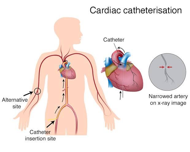
Cardiac catheterization (CC) and electrophysiology (EP) testing are cornerstones of modern cardiovascular diagnostics and intervention. While both procedures involve minimally invasive access to the heart via peripheral vasculature under fluoroscopic guidance, they serve distinct purposes: CC primarily assesses the structural integrity, pressures, and blood flow (hemodynamics) of the heart and coronary arteries, while EP testing specializes in mapping and evaluating the electrical conduction system.
Defining the Procedures
A. Cardiac Catheterization (CC)
Cardiac catheterization involves inserting a thin, flexible tube (catheter) into a peripheral artery or vein (most commonly the radial or femoral artery) and guiding it under fluoroscopy into the heart chambers, pulmonary artery, or coronary arteries.
1. Diagnostic Cardiac Catheterization (Angiography): This involves injecting contrast dye to visualize the coronary arteries (coronary angiography) to identify atherosclerotic blockages. It also allows for the assessment of chamber pressures, cardiac output, shunt detection, and valvular function.
2. Interventional Cardiac Catheterization: If significant blockages are identified, the procedure may transition immediately into intervention, such as percutaneous coronary intervention (PCI). This includes balloon angioplasty to compress plaque and stent placement to maintain vessel patency.
B. Electrophysiology (EP) Testing
Electrophysiology testing is a specialized diagnostic procedure used to assess the heart’s electrical system, often referred to as the “wiring.” Multiple electrode catheters are positioned strategically within the heart chambers.
The primary function of EP testing is to induce and analyze arrhythmias. Controlled bursts of electrical stimulation are delivered to identify the source of the dysrhythmia, map the specific conduction pathways (accessory pathways), and determine the appropriate therapeutic approach. If an abnormal electrical pathway is found, the procedure often evolves into a curative catheter ablation, where radiofrequency energy or cryotherapy is used to destroy the tissue responsible for the abnormal signals.
Indications for Cardiac Catheterization and Electrophysiology Testing
A. Indications for Cardiac Catheterization
Cardiac catheterization is indicated when non-invasive tests (e.g., stress tests, echocardiograms) suggest significant structural or flow abnormalities, or in acute life-threatening situations.
| Category | Specific Clinical Indications |
|---|---|
| Coronary Artery Disease (CAD) | Evaluation of chest pain (angina pectoris), suspicion of significant coronary stenosis, and assessment of bypass graft patency. |
| Acute Coronary Syndromes (ACS) | Definitive diagnosis and immediate intervention (PCI) for patients presenting with STEMI (ST-segment elevation myocardial infarction) or high-risk NSTEMI. |
| Valvular Heart Disease | Assessment of pressure gradients across stenotic valves, quantification of regurgitation severity, and preparation for transcatheter valve replacement (TAVR, TMVR). |
| Heart Failure | Right heart catheterization (RHC) to assess intracardiac and pulmonary pressures (PCWP) to guide medical management. |
| Congenital Heart Disease | Assessment of shunts and pulmonary pressures, and interventional closure of defects (e.g., patent foramen ovale, atrial septal defect). |
B. Indications for Electrophysiology Testing
EP testing is indicated when symptoms suggest an underlying electrical instability that cannot be fully characterized by standard surface electrocardiograms (ECG).
| Category | Specific Clinical Indications |
|---|---|
| Complex Tachyarrhythmias | Diagnosis and mapping of sustained, poorly tolerated supraventricular tachycardias (SVT), atrial fibrillation/flutter, and ventricular tachycardia (VT). |
| Symptomatic Bradycardia/Syncope | Evaluation of unexplained syncope or near-syncope, especially if standard testing suggests intermittent heart block or sick sinus syndrome, determining the need for a pacemaker. |
| Risk Stratification | Assessment of patients with structural heart disease (e.g., post-MI or heart failure) who are at high risk for sudden cardiac death, often guiding the decision for an implantable cardioverter-defibrillator (ICD). |
| Guidance for Ablation | Precisely identifying and locating the aberrant electrical focus prior to catheter ablation. |
Nursing Management of Cardiac Catheterization and Electrophysiology Testing
Effective nursing management is critical to patient safety and procedural success, spanning the pre-procedural education phase, vigilant intra-procedural monitoring, and crucial post-procedural recovery.
A. Pre-Procedure Nursing Management
The goal of pre-procedure care is patient preparation, risk mitigation, and education.
- Patient Education and Consent:
- Explain the procedure, the expected duration, and the location of the access site (e.g., radial or femoral).
- Educate the patient on common sensations, such as transient flushing or a warm feeling upon contrast injection (CC), or palpitations/dizziness during induced arrhythmias (EP).
- Ensure informed consent is signed and understood by the patient prior to sedation.
- Assessment and Laboratory Review:
- Obtain complete baseline vital signs, pedal (or radial) pulses, and capillary refill for comparison post-procedure.
- Review Coagulation Studies (PT, PTT, INR) to assess bleeding risk.
- Review Renal Function (BUN and Creatinine) to assess the risk of Contrast-Induced Nephropathy (CIN), especially for CC. Hydration protocols may be initiated if renal function is suboptimal.
- Verify NPO status (typically 6-8 hours) to minimize aspiration risk during sedation.
- Medication Management:
- Clarify which medications must be held (e.g., anticoagulants, certain diabetes medications like metformin due to interaction with contrast dye) and which must be taken (e.g., cardiac medications).
- Assess for allergies, vitalizing contrast media allergy (iodine/shellfish) and latex. Pre-medication (steroids or antihistamines) may be needed.
B. Intra-Procedure Nursing Management
During the procedure, the nurse functions as the patient advocate, continuously monitoring physiological status and maintaining safety.
- Monitoring and Safety:
- Continuous cardiac monitoring (ECG) and frequent non-invasive blood pressure assessments.
- Administer and monitor sedation (conscious sedation) and analgesia, ensuring the patient remains cooperative but comfortable. Assess airway patency and oxygen saturation frequently.
- Maintain the sterile field and assist the physician with equipment needs.
- Specific Intra-CC Concerns: Monitor for signs of contrast reaction (urticaria, wheezing, hypotension) and assess for signs of myocardial ischemia (ST segment changes, chest discomfort).
- Specific Intra-EP Concerns: Observe closely during arrhythmia induction, ensuring immediate availability of a defibrillator/external pacing capabilities. Document all induced arrhythmias and patient tolerance.
C. Post-Procedure Nursing Management (The Critical Recovery Phase)
The immediate post-procedure period is high-risk, focusing primarily on managing the vascular access site and preventing complications.
- Vascular Access Site Management (Most Critical Step):
- Bleeding Assessment: Frequently inspect the access site (femoral or radial) for signs of bleeding, hematoma, or swelling. If bleeding occurs, immediate, sustained, manual pressure must be applied (often requiring two nurses or physician assistance), and the provider notified.
- Femoral Access: Maintain strict bed rest with the head of the bed elevated no more than 30 degrees (depending on facility policy and closure device used). The extremity must remain straight and immobilized for the prescribed time (typically 4–6 hours).
- Radial Access: The patient may ambulate sooner, but the wrist band/device must remain in place and be monitored frequently for distal perfusion.
- Peripheral Pulse Assessment:
- Perform frequent circulatory checks distal to the access site (the 5 Ps: Pain, Pallor, Pulses, Paresthesia, Paralysis). A diminished or absent pulse, coolness, or loss of sensation can indicate arterial occlusion, requiring immediate intervention.
- Hydration and Renal Function:
- Encourage liberal oral fluid intake (if not contraindicated) to facilitate the excretion of contrast dye.
- Monitor hourly urine output to assess kidney perfusion and watch for signs of Contrast-Induced Nephropathy (CIN), particularly in high-risk patients.
- Vital Signs and Pain Management:
- Monitor vital signs every 15 minutes for the first hour, then every 30 minutes for the next hour, increasing frequency if hypotension or tachycardia occurs.
- Assess for generalized discomfort and specific complications, such as reoccurring chest pain (indicating potential coronary re-occlusion post-stenting) or back pain (which can signal retroperitoneal bleeding).
- EP Specific Management:
- Post-ablation, monitoring must prioritize ruling out pericardial effusion/tamponade (signs include muffled heart sounds, hypotension, or paradoxic pulse) and ensuring the new electrical rhythm is stable.
In summary, both cardiac catheterization and electrophysiology testing provide indispensable diagnostic and therapeutic insight into cardiovascular function. While CC focuses on structural and hemodynamic assessment, EP testing offers detailed mapping of the electrical pathways. The professional nurse plays an indispensable role by ensuring comprehensive patient education, vigilant monitoring, and meticulous post-procedural care essential for optimizing outcomes and preventing high-risk complications related to vascular access and organ perfusion.
References
- American Heart Association (AHA). (2021). Cardiac Catheterization: What to Expect. Retrieved from Heart.org.
- Hickey, M., & Taylor, S. (2018). The Cardiac Catheterization Handbook. Springer Publishing.
- Ignatavicius, D. D., Workman, M. L., & Rebar, C. R. (2021). Medical-Surgical Nursing: Concepts for Interprofessional Collaborative Care (10th ed.). Elsevier.
- Zipes, D. P., Libby, P., Bonow, R. O., Mann, D. L., Tomaselli, G. F., & Braunwald, E. (2018). Braunwald’s Heart Disease: A Textbook of Cardiovascular Medicine (11th ed.). Elsevier.


