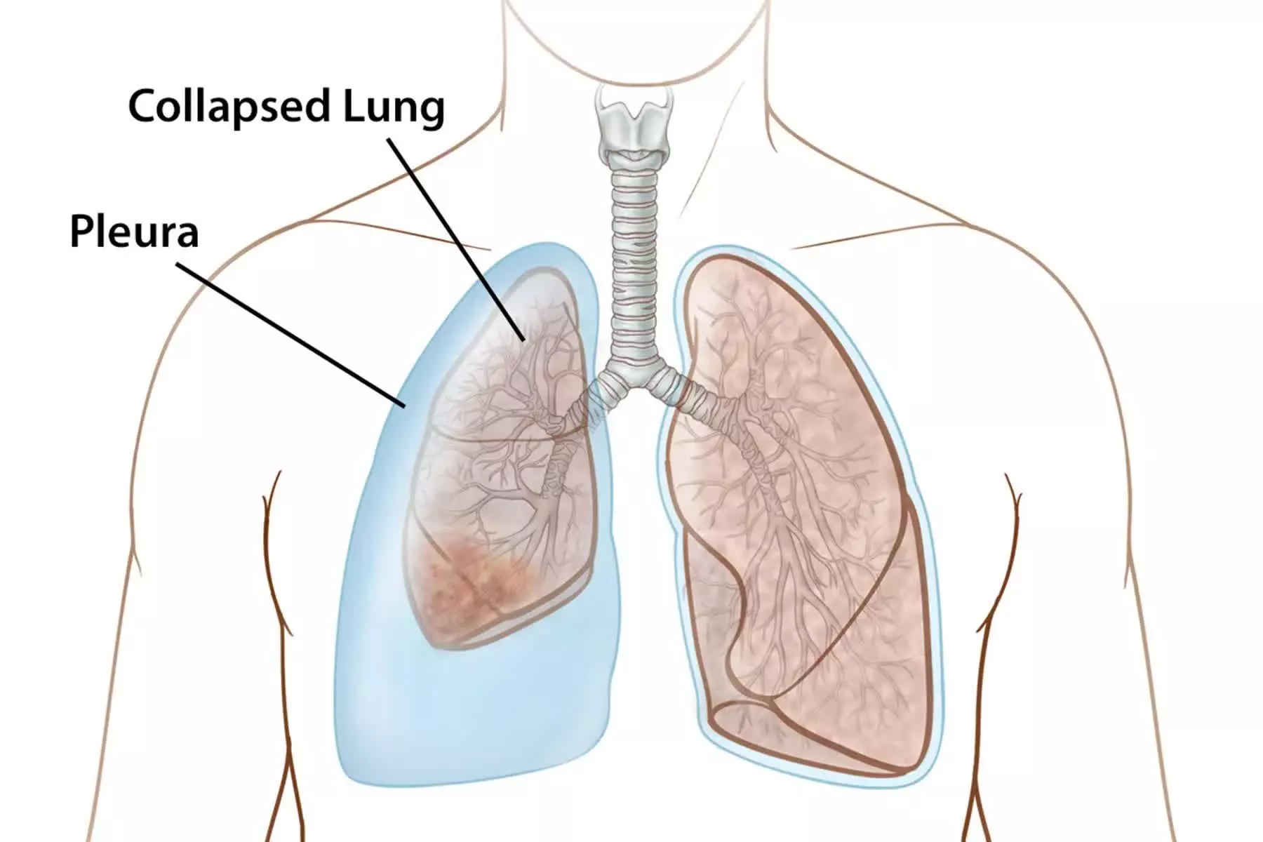
Atelectasis, derived from the Greek words ateles (incomplete) and ektasis (expansion), is a common yet potentially serious respiratory condition characterized by the collapse of part or all of a lung. This collapse involves the deflation of alveoli—the tiny, elastic air sacs in the lungs where the exchange of oxygen and carbon dioxide takes place. When these alveoli are not filled with air, they cannot participate in gas exchange, leading to a reduction in the lung’s functional capacity. Understanding the nuances of atelectasis, from its underlying causes to its comprehensive management, is critical for healthcare professionals aiming to prevent complications and optimize patient outcomes.
Understanding and Discussing Atelectasis
The pathophysiology of atelectasis can be categorized into several types, with the most common being obstructive (or resorptive) atelectasis. This occurs when an obstruction in an airway, such as a mucus plug, foreign body, or tumor, prevents air from reaching the distal alveoli. The air already present in these trapped alveoli is gradually absorbed into the bloodstream, but no new air can enter to replace it, causing the alveoli to collapse.
Non-obstructive atelectasis encompasses several other mechanisms:
- Compression Atelectasis: Occurs when an external force presses on the lung tissue, such as a pleural effusion (fluid in the pleural space), pneumothorax (air in the pleural space), or a large tumor.
- Contraction (Cicatrization) Atelectasis: This is caused by fibrotic changes in the lung or pleura that prevent full expansion. Conditions like pulmonary fibrosis or tuberculosis can lead to scarring that pulls on and collapses lung tissue.
- Adhesive Atelectasis: Results from a deficiency or inactivation of surfactant, a substance that reduces surface tension within the alveoli and prevents them from collapsing at the end of expiration. This is commonly seen in acute respiratory distress syndrome (ARDS) and in premature infants.
- Postoperative Atelectasis: This is the most frequently encountered clinical form, resulting from a combination of factors including the effects of anesthesia, shallow breathing due to pain, immobility, and retained secretions.
Clinical Manifestations of Atelectasis
The signs and symptoms of atelectasis are highly variable and depend on the extent of the lung collapse and the speed at which it develops. Small, patchy areas of atelectasis may be entirely asymptomatic or produce only minor symptoms. However, when a larger area of the lung is involved, especially in an acute setting, the clinical manifestations can be significant.
Common signs and symptoms include:
- Dyspnea (Shortness of Breath): This is a primary symptom, occurring because the reduced lung volume decreases the surface area available for gas exchange, leading to a ventilation-perfusion (V/Q) mismatch.
- Tachypnea (Rapid Breathing): The body attempts to compensate for the decreased gas exchange by increasing the respiratory rate.
- Cough: Often a dry, non-productive cough initially, but it can become productive if associated with retained secretions or a developing infection.
- Chest Pain: Patients may experience pleuritic chest pain, which is sharp and worsens with deep inspiration or coughing.
- Low-Grade Fever: A mild fever is common within the first 24-48 hours, resulting from the inflammatory response to collapsed tissue and retained secretions.
- Tachycardia and Cyanosis: In cases of severe atelectasis with significant hypoxemia, the heart rate increases to compensate, and a bluish discoloration of the skin and mucous membranes (cyanosis) may appear due to low oxygen saturation.
Assessment and Diagnostic Findings
A thorough clinical assessment combined with diagnostic imaging is essential for confirming atelectasis.
Physical Assessment:
- Inspection: Decreased or asymmetrical chest wall movement on the affected side may be visible.
- Palpation: Tactile fremitus (vibrations felt on the chest wall) is typically decreased or absent over the atelectatic area. A key finding in significant collapse is tracheal deviation towards the affected side due to the loss of lung volume.
- Percussion: Tapping on the chest over the affected area will produce a dull sound, as opposed to the normal resonant sound of air-filled lungs.
- Auscultation: Breath sounds will be diminished or completely absent over the collapsed lung tissue. Fine crackles (rales) may be heard during late inspiration as some alveoli pop open.
Diagnostic Studies:
- Chest X-ray (CXR): This is the primary diagnostic tool. It can reveal opacification (whitening) in the affected area, signs of volume loss such as elevation of the hemidiaphragm, and displacement (shift) of the mediastinum or trachea toward the side of the collapse.
- Pulse Oximetry and Arterial Blood Gas (ABG): Pulse oximetry will often show decreased oxygen saturation (SpO2). An ABG analysis can provide a more detailed picture, typically revealing hypoxemia (low PaO2).
- Computed Tomography (CT) Scan: A CT scan offers a more detailed view of the lungs and can help identify the underlying cause of the atelectasis, such as an endobronchial lesion or pleural disease.
- Bronchoscopy: This procedure allows for direct visualization of the airways and can be both diagnostic (to identify an obstruction) and therapeutic (to remove a mucus plug or foreign body).
Complications of Acute Atelectasis
If left untreated, acute atelectasis can lead to several severe complications:
- Hypoxemia: This is the most immediate danger. A significant V/Q mismatch can lead to inadequate oxygenation of the blood, potentially causing respiratory failure.
- Pneumonia: The stagnant secretions trapped in the collapsed airways provide an ideal breeding ground for bacteria, leading to a secondary infection. Atelectasis is a major risk factor for developing hospital-acquired pneumonia.
- Respiratory Failure: If a large portion of the lung collapses or if the underlying lung function is already compromised, the patient may be unable to maintain adequate gas exchange, necessitating mechanical ventilation.
- Sepsis: A severe lung infection can progress to sepsis, a life-threatening systemic inflammatory response.
Medical Management of Atelectasis
The primary goals of medical management are to re-expand the collapsed lung tissue and treat the underlying cause. Strategies are often multifaceted and focus on improving lung volumes and clearing secretions.
- Secretion Management: Measures to mobilize and remove airway secretions are crucial. This includes chest physiotherapy (CPT), which involves percussion, vibration, and postural drainage, as well as nebulized treatments with bronchodilators (e.g., albuterol) to open airways and mucolytics (e.g., acetylcysteine) to thin secretions.
- Lung Expansion Maneuvers: The cornerstone of treatment is encouraging deep breathing. Incentive spirometry provides visual feedback to patients to encourage slow, sustained maximal inspirations. Positive pressure ventilation, either non-invasively via Continuous Positive Airway Pressure (CPAP) or invasively via mechanical ventilation with Positive End-Expiratory Pressure (PEEP), can be used to physically stent the alveoli open.
- Treating the Cause: If an obstruction is present, a bronchoscopy may be performed to remove it. If the cause is compression from a pleural effusion or pneumothorax, a thoracentesis or chest tube placement may be required to drain the fluid or air.
Nursing Management of Atelectasis
Nurses play a pivotal role in the prevention, identification, and management of atelectasis, particularly in at-risk populations like postoperative patients.
Prevention:
- Early Mobilization: Encouraging patients to ambulate as soon as possible after surgery stimulates deep breathing and helps mobilize secretions.
- Frequent Repositioning: Turning patients who are confined to bed at least every two hours helps ventilate different lung regions and prevent the pooling of secretions.
- Deep Breathing and Coughing: Proactively teaching and encouraging patients to perform directed coughing and deep breathing exercises is a fundamental preventive measure.
- Adequate Pain Management: Effective analgesia is critical, as uncontrolled pain prevents patients from taking deep breaths or coughing effectively.
- Patient Education: Instructing patients on the correct use of an incentive spirometer and the importance of all preventive measures is key to adherence and success.
Interventions and Monitoring:
- Respiratory Assessment: Nurses must perform frequent assessments of respiratory rate, effort, breath sounds, and oxygen saturation to detect early signs of atelectasis.
- Administering Treatments: This includes administering supplemental oxygen, nebulizer treatments, and assisting with CPT as ordered.
- Hydration: Ensuring the patient is well-hydrated helps keep respiratory secretions thin and easier to expectorate.
- Collaboration: Nurses work closely with respiratory therapists, physicians, and physical therapists to implement a coordinated care plan aimed at optimizing respiratory function.
In conclusion, atelectasis is a prevalent and preventable condition that ranges from an incidental finding to a life-threatening emergency. A thorough understanding of its pathophysiology, clinical signs, and risk factors allows for prompt diagnosis. Effective management relies on a multidisciplinary approach focused on treating the root cause, clearing secretions, and implementing lung expansion therapies, with nursing care at the forefront of prevention and patient education.
References
- Hinkle, J. L., & Cheever, K. H. (2018). Brunner & Suddarth’s Textbook of Medical-Surgical Nursing (14th ed.). Wolters Kluwer.
- Peroni, D. G., & Boner, A. L. (2000). Atelectasis: mechanisms, diagnosis and management. Paediatric Respiratory Reviews, 1(3), 274–278. https://doi.org/10.1053/prrv.2000.0071
- Mavros, M. N., Velmahos, G. C., & Falagas, M. E. (2011). Atelectasis as a cause of postoperative fever: where is the clinical evidence? Chest, 140(2), 418–424. https://doi.org/10.1378/chest.11-0127
- Woodring, J. H., & Reed, J. C. (1996). Types and mechanisms of pulmonary atelectasis. Journal of Thoracic Imaging, 11(2), 92–108.


