Orbital compartment syndrome (OCS) represents a true ophthalmic emergency, characterized by rapidly escalating pressure within the confined bony orbit that threatens central retinal artery perfusion and, consequently, vision. When conservative measures fail or are insufficient, an urgent surgical intervention known as a lateral canthotomy with inferior cantholysis is required to decompress the orbit and preserve sight. This procedure, while seemingly straightforward, demands precise anatomical understanding, sterile technique, and prompt execution by trained medical professionals.
Introduction to Orbital Compartment Syndrome (OCS) and Lateral Canthotomy
The orbit is a rigid, bony structure that houses the globe, optic nerve, extraocular muscles, fat, and vasculature. Any rapid increase in volume within this confined space, such as from retrobulbar hemorrhage (often post-trauma or surgery), orbital cellulitis, orbital emphysema, or rapidly expanding tumors, can lead to OCS. As pressure rises, it compresses the optic nerve and, critically, the central retinal artery, impairing blood flow to the retina. If not relieved swiftly, this can lead to irreversible ischemic damage and permanent vision loss within hours.
A lateral canthotomy with inferior cantholysis is a sight-saving procedure designed to rapidly decompress the orbit by releasing the lateral canthal tendon, thereby allowing the eyelids to separate and the globe to prolapse slightly forward, reducing intraorbital pressure. This extra space alleviates pressure on the optic nerve and retinal blood supply.
Indications for Lateral Canthotomy
The primary indication for lateral canthotomy is vision loss or impending vision loss due to elevated intraorbital pressure, confirmed by clinical signs and symptoms of OCS. These include:
- Acute vision loss: Decreased visual acuity, often progressing rapidly.
- Afferent pupillary defect (APD): A diminished pupillary response to light in the affected eye, indicating optic nerve dysfunction.
- Proptosis: Forward displacement of the globe, often tense and firm to palpation.
- Ophthalmoplegia: Restricted extraocular movements, leading to diplopia.
- Periorbital ecchymosis and edema: Swelling and bruising around the eye.
- Elevated intraocular pressure (IOP): Though not always directly correlative to intraorbital pressure, a significantly high IOP (e.g., >40 mmHg or > an increase of 10 mmHg above contralateral eye, especially with vision loss) is a strong indicator.
- Chelosis (tightness of eyelids): Inability to open the eyelids due to extreme tension.
- Cherry red spot on fundoscopy: A late, ominous sign of central retinal artery occlusion.
Contraindications (Relative)
While the urgency of OCS often outweighs most relative contraindications, extreme caution is advised in cases of:
- Suspected Globe Rupture: If a ruptured globe is suspected (e.g., irregular pupil, hyphema, exposed uveal tissue, positive Seidel test), performing a lateral canthotomy could potentially worsen the rupture or cause extrusion of intraocular contents. However, if OCS is also present and vision is threatened, slow and careful decompression may still be necessary, often guided by an ophthalmologist.
- Coagulopathy: While not a contraindication in an emergency, it increases the risk of bleeding and requires careful hemostasis.
Anatomy Review
Understanding the anatomy of the lateral canthus is paramount for safe and effective canthotomy:
- Lateral Canthus: The outer corner of the eye where the upper and lower eyelids meet.
- Lateral Canthal Tendon (LCT): A fibrous band that anchors the eyelids to the lateral orbital rim. It originates from the tarsal plates of both eyelids, extends laterally, and inserts into the lateral orbital tubercle of Whitnall on the zygomatic bone.
- Crura of the LCT: The LCT has superior and inferior components (crura) corresponding to the upper and lower eyelids. The inferior crus is the primary target for release in a canthotomy, as its severance allows the lower eyelid to move away from the globe, creating decompression.
- Orbicularis Oculi Muscle: A circular muscle surrounding the orbit, responsible for eyelid closure.
- Conjunctiva: The mucous membrane lining the inner surface of the eyelids (palpebral conjunctiva) and covering the anterior sclera (bulbar conjunctiva).
Equipment
A sterile tray containing the following equipment should be prepared:
- Sterile gloves, gown, and drapes
- Antiseptic solution (e.g., povidone-iodine, chlorhexidine)
- Local anesthetic (e.g., 1-2% lidocaine with epinephrine)
- Syringe (3-5 mL) and 25-27 gauge needle
- Straight iris scissors (blunt-tipped or tenotomy scissors preferred)
- Toothed forceps (e.g., Adson-Brown forceps)
- Mosquito hemostats (straight or curved) for clamping and blunt dissection
- Gauze sponges
- Needle driver and fine absorbable sutures (e.g., 6-0 Vicryl) for potential bleeding control
- Protective eyewear for the operator
- Good light source (headlamp is ideal)
Procedure
I. Preparation
- Consent: If the patient is conscious and time permits, briefly explain the procedure, its necessity, and potential risks. In an emergency, implied consent is assumed, but document the situation.
- Patient Positioning: Position the patient supine with their head stabilized and slightly elevated if possible.
- Analgesia/Anesthesia: If the patient is conscious, administer a local anesthetic by infiltrating 1-2 mL of lidocaine (with epinephrine, if no contraindications) into the skin and deeper tissues of the lateral canthus. Allow a few minutes for the anesthetic to take effect. Systemic analgesia/sedation may also be considered, especially in anxious patients or children.
- Sterile Field: Don sterile gloves. Prepare the area by cleaning the lateral canthus and surrounding skin with an antiseptic solution (e.g., povidone-iodine) and drape the area.
- Identify Anatomy: Visually identify the lateral canthus and mentally map the course of the lateral canthal tendon.
- Protective Measures: Ensure the operator is wearing protective eyewear.
II. The Canthotomy (Lateral Incision)
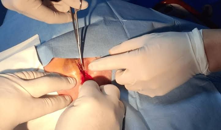
- Isolate the Lateral Canthus: Using the non-dominant hand, gently pull the eyelid skin taut laterally and away from the globe. Some prefer to use forceps to grasp the lateral canthus.
- Make the Incision: With the dominant hand, use the straight iris scissors to make a horizontal incision approximately 1-2 cm long at the lateral canthus, extending laterally from the angle formed by the upper and lower eyelids. The incision should cut through the skin and conjunctiva. Crucially, direct the scissors away from the globe to avoid inadvertent injury. You may hear a “snip” sound as the tissues are cut. This initial incision will cause the canthus to gape slightly.
III. The Cantholysis (Tendon Release, the Critical Step)
- Locate the Lateral Canthal Tendon:
- Place the inferior blade of the scissors into the conjunctival sac, behind the lower eyelid, aiming towards the lateral orbital rim.
- With the other hand, pull the lower eyelid laterally and slightly inferiorly. This will make the inferior crus of the lateral canthal tendon taut and palpable.
- Alternatively, you can use toothed forceps to grasp the freed lower eyelid and pull it away from the globe, making the tendon more accessible.
- Perform Inferior Cantholysis:
- Advance the scissors into the incision, aiming towards the lateral orbital rim.
- Palpate the taut inferior crus of the lateral canthal tendon with the tips of the scissors or a finger.
- Position the open blades of the scissors around the inferior crus of the lateral canthal tendon, ensuring the globe is protected (the inferior blade should be behind the lower eyelid, between the eyelid and the globe).
- Crucially, direct the scissors slightly inferiorly and posteriorly, away from the globe.
- Snip the inferior crus of the lateral canthal tendon. You should feel a distinct “give” or “pop” as the tendon is severed, and the lower eyelid will suddenly become much more mobile and slack, moving inferiorly away from the globe.
- Confirmation: If the lower eyelid does not become significantly more mobile, the tendon may not have been completely severed. Repeat the maneuver, exploring slightly more inferiorly and posteriorly.
IV. Assessing Efficacy and Post-Procedure Care
- Assess Decompression:
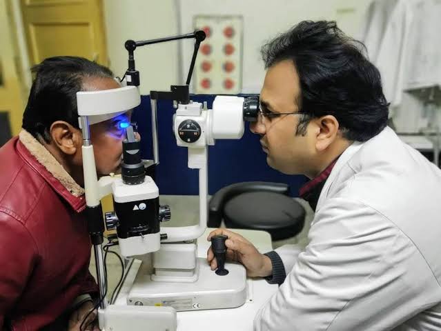
- Immediately re-evaluate the patient’s vision, pupillary response (checking for resolution of APD), and intraocular pressure.
- The globe should appear less proptotic and the eyelids should appear less tense.
- If there is insufficient improvement in vision or IOP, or if the eyelid remains taut, the superior crus of the lateral canthal tendon may also need to be cut. This is less common but can be performed by repeating the cantholysis step, aiming superiorly.
- Hemostasis: Apply direct pressure with gauze to the incision site to control any bleeding. Cautery or fine sutures may be used if bleeding is persistent, but usually, direct pressure is sufficient.
- Dressing and Antibiotics: Apply a sterile, non-adherent dressing to the incision site. Begin broad-spectrum topical and/or systemic antibiotics to prevent infection.
- Ophthalmology Consultation: Urgent ophthalmology consultation is mandatory for definitive management of the underlying cause of OCS and ongoing monitoring.
- Monitoring: Continue to monitor vision, IOP, and the general condition of the eye closely.
Potential Complications
While lateral canthotomy is a sight-saving procedure, potential complications include:
- Hemorrhage: Bleeding from the incision site.
- Infection: Localized cellulitis or orbital infection.
- Globe Perforation: Though rare, inadvertent damage to the globe or surrounding structures (e.g., lacrimal gland, extraocular muscles) can occur if the scissors are directed improperly.
- Incomplete Decompression: Failure to adequately relieve pressure, requiring repeat cantholysis or further intervention.
- Cosmetic Deformity: Notching or scarring of the lateral canthus, rounding of the lateral canthus, or lower eyelid malposition (ectropion) can occur, often requiring reconstructive surgery later.
- Recurrence of OCS: If the underlying cause is not addressed.
Important Considerations and Pearls
- Timeliness: This is a time-critical procedure. Delays can lead to irreversible vision loss.
- Protection of the Globe: Always be acutely aware of the globe’s position and direct instruments away from it. The inferior blade of the scissors should slide along the posterior aspect of the lower eyelid, between the eyelid and the globe.
- Adequate Anesthesia: Ensure sufficient local anesthesia for patient comfort if conscious.
- The “Pop”: Feeling or hearing the distinct “pop” or “snap” as the inferior crus of the lateral canthal tendon is severed is a key indicator of successful cantholysis.
- Post-Op Care: Close follow-up with ophthalmology is essential for management of the underlying cause, wound care, and potential reconstructive surgery.
Conclusion
Lateral canthotomy with inferior cantholysis is a crucial emergency procedure that can prevent permanent vision loss in patients suffering from orbital compartment syndrome. Its successful execution relies on a clear understanding of the relevant anatomy, meticulous sterile technique, and prompt, decisive action. While this guide provides a detailed educational overview, it underscores the necessity of hands-on training and supervised clinical experience for any practitioner involved in emergency ophthalmic care. The preservation of vision hinges on the clinician’s ability to recognize OCS and intervene effectively using this vital technique.
References
- Stone, R. T., & De Silva, S. R. (2018). Lateral Canthotomy and Cantholysis. StatPearls. Available from: https://www.ncbi.nlm.nih.gov/books/NBK499876/
- Desai, V. A., & Sires, B. S. (2018). Orbital Compartment Syndrome (Lateral Canthotomy and Cantholysis). Ophthalmic Plastic and Reconstructive Surgery, 34(5), S116-S122.
- American College of Emergency Physicians. (2019). Clinical Policy: Critical Issues in the Evaluation and Management of Adult Patients With Minor Head Injury. Annals of Emergency Medicine, 74(3), 434-453.
- Lauer, B. A., & Belson, D. L. (2017). Lateral Canthotomy and Cantholysis. The Journal of Emergency Medicine, 52(3), 406-407.
- Karcioglu, Z. A. (2019). Orbital compartment syndrome: A review of diagnosis and management. Seminars in Ophthalmology, 34(4), 211-218.


