Postpartum hemorrhage PPH care baby loss Prevent therapy problem vagina period Lochia mother uterus trauma previa atony labor blood women birth treat Risk
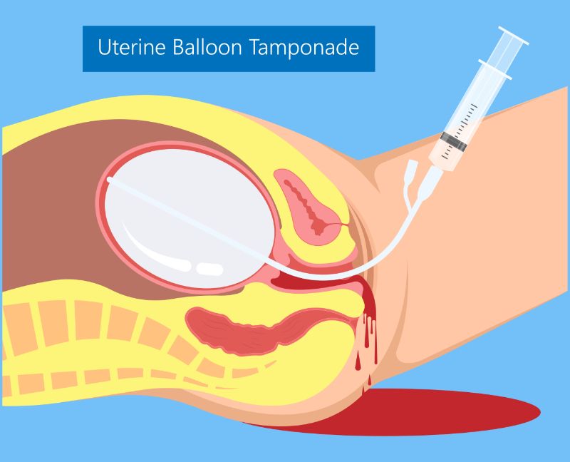
Postpartum hemorrhage (PPH) remains a leading cause of maternal mortality worldwide, with uterine atony—the failure of the uterus to contract adequately after childbirth—being the most common etiology. Prompt and effective management of PPH is critical, and while uterotonic medications and bimanual uterine massage are first-line interventions, a significant proportion of cases require additional measures. Uterine Balloon Tamponade (UBT) is a highly effective, minimally invasive, and life-saving mechanical intervention used to control PPH primarily due to uterine atony, by applying continuous internal pressure against the bleeding uterine wall.
Understanding Uterine Balloon Tamponade
UBT functions by creating an intracavitary pressure that exceeds the systolic arterial pressure within the uterine blood vessels, thereby compressing the vessels and facilitating clot formation. It serves as a bridge to definitive treatment, allowing time for resuscitation, coagulation correction, and preparation for potential further surgical interventions if UBT fails.
Indications and Contraindications
Indications for UBT:
- Persistent PPH due to uterine atony: When initial measures like uterine massage and administration of uterotonic agents (oxytocin, methylergonovine, carboprost, misoprostol) have failed to control bleeding.
- Abnormal placentation: After manual removal of the placenta or curettage for retained products, if bleeding persists.
- Prior to transfer to a higher-level facility: As a stabilizing measure.
- As part of a PPH management algorithm: Often recommended after medical management failure and before surgical options like B-Lynch sutures, uterine artery ligation, or hysterectomy.
Contraindications for UBT:
- Known uterine rupture: UBT can exacerbate bleeding and potentially cause further damage.
- Cervical lacerations or significant lower genital tract trauma: The primary source of bleeding may not be uterine, and UBT will not address this. These require direct surgical repair.
- Uterine anomaly precluding proper balloon placement.
- Active infection: Endometritis or chorioamnionitis, though not an absolute contraindication, requires careful consideration and concomitant antibiotic therapy.
- Retained placental fragments: While UBT might temporarily control bleeding, the retained tissue should be removed as it is the underlying cause. UBT can be used after removal if bleeding persists.
- Coagulopathy: While not an absolute contraindication, a severe coagulopathy might limit the effectiveness of UBT in achieving hemostasis and requires concurrent correction.
Types of UBT Devices
While several commercial UBT devices exist, such as the Bakri balloon, Sengstaken-Blakemore tube (modified for uterine use), and Rusch balloon, simple and effective non-commercial devices can also be improvised. The most common commercial device is the Bakri balloon, designed specifically for uterine tamponade. Improvised devices, like a condom catheter attached to a Foley catheter and inflated with saline, are crucial in resource-limited settings. The principles of insertion and inflation remain largely consistent across devices.
Preparation for UBT Insertion
Successful UBT deployment requires a calm, coordinated, and rapid response from a multidisciplinary team.
- Assemble the Team: Clearly define roles for obstetrician/midwife, anesthesiologist, nurses, and laboratory personnel. Designate a team leader.
- Patient Resuscitation:
- Establish IV access: Ensure at least two large-bore (14-16 gauge) intravenous lines are patent.
- Fluid resuscitation: Administer crystalloids and colloids rapidly to maintain hemodynamic stability.
- Blood products: Type and cross-match blood immediately. Administer packed red blood cells, fresh frozen plasma, and platelets as indicated by blood loss, laboratory results, and clinical status.
- Oxygen therapy: Administer supplemental oxygen via face mask.
- Monitoring: Continuous monitoring of vital signs (heart rate, blood pressure, respiratory rate, oxygen saturation), urine output (via Foley catheter), and estimated blood loss.
- Optimize Uterotonic Use: Ensure all appropriate uterotonic agents have been administered and continue infusions of oxytocin as indicated.
- Equipment Readiness:
- UBT kit: Bakri balloon or equivalent, pre-checked for integrity.
- Sterile saline: Typically 250-750 mL bags (or bottles) for balloon inflation.
- Large syringe: 60 mL syringe for initial inflation.
- Pressure bag or IV pole: For continuous saline infusion into the balloon.
- Sterile gloves and gown: For the operator.
- Antiseptic solution: Povidone-iodine or chlorhexidine for perineal and vaginal preparation.
- Speculum and ring forceps/sponge sticks: For visualizing the cervix and grasping the balloon.
- Foley catheter with drainage bag: If not already in place, to monitor urine output and keep the bladder empty.
- Sutures/tape: To secure the UBT catheter to the thigh.
- Analgesia: Fentanyl, morphine, or regional anesthesia if appropriate and available.
- Patient Assessment: Re-evaluate estimated blood loss, uterine tone, and rule out other causes of PPH (lacerations, retained placental fragments).
- Consent: If the patient is conscious and time permits, briefly explain the procedure and obtain consent.
Procedure for Uterine Balloon Tamponade Insertion
The success of UBT depends on meticulous technique and proper placement.
- Patient Positioning: Place the patient in the dorsal lithotomy position, with legs supported in stirrups.
- Aseptic Technique: Perform thorough perineal and vaginal cleansing with an antiseptic solution. The operator and assistant should wear sterile gloves and gowns, and a sterile field should be draped.
- Inspect the Uterus and Cervix: Manually explore the uterine cavity to ensure no retained placental fragments are present. Systematically inspect the cervix and vagina for lacerations that may be contributing to bleeding. If lacerations are found, repair them before or concurrently with UBT insertion.
- Insert the UBT Device:
- Empty the bladder: Ensure the patient’s bladder is empty, preferably with a Foley catheter in place, as a full bladder can displace the uterus and interfere with effective tamponade.
- Guide the balloon: With one hand palpating the fundus transabdominally, grasp the distal end of the UBT catheter with ring forceps or a sponge stick (or guide manually). Carefully insert the deflated balloon through the cervix and into the uterine cavity, ensuring the balloon tip passes beyond the internal os and into the fundus. The entire balloon portion of the device must be within the uterine cavity; if any part remains in the cervix, it will not tamponade effectively and may cause cervical trauma.
- Confirm placement: When using a Bakri balloon, the inflation port is typically marked to indicate that the balloon segment is completely within the uterus.
- Inflate the Balloon:
- Sterile Saline: Connect a 60 mL syringe filled with sterile saline to the inflation port of the UBT catheter.
- Slow, Controlled Inflation: Slowly inject saline into the balloon. For a Bakri balloon, typically 250-500 mL of saline is injected initially. Continue injecting saline until resistance is felt or, more importantly, until uterine bleeding from the cervix visibly slows or stops. The maximum recommended volume for a Bakri balloon is usually 500-750 mL, but this can vary by manufacturer and clinical judgment.
- Assess Uterine Tone: While inflating, continuously palpate the abdomen to assess uterine tone and size. The uterus should feel firm, and the balloon should fill the cavity without overdistending it.
- Drainage Check: The drain port of the UBT (if present, like on a Bakri balloon) allows observation of ongoing uterine bleeding, which should decrease significantly or stop after adequate inflation.
- Secure the Catheter: Once adequate tamponade is achieved and bleeding is controlled, gently pull the catheter downward slightly to ensure it is snugly against the internal os. Secure the catheter to the patient’s inner thigh with tape, ensuring sufficient tension to maintain pressure. Attach the drainage port (if applicable) to a urine drainage bag to monitor ongoing blood loss from the uterus (which should be minimal).
- Apply External Pressure (Optional but often recommended): For continuous optimal pressure, after initial inflation, connect an IV bag of saline to the inflation port and use a pressure infuser bag to maintain a constant pressure, typically around 300 mmHg. This ensures sustained tamponade as the uterus contracts and relaxes.
Post-Insertion Management
Following successful UBT insertion, vigilant monitoring and supportive care are crucial.
- Continuous Monitoring:
- Vital Signs: Monitor vital signs every 5-15 minutes initially, then hourly once stable.
- Blood Loss: Closely monitor external blood loss from the vagina and blood collected in the UBT drainage bag (if applicable).
- Uterine Tone: Continuously assess uterine tone and fundal height.
- Urine Output: Monitor urine output via the Foley catheter.
- Hemoglobin/Hematocrit: Obtain serial laboratory measurements to assess for ongoing blood loss or resolution.
- Adjunctive Therapies: Continue administration of uterotonic agents as prescribed. Continue fluid resuscitation and blood product transfusions as needed.
- Analgesia: Administer appropriate analgesia to manage discomfort from the balloon.
- Antibiotics: Prophylactic broad-spectrum antibiotics are often considered due to the presence of a foreign body in the uterus, although evidence for this is debated.
- Documentation: Meticulously document the time of insertion, type of device, volume of saline used for inflation, cessation of bleeding, vital signs, and all interventions.
Removal of the UBT Device
The UBT device is typically left in place for 12 to 24 hours after PPH has been controlled and the patient is hemodynamically stable, off vasoactive medications, and no longer actively bleeding.
- Gradual Deflation: The balloon should be deflated gradually over several hours (e.g., remove 50-100 mL every 1-2 hours) while continuously monitoring for recurrent bleeding. This allows the uterus to gradually resume its normal tone and clotting to further consolidate.
- Monitor for Rebound Bleeding: If fresh bleeding resumes during deflation, reinflate the balloon immediately to the previous volume and reassess. This indicates that the uterus is not yet ready for removal, and the balloon may need to remain in place for longer or alternative interventions may be necessary.
- Complete Removal: Once the balloon is completely deflated and there is no evidence of recurrent bleeding for at least 2-4 hours, the catheter can be gently removed.
- Post-Removal Observation: Continue close observation of the patient for several hours after complete removal of the device to ensure sustained hemostasis.
Potential Complications
While UBT is generally safe, potential complications include:
- Uterine perforation: Can occur during insertion, especially if excessive force is used or if the uterus is thinned.
- Infection: Risk of endometritis or ascending infection, though rare with proper technique and often prevented by prophylactic antibiotics.
- Balloon malposition or expulsion: If not properly secured or if the uterus contracts strongly, the balloon can migrate or be expelled.
- Failure of tamponade: If bleeding persists despite maximal inflation, it suggests other etiologies (e.g., severe coagulopathy, undetected lacerations, retained products) or that UBT alone is insufficient, necessitating further surgical interventions.
- Uterine ischemia: Rarely, excessive pressure for prolonged periods could theoretically lead to uterine tissue ischemia, although this is very uncommon.
Key Considerations and Best Practices
- Early Recognition: The most critical step in managing PPH is early recognition and initiation of treatment.
- Multidisciplinary Approach: PPH management, especially with mechanical interventions, requires a coordinated effort.
- Continuous Assessment: Never assume the patient is stable simply because bleeding has reduced. Continuous monitoring is paramount.
- Training and Simulation: Regular training and simulation exercises help teams become proficient and confident in PPH management and UBT insertion.
- Escalation Plan: Always have a clear escalation plan if UBT fails, including preparation for surgical interventions (e.g., uterine artery embolization, B-Lynch sutures, hysterectomy).
Conclusion
Uterine Balloon Tamponade is a highly effective, low-cost, and relatively simple intervention for controlling severe postpartum hemorrhage due to uterine atony when uterotonics fail. Its timely and correct application can prevent life-threatening complications, reduce the need for more invasive surgical procedures, and significantly improve maternal outcomes. By adhering to a systematic, step-by-step approach and maintaining vigilant patient monitoring, healthcare providers can confidently utilize UBT as a crucial component of their PPH management armamentarium.
References
- American College of Obstetricians and Gynecologists (ACOG). (2017). Practice Bulletin No. 183: Postpartum Hemorrhage. Obstetrics & Gynecology, 130(4), e168-e186.
- World Health Organization (WHO). (2012). WHO recommendations for the prevention and treatment of postpartum haemorrhage. Geneva: WHO Press.
- Bakri, Y. N. (2001). Uterine tamponade with the Bakri Postpartum Balloon for intractable postpartum hemorrhage. International Journal of Gynaecology and Obstetrics, 72(1), 139-142.
- Dildy, G. A., & Belfort, M. A. (2023). Uterine balloon tamponade for postpartum hemorrhage. UpToDate. Retrieved from www.uptodate.com.
- Doumouchtsis, S. K., & Papageorghiou, A. T. (2017). Surgical management of postpartum hemorrhage: New insights. Best Practice & Research Clinical Obstetrics & Gynaecology, 40, 12-25.


