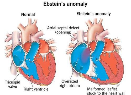
Ebstein anomaly is a rare, complex congenital heart defect characterized by an apical displacement of the septal and posterior leaflets of the tricuspid valve, leading to a varying degree of atrialization of the morphological right ventricle (RV) and tricuspid regurgitation.
Etiology and Clinical Presentation of Ebstein Anomaly
Etiology: Ebstein anomaly arises from an abnormal development of the tricuspid valve during the delamination process of the endocardial cushions, typically occurring between the 8th and 12th weeks of gestation. Instead of detaching normally from the annular ring, the septal and posterior leaflets remain partially tethered to the underlying RV myocardium. This results in their displacement into the RV cavity, creating an “atrialized” portion of the right ventricle above the displaced leaflets and a smaller, functional RV below. The anterior leaflet, while usually less displaced, is often hypertrophied, redundant, and sail-like, frequently tethered to the RV free wall.
While the exact etiology is largely unknown, most cases are sporadic. However, several factors have been implicated:
- Genetic Factors: Familial cases suggest a genetic predisposition, with mutations in genes such as NKX2.5, GATA4, and NOTCH1 occasionally observed, though no single gene mutation accounts for a significant proportion of cases. It is often considered a polygenic or multifactorial condition.
- Maternal Drug Exposure: Maternal lithium exposure during pregnancy, particularly in the first trimester, has been associated with an increased risk of Ebstein anomaly, although this link remains a subject of ongoing research and is not universally accepted as a direct causation for all cases.
- Associated Conditions: Ebstein anomaly frequently co-occurs with other congenital heart defects, most notably an atrial septal defect (ASD) or patent foramen ovale (PFO), which is present in over 80% of cases and often serves as a pathway for right-to-left shunting and cyanosis. Wolff-Parkinson-White (WPW) syndrome, a pre-excitation syndrome, is also found in a significant proportion (5-25%) of patients, predisposing them to supraventricular tachyarrhythmias (SVTs).
Clinical Presentation: The clinical presentation of Ebstein anomaly is remarkably variable, ranging from asymptomatic individuals diagnosed incidentally in adulthood to severely cyanotic neonates with profound heart failure and early mortality. The severity of symptoms depends primarily on the degree of tricuspid valve displacement and regurgitation, the size and function of the functional RV, and the presence and direction of intracardiac shunting (e.g., across an ASD/PFO).
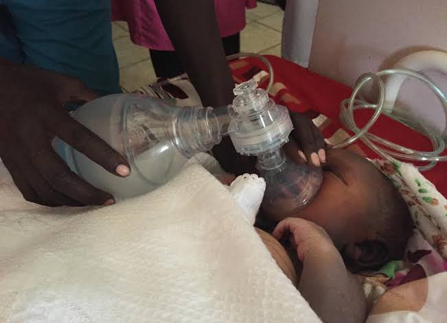
- Neonatal Period: Severe forms typically present in the neonatal period. Profound cyanosis due to significant right-to-left shunting across an ASD/PFO and severe tricuspid regurgitation (TR) leading to right ventricular outflow tract obstruction and reduced pulmonary blood flow is common. Symptoms of right-sided heart failure (tachypnea, poor feeding, hepatomegaly) may also be present. The prognosis in this group is often guarded, with high mortality rates without intervention.
- Childhood and Adolescence: Less severe cases may present later. Symptoms often include fatigue, dyspnea on exertion, palpitations, and mild, intermittent cyanosis, especially with activity. Exercise intolerance is a common complaint. Arrhythmias, particularly supraventricular tachycardias (SVT) related to accessory pathways (WPW), atrial flutter, or atrial fibrillation, can become prominent and symptomatic, causing dizziness, syncope, or chest discomfort.
- Adulthood: Patients who reach adulthood without significant symptoms may still experience progressive worsening of TR, RV dysfunction, and increasing arrhythmias. Dyspnea, exercise intolerance, and chronic fatigue become more pronounced. Right-sided heart failure symptoms (peripheral edema, ascites, jugular venous distension) may develop. Paradoxical emboli through an ASD/PFO are a rare but serious complication.
Physical Examination Findings:
- Cyanosis: May be absent, mild, or severe.
- Cardiomegaly: Often evidenced by a diffuse apical impulse or a prominent right ventricular heave.
- Heart Sounds: A widely split S1 (due to delayed closure of the displaced tricuspid valve) is characteristic. A holosystolic or pansystolic murmur of tricuspid regurgitation is typically heard best at the lower left sternal border and increases with inspiration. Additional sounds like an S3 or S4 gallop may be present. A “sail sound” or “scratchy” late systolic murmur related to the abnormal anterior leaflet can also be heard.
- Jugular Venous Pulsations: May show prominent ‘a’ waves if severe TR leads to high right atrial pressures.
Diagnostic Workup for Ebstein Anomaly
A thorough diagnostic workup is crucial for accurately assessing the severity of Ebstein anomaly and guiding management.
- Electrocardiogram (ECG):
- Right Atrial Enlargement: P-waves are often tall and broad (“P congenitale” or “Himalayan P waves”).
- Right Bundle Branch Block (RBBB): A characteristic incomplete or complete RBBB pattern, often described as an “rsR'” pattern in V1, indicative of RV conduction delay.
- Prolonged PR Interval: Common finding.
- Axis Deviation: Can show right axis deviation.
- Arrhythmias: Evidence of pre-excitation (short PR interval, delta wave) suggestive of Wolff-Parkinson-White syndrome. Ectopic atrial rhythms, SVT, atrial flutter, or atrial fibrillation may be captured or suggested.
- Chest X-ray (CXR):
- Cardiomegaly: Often striking, with the heart appearing globular or “box-shaped” (the “walrus heart” appearance), due to massive right atrial enlargement.
- Pulmonary Vascular Markings: May be decreased in severe cases with reduced pulmonary blood flow due to right-to-left shunt.
- Echocardiography (Transthoracic and Transesophageal):
- Cornerstone of Diagnosis: This is the most critical imaging modality.
- Key Diagnostic Criteria: Apical displacement of the septal and posterior tricuspid valve leaflets from the annulus into the right ventricle (typically >8 mm/m² body surface area).
- Leaflet Morphology: Evaluation of leaflet dysplasia, tethering, and fenestrations of all three leaflets. The anterior leaflet is often large and sail-like.
- Atrialization of the RV: Quantification of the extent of the atrialized portion of the RV and the size and function of the true, functional RV.
- Tricuspid Regurgitation (TR): Assessment of severity (color Doppler, vena contracta, PISA), jet characteristics, and impact on RV function.
- Right Atrial Enlargement: Evaluation of the degree of RA dilation.
- Associated Lesions: Identification of an atrial septal defect (ASD) or patent foramen ovale (PFO), quantification of right-to-left or left-to-right shunting. Assessment of other cardiac structures, including left ventricular function and pulmonary outflow tract.
- Prognostic Scoring: The Celermajer Index (or “Great Ormond Street Score”) uses echocardiographic measurements (ratio of combined areas of the right atrium and atrialized right ventricle to the area of the functional right ventricle and left atrium/left ventricle) to stratify severity and predict outcomes.
- Cardiac Magnetic Resonance Imaging (CMR):
- Detailed Anatomy and Function: Provides exquisite anatomical detail of the RV, tricuspid valve, and surrounding structures, especially valuable in cases with suboptimal echocardiographic windows or complex anatomy.
- RV Volume and Function: Gold standard for accurate quantification of right ventricular volumes, ejection fraction, and myocardial mass, which is crucial for serial follow-up and surgical planning.
- Flow Assessment: Can quantify shunts and regurgitant volumes.
- Myocardial Fibrosis: Late gadolinium enhancement can detect myocardial fibrosis, which may have prognostic implications.
- Cardiac Catheterization and Electrophysiology Study (EPS):
- Diagnostic Catheterization: Rarely needed for primary diagnosis but may be performed to assess pulmonary artery pressures, differentiate from other causes of right heart failure, or quantify shunts that are difficult to assess non-invasively. Characteristic findings include simultaneous pressure tracings from the right atrium and the atrialized RV portion.
- Electrophysiology Study (EPS): Indicated in patients with symptomatic arrhythmias (especially recurrent SVTs or suspected WPW) to map accessory pathways or arrhythmogenic foci, often prior to catheter ablation.
- Fetal Echocardiography: Diagnosis in utero is possible and provides crucial information for prenatal counseling, delivery planning, and early management strategies.
Management for Ebstein Anomaly
Management of Ebstein anomaly is complex and highly individualized, ranging from conservative medical therapy to complex surgical intervention or heart transplantation, depending on the patient’s age, symptom severity, RV function, and the specific anatomical features of the anomaly.
1. Conservative/Medical Management:
- Monitoring: Asymptomatic or mildly symptomatic patients with good RV function are managed conservatively with regular clinical follow-up, including ECG and echocardiography, to monitor for progression of TR, RV dysfunction, and arrhythmias.
- Arrhythmias:
- Antiarrhythmic Drugs: Beta-blockers or other antiarrhythmic medications may be used for symptomatic supraventricular tachycardias or atrial fibrillation.
- Catheter Ablation: For symptomatic Wolff-Parkinson-White syndrome or other re-entrant tachycardias, catheter-based radiofrequency or cryoablation of accessory pathways or arrhythmogenic foci is highly effective and often preferred over chronic drug therapy, especially due to the high success rates and potential for cure.
- Heart Failure: Diuretics (e.g., furosemide) are used to manage volume overload and symptoms of right-sided heart failure. ACE inhibitors or angiotensin receptor blockers may be considered, though their role in predominantly right-sided failure needs careful consideration. Digoxin can be used for rate control in atrial fibrillation but should be used cautiously as patients with Ebstein anomaly may be more sensitive to its effects.
- Cyanosis: In severely cyanotic neonates, prostaglandin E1 infusion may be initiated to maintain patency of the ductus arteriosus, improving pulmonary blood flow and systemic oxygenation, buying time for stabilization or intervention. Oxygen therapy may also be used.
- Endocarditis Prophylaxis: Generally not recommended unless there is a prosthetic valve, previous infective endocarditis, or uncorrected cyanotic congenital heart disease.
2. Surgical Management: Surgery is the definitive treatment for symptomatic or progressive Ebstein anomaly. Indications for Surgery:
- Severe symptoms: Progressive cyanosis, severe dyspnea, fatigue, exercise intolerance.
- Progressive cardiomegaly on CXR.
- Worsening right ventricular dysfunction or dilation.
- Severe tricuspid regurgitation.
- Significant right-to-left shunting through an ASD/PFO causing hypoxemia.
- Recurrent, intractable arrhythmias refractory to medical/ablation therapy.
- Sudden cardiac death risk (though difficult to predict).
Surgical Goals: The primary goals of surgery are to improve tricuspid valve function (reduce TR), decrease the size of the right atrium, restore optimal right ventricular geometry and function, and close any associated shunts.
Types of Surgical Repair:
- Cone Reconstruction (Carpentier-Chauvaud-Brom Procedure): This is the most widely adopted and preferred repair technique. It involves detaching the dysplastic anterior leaflet from the annulus, mobilizing the septal and posterior leaflets, and then reshaping and suturing these leaflets to form a “cone” that functions as a competent monocuspid or bicuspid valve. The atrialized portion of the RV is plicated and excluded from the functional RV, and the tricuspid annulus is reduced. This technique aims to create a highly functional valve and remodel the RV, demonstrating excellent long-term outcomes.
- Dall-Treguenda Repair: Another reconstructive technique involving annuloplasty and plication of the atrialized portion of the RV, often combined with leaflet mobilization or repair.
- Tricuspid Valve Replacement: If the native valve tissue is severely dysplastic or insufficient for repair, valve replacement with a bioprosthetic or mechanical valve may be necessary. This is generally less favored, especially in children, due to issues with growth, calcification, and the need for anticoagulation with mechanical valves.
- Biventricular Repair: The primary goal is always to achieve a biventricular circulation if possible.
- Single-Ventricle Palliation (e.g., Glenn or Fontan Procedure): For patients with exceptionally severe Ebstein anomaly, a hypoplastic functional RV, or significant associated RV outflow tract obstruction where biventricular repair is not feasible, single-ventricle palliation (e.g., superior cavopulmonary anastomosis [Glenn shunt] or total cavopulmonary connection [Fontan procedure]) may be performed to establish a staged circulation. This diverts systemic venous return directly to the pulmonary arteries, bypassing the right ventricle entirely. This is considered a last resort for complex cases.
- Heart Transplantation: Reserved for end-stage right heart failure with intractable symptoms despite other interventions, or when other surgical options are exhausted or contraindicated.
3. Interventional Catheterization:
- Balloon Atrial Septostomy: In severely cyanotic neonates with a highly restrictive interatrial communication (PFO/ASD), balloon atrial septostomy can be performed as an emergency palliative procedure to create a larger opening, allowing better mixing of blood and improving systemic oxygenation, thus “bridging” to definitive surgery.
- Catheter Ablation: As mentioned, for symptomatic arrhythmias, particularly WPW syndrome, catheter ablation is highly effective.
Follow-up: Patients with Ebstein anomaly require lifelong follow-up by a congenital heart disease specialist. Regular monitoring includes clinical assessment, ECGs, and echocardiograms (and possibly CMR) to assess valve function, RV size and function, development of arrhythmias, and any signs of progressive heart failure. Women of childbearing age require careful counseling regarding pregnancy, as it can place significant strain on the heart.
In conclusion, Ebstein anomaly is a fascinating and challenging congenital heart defect with a wide spectrum of clinical presentations. A precise diagnosis, largely driven by echocardiography and CMR, is essential for risk stratification. Management requires a multidisciplinary approach, with individualized medical, interventional, and surgical strategies aimed at optimizing cardiac function, managing symptoms, and improving long-term outcomes for these complex patients.
References:
- Attenhofer Jost CH, Connolly HM, Dearani JA, et al. Ebstein’s Anomaly. Circulation. 2007;115(15):2072-2081. doi:10.1161/CIRCULATIONAHA.106.619152
- Celermajer DS, Bull C, Till JA, et al. Ebstein’s anomaly: presentation and outcome from fetus to adult. J Am Coll Cardiol. 1994;23(1):110-116. doi:10.1016/0735-1097(94)90522-3
- Carpentier A, Chauvaud S, Mace L, et al. A new reconstructive operation for Ebstein’s anomaly of the tricuspid valve. J Thorac Cardiovasc Surg. 1988;96(1):92-101.
- Warnes CA, Williams RG, Bashore TM, et al. Guidelines for the management of adults with congenital heart disease: a report of the American College of Cardiology/American Heart Association Task Force on Practice Guidelines (Writing Committee to Develop Guidelines on the Management of Adults With Congenital Heart Disease). J Am Coll Cardiol. 2008;52(23):e143-e263. doi:10.1016/j.jacc.2008.10.001
- Mayo Clinic. Ebstein’s anomaly. Available at: https://www.mayoclinic.org/diseases-conditions/ebsteins-anomaly/symptoms-causes/syc-20351720 (Accessed [Current Date])
- Dearani JA, Mora BN, Fraser CD Jr, et al. Cone reconstruction for Ebstein’s anomaly: an established surgical therapy. Ann Thorac Surg. 2016;101(6):2359-2365. doi:10.1016/j.athoracsur.2016.01.006
- Lin Y, Li H, Chen Z, et al. Diagnostic value of cardiac magnetic resonance in Ebstein’s anomaly. Quant Imaging Med Surg. 2021;11(11):4759-4770. doi:10.21037/qims-21-209


