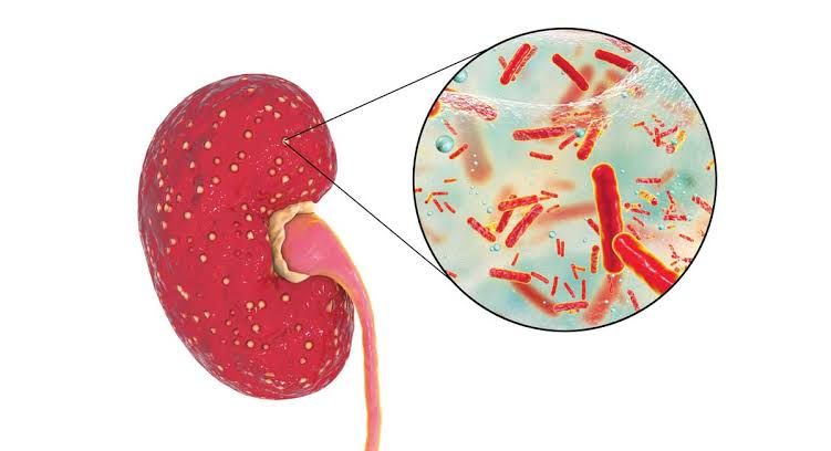
Acute pyelonephritis, a severe infection of the renal parenchyma and collecting system, represents a significant clinical challenge due to its potential for serious complications, including sepsis, renal abscess formation, and chronic kidney damage. Timely and accurate diagnosis combined with an appropriate management strategy is paramount to optimizing patient outcomes.
Diagnostic Workup for Acute Pyelonephritis
Diagnosing acute pyelonephritis hinges on a combination of clinical presentation, laboratory findings, and, in selected cases, imaging studies. The workup aims to confirm the infection, identify the causative pathogen, assess severity, and rule out complications or underlying anatomical abnormalities.
1. Clinical Presentation
Patients typically present with a characteristic symptom constellation:
- Fever and Chills: Often sudden onset and high-grade, indicating systemic inflammation.
- Flank Pain: Unilateral or bilateral pain in the costovertebral angle (CVA), which can radiate to the abdomen or groin. Tenderness to palpation or percussion over the CVA (Murphy’s punch sign) is a classic finding.
- Nausea and Vomiting: Common, often contributing to dehydration and electrolyte imbalances.
- Lower Urinary Tract Symptoms (LUTS): Dysuria (painful urination), urgency, and frequency may precede or accompany the flank pain, but can be absent in some cases.
- Malaise and Fatigue: General signs of systemic illness.
A detailed medical history is crucial, including prior urinary tract infections (UTIs), comorbidities (e.g., diabetes, kidney stones, immunocompromised status), recent instrumentation, and current medications. Physical examination should assess vital signs, hydration status, abdominal tenderness, and CVA tenderness.
2. Laboratory Tests
Laboratory investigations are essential for confirming infection, identifying the pathogen, and evaluating systemic impact.
- Urinalysis (UA): The initial rapid diagnostic test. Key findings include:
- Pyuria: Presence of white blood cells (WBCs) in urine (typically >10 WBCs/HPF), indicating inflammation.
- Bacteriuria: Presence of bacteria (often visualized on microscopy).
- Nitrite Test: Positive nitrites suggest the presence of Gram-negative bacteria that convert nitrates to nitrites (e.g., E. coli).
- Leukocyte Esterase Test: Positive, indicating WBCs in urine.
- Hematuria: Microscopic or macroscopic blood may be present, often due to inflammation.
- Proteinuria: Mild proteinuria can be seen due to inflammation. A clean-catch midstream urine sample is critical to minimize contamination.
- Urine Culture and Sensitivity (C&S): This is the gold standard for identifying the causative pathogen and determining its antibiotic susceptibility profile. A positive culture is typically defined as a single organism count of ≥10^4 colony-forming units (CFU)/mL in a symptomatic patient. Prompt collection is vital, ideally before initiating antibiotic therapy, as treatment can sterilize the urine and confound results. Escherichia coli is the most common pathogen, but Klebsiella pneumoniae, Proteus mirabilis, Enterococcus faecalis, and Pseudomonas aeruginosa can also be implicated.
- Blood Tests:
- Complete Blood Count (CBC) with Differential: Typically shows leukocytosis with a “left shift” (increased band neutrophils), reflecting a bacterial infection.
- Inflammatory Markers: C-reactive protein (CRP) and erythrocyte sedimentation rate (ESR) are usually elevated, indicating systemic inflammation.
- Renal Function Tests: Blood urea nitrogen (BUN) and creatinine should be monitored to assess baseline kidney function and detect any acute kidney injury (AKI) potentially caused by severe infection or dehydration.
- Electrolytes: To assess for dehydration or electrolyte imbalances, especially if there is significant vomiting.
- Blood Cultures: Recommended for patients who appear severely ill, are hospitalized, immunocompromised, have signs of sepsis or septic shock, or fail to respond to initial therapy. Positive blood cultures indicate bacteremia and are associated with increased morbidity and mortality.
- Procalcitonin: Can be useful in differentiating bacterial infections from non-bacterial inflammatory conditions and may correlate with severity, but its routine use in uncomplicated pyelonephritis is debated.
3. Imaging Studies
Imaging is not routinely required for all patients with acute pyelonephritis, especially if the presentation is typical and the patient responds well to empiric therapy. However, it is essential in specific scenarios:
- Failure to Respond to Empiric Antibiotics: Persistent fever or symptoms after 48-72 hours of appropriate antibiotic therapy.
- Severe Sepsis or Septic Shock: To rule out complications like obstruction or abscess.
- Immunocompromised Patients: (e.g., diabetics, transplant recipients, HIV patients) due to higher risk of complications.
- Recurrent Pyelonephritis: To identify underlying anatomical abnormalities.
- Known Urological Abnormalities: (e.g., history of kidney stones, recent urinary tract surgery, congenital anomalies).
- Male Patients: Given a lower incidence in men, an underlying anatomical or functional abnormality should be suspected.
- Pregnancy: Imaging is often considered due to altered anatomy and potential for complications, though with radiation precautions.
- Renal Ultrasound (US): Often the initial imaging modality due to its non-invasiveness and lack of radiation exposure. It is excellent for detecting hydronephrosis (indicating obstruction), renal or perinephric abscesses, and renal calculi. However, it has limited sensitivity for detecting subtle parenchymal inflammation or small abscesses.
- Computed Tomography (CT) Scan: The gold standard for identifying the extent of renal inflammation, abscess formation, renal calculi, urinary tract obstruction, and other complications. Contrast-enhanced CT typically shows characteristic wedge-shaped or streaky areas of decreased enhancement in the inflamed renal parenchyma, and can delineate abscesses or gas-forming infections (emphysematous pyelonephritis). It provides superior anatomical detail compared to ultrasound.
- Magnetic Resonance Imaging (MRI): Can be used as an alternative to CT, especially in pregnant women or patients with contraindications to iodinated contrast. Similar to CT, it can demonstrate renal inflammation, abscesses, and obstruction, though it may be less readily available and more expensive.
- Dimercaptosuccinic Acid (DMSA) Scintigraphy: Primarily used in pediatric populations or in follow-up to detect renal scarring, but not typically in the acute diagnostic phase.
Management Approach for Acute Pyelonephritis
The management of acute pyelonephritis involves prompt initiation of appropriate antibiotics, supportive care, and addressing any underlying complications or predisposing factors.
1. Initial Assessment and Risk Stratification
The decision to manage a patient on an outpatient or inpatient basis depends on disease severity, patient comorbidities, and ability to tolerate oral intake.
- Outpatient Management: Suitable for otherwise healthy, non-pregnant adults with mild-to-moderate symptoms, no signs of sepsis, normal renal function, and ability to tolerate oral medications and fluids. Adequate social support and reliable follow-up are critical.
- Inpatient Management: Indicated for patients with severe symptoms (e.g., high fever, unrelenting pain, persistent vomiting preventing oral intake), signs of sepsis/septic shock, immunocompromised status, pregnancy, suspected urinary tract obstruction, rapid deterioration, uncertain diagnosis, or failed outpatient therapy.
2. Empiric Antibiotic Therapy
Initial antibiotic therapy is empiric, meaning it is started before culture results are available, and should be guided by local resistance patterns, patient comorbidities, and severity of illness.
- Outpatient Therapy:
- Fluoroquinolones (e.g., Ciprofloxacin, Levofloxacin): Highly effective against common uropathogens and achieve high tissue concentrations in the kidney. Often considered first-line, but increasing resistance rates may limit their use. Duration typically 5-7 days for uncomplicated cases.
- Trimethoprim-Sulfamethoxazole (TMP-SMX): Another effective oral option, but resistance is increasing. Should only be used if local resistance rates are known to be low (<20%) or if the pathogen is known to be susceptible. Duration typically 14 days.
- Oral Beta-Lactams (e.g., Amoxicillin-Clavulanate, Cefdinir, Cefpodoxime): Generally less effective than fluoroquinolones or TMP-SMX for empiric pyelonephritis due to lower renal tissue penetration and higher resistance rates for E. coli. May be considered if other options are contraindicated or resistance patterns favor them, often preceded by a single intravenous (IV) dose of a long-acting parenteral agent (e.g., Ceftriaxone or an aminoglycoside). Duration typically 10-14 days.
- Inpatient Therapy (Intravenous):
- Fluoroquinolones (e.g., Ciprofloxacin, Levofloxacin): Excellent choice for empiric IV therapy, particularly if local resistance patterns allow.
- Extended-Spectrum Cephalosporins (e.g., Ceftriaxone, Cefotaxime): Broad-spectrum and effective against many Gram-negative uropathogens.
- Aminoglycosides (e.g., Gentamicin, Amikacin): Effective, especially in conjunction with other agents (e.g., beta-lactams) for severe infections or when Gram-positive coverage is needed. Nephrotoxicity and ototoxicity are concerns, requiring careful monitoring.
- Piperacillin-Tazobactam or Carbapenems (e.g., Ertapenem, Meropenem): Reserved for severe cases, patients with risk factors for multidrug-resistant (MDR) organisms (e.g., recent hospitalization, healthcare-associated infection, prior MDR infections), or those with suspected urosepsis.
- Vancomycin: Should be considered if Gram-positive cocci (e.g., Enterococcus, MRSA) are suspected, especially in severely ill or immunocompromised patients.
Antibiotic therapy should be initiated promptly. Patients started on IV antibiotics can usually be transitioned to oral therapy once afebrile for 24-48 hours and clinically improving, to complete a total course of 7-14 days, depending on the agent and severity.
3. Definitive Therapy
Once urine culture and sensitivity results are available (usually within 24-72 hours), empiric antibiotic therapy should be narrowed to the most appropriate, narrow-spectrum agent with proven efficacy against the identified pathogen. This de-escalation helps reduce the risk of antibiotic resistance and adverse effects. The typical duration of therapy ranges from 7 to 14 days, with some guidelines suggesting 5-7 days for fluoroquinolones in uncomplicated cases.
4. Supportive Care
- Hydration: Oral or intravenous fluids are crucial, especially in patients with fever, vomiting, or signs of dehydration.
- Antipyretics and Analgesics: Acetaminophen or NSAIDs (unless contraindicated) can help manage fever and flank pain.
- Antiemetics: For patients with significant nausea and vomiting, to improve comfort and allow oral intake.
5. Management of Complications
- Urosepsis/Septic Shock: Requires aggressive management in an intensive care setting, including fluid resuscitation, vasopressors, broad-spectrum IV antibiotics, and source control.
- Renal Abscess: May require percutaneous drainage guided by ultrasound or CT, in addition to prolonged courses (4-6 weeks) of appropriate antibiotics.
- Obstructive Pyelonephritis: This is a urological emergency. Urgent relief of obstruction (e.g., via ureteral stent placement or percutaneous nephrostomy) is critical to prevent permanent kidney damage and reduce bacterial load, in addition to antibiotic therapy.
- Emphysematous Pyelonephritis: A severe, necrotizing infection characterized by gas formation within the renal parenchyma or collecting system, often seen in diabetics. Management typically involves aggressive IV antibiotics, fluid resuscitation, and often surgical intervention (e.g., nephrectomy or percutaneous drainage) to remove infected necrotic tissue.
6. Follow-up
- Clinical Monitoring: Patients should be monitored for clinical improvement (decreasing fever, pain, and improved general well-being).
- Repeat Urine Culture: Not routinely recommended for all patients who respond well to therapy. However, it is indicated for pregnant women (test of cure), patients with persistent symptoms, recurrent infections, or those with known structural abnormalities to ensure eradication.
- Imaging Follow-up: If initial imaging revealed complications (e.g., abscess, hydronephrosis), follow-up imaging may be necessary to ensure resolution.
- Identification of Underlying Causes: For male patients, recurrent episodes, or those with complicated pyelonephritis, a urological workup to identify and address underlying anatomical or functional abnormalities (e.g., stones, congenital anomalies, vesicoureteral reflux) is essential.
7. Special Populations
- Pregnancy: All pregnant women with pyelonephritis require hospitalization and intravenous antibiotics (e.g., beta-lactams like cephalosporins; fluoroquinolones, tetracyclines, and TMP-SMX are generally avoided due to fetal risks). Close monitoring and a “test of cure” urine culture are imperative due to the high risk of preterm labor and recurrent UTIs.
- Men: Acute pyelonephritis in men is less common and often indicative of an underlying urological abnormality (e.g., benign prostatic hyperplasia, strictures, stones). A urological investigation is almost always warranted.
- Immunocompromised Patients: (e.g., diabetes mellitus, HIV, transplant recipients) are at higher risk for severe disease and complications. They often require hospitalization, broader empiric antibiotic coverage, and a lower threshold for imaging.
Conclusion
Acute pyelonephritis demands a methodical and timely approach for diagnosis and management. A thorough clinical evaluation complemented by judicious laboratory testing (especially urinalysis and urine culture) and targeted imaging forms the cornerstone of accurate diagnosis. Management involves swift initiation of appropriate empiric antibiotics, tailored to local resistance patterns and patient characteristics, followed by de-escalation based on culture sensitivities. Supportive care and aggressive management of complications are critical. Recognizing specific patient populations and underlying predisposing factors allows for a personalized and comprehensive strategy, ultimately improving patient outcomes and preventing long-term sequelae.
References
- Hooton, T. M., & Gupta, K. (2022). Acute pyelonephritis in adults. UpToDate. Retrieved from https://www.uptodate.com/contents/acute-pyelonephritis-in-adults (A comprehensive and frequently updated resource covering diagnosis and management guidelines).
- Nicolle, L. E., Drekonja, D., Vergis, E. N., Walker, S., Smietana, M., & Taylor, S. N. (2019). Clinical Practice Guideline for the Management of Asymptomatic Bacteriuria: 2019 Update by the Infectious Diseases Society of America. Clinical Infectious Diseases, 68(10), e83-e91. (While focused on asymptomatic bacteriuria, this IDSA guideline provides context on UTI management and associated conditions).
- National Institute for Health and Care Excellence (NICE). (2018). Urinary tract infection (lower) – women: antimicrobial prescribing. NICE guideline [NG112]. Retrieved from https://www.nice.org.uk/guidance/ng112 (Provides UK-based guidelines on UTI management, including acute pyelonephritis aspects).
- Gupta, K., Hooton, T. M., Naber, K. G., Wullt, B., Colgan, R., Miller, G. G., … & Svanborg, C. (2011). International Clinical Practice Guidelines for the Treatment of Acute Uncomplicated Cystitis and Pyelonephritis in Women: A 2010 Update by the Infectious Diseases Society of America and the European Society for Microbiology and Infectious Diseases. Clinical Infectious Diseases, 52(5), e103-e120. (Though published in 2011, this remains a foundational guideline for uncomplicated cases, with principles still relevant).
- Vasudevan, B. (2020). Acute pyelonephritis: an update. British Journal of Hospital Medicine, 81(11), 1-6. (A recent review article providing a concise update on current diagnostic and management strategies).


