The human vascular system is a marvel of biological engineering, designed to maintain the fluidity of blood while simultaneously possessing an intricate capacity to halt bleeding in response to injury. This delicate equilibrium, known as hemostasis, is governed by a finely tuned balance between procoagulant (clot-forming) and anticoagulant (clot-inhibiting) forces. A crucial component of this balance is the system of intravascular anticoagulants, which act continuously within the bloodstream to prevent aberrant or excessive clot formation, thereby ensuring unimpeded blood flow in normal physiological conditions. Among these vital regulators, Protein C and Protein S stand out as central figures, forming a formidable multi-component pathway essential for maintaining vascular patency and preventing pathological thrombosis.
The Role of Intravascular Anticoagulants: Maintaining Vascular Patency
Intravascular anticoagulants are a diverse group of proteins and substances that circulate within the blood or are expressed on the surface of endothelial cells, the lining of blood vessels. Their primary role is to modulate or inhibit the coagulation cascade, a complex sequence of enzymatic reactions that ultimately leads to the formation of a fibrin clot. Without these intrinsic checks and balances, the procoagulant stimuli inherent in the blood, or even minor vascular perturbations, could trigger widespread and life-threatening thrombosis.
The necessity of this constant anticoagulant activity stems from several factors:
- Prevention of Spontaneous Clotting: Even in the absence of overt injury, the blood contains various clotting factors that, if unchecked, could spontaneously activate. Intravascular anticoagulants provide a basal level of inhibition.
- Localization of Clot Formation: When an injury occurs, the goal is to form a localized clot at the site of damage, not throughout the entire circulatory system. Anticoagulants act to contain the clotting process, preventing its spread from the site of injury into the healthy vasculature.
- Regulation of Inflammatory Responses: Many components of the coagulation cascade are intertwined with inflammatory pathways. Intravascular anticoagulants, particularly the Protein C system, also possess anti-inflammatory and cytoprotective properties, contributing to overall vascular health.
- Inactivation of Active Factors: Once coagulation factors are activated, they must be swiftly inactivated to prevent runaway amplification. Anticoagulants achieve this through various mechanisms, including direct inhibition, proteolytic degradation, or acting as cofactors for other inhibitory processes.
Key players in the intravascular anticoagulant system include Antithrombin (a potent inhibitor of thrombin and other activated clotting factors), Tissue Factor Pathway Inhibitor (TFPI, which inhibits the initiation phase of coagulation), and the focus of this discussion, the Protein C system. Together, these systems represent a multi-layered defense against generalized thrombosis, adapting dynamically to maintain vascular homeostasis.
The Coagulation Cascade: A Brief Précis for Context
To fully appreciate the roles of Protein C and Protein S, a brief understanding of the coagulation cascade is beneficial. The cascade is traditionally described through extrinsic and intrinsic pathways, both converging into a common pathway.
- The extrinsic pathway is initiated upon vascular injury by the exposure of Tissue Factor (TF) to Factor VII (FVII). The TF-FVIIa complex then activates Factor X (FX) to FXa and Factor IX (FIX) to FIXa.
- The intrinsic pathway involves factors XII, XI, IX, and VIII, typically activated by contact with negatively charged surfaces or by thrombin. FIXa, along with its cofactor Factor VIIIa (FVIIIa), forms the “tenase complex” which activates FX to FXa.
- The common pathway starts with FXa, which, along with its cofactor Factor Va (FVa), forms the “prothrombinase complex.” This complex converts prothrombin (Factor II) into thrombin (Factor IIa). Thrombin then cleaves fibrinogen into fibrin monomers, which polymerize and are cross-linked by Factor XIIIa to form a stable fibrin clot. Thrombin is a central protease, not only forming fibrin but also activating platelets and several other coagulation factors (V, VIII, XI, XIII), thereby amplifying the clotting response. It is precisely this amplification that the Protein C system is designed to control.
Protein C: The Master Regulator of Procoagulant Cofactors
Protein C is a vitamin K-dependent plasma glycoprotein, synthesized in the liver, that circulates as an inactive zymogen. Its pivotal role in preventing excessive blood clotting lies in its ability, once activated, to proteolytically inactivate the essential cofactors Factor Va and Factor VIIIa, thereby dampening the amplification phase of the coagulation cascade.
The activation of Protein C is a carefully orchestrated process occurring primarily on the surface of intact endothelial cells, representing a critical interface between procoagulant and anticoagulant forces:
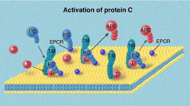
- Thrombin-Thrombomodulin Complex: When thrombin is generated in the bloodstream, instead of solely acting as a procoagulant, it can bind to thrombomodulin (TM), a transmembrane glycoprotein expressed on the surface of healthy endothelial cells. This binding dramatically alters thrombin’s substrate specificity. The thrombin-TM complex loses its procoagulant activity (e.g., cleaving fibrinogen, activating platelets) and gains potent anticoagulant activity.
- Protein C Activation: The thrombin-TM complex then efficiently binds and proteolytically cleaves Protein C, converting it into its active form, Activated Protein C (APC). This endothelial-dependent activation mechanism strategically localizes APC generation to areas where clot formation needs to be contained or prevented, rather than at the immediate site of vascular injury where procoagulant activity is paramount.
- Endothelial Protein C Receptor (EPCR): Another endothelial cell surface receptor, the Endothelial Protein C Receptor (EPCR), further enhances the efficiency of Protein C activation by binding Protein C and presenting it to the thrombin-TM complex. EPCR also plays a role in the cytoprotective and anti-inflammatory functions of APC.
Once generated, Activated Protein C (APC) is a serine protease that exerts its anticoagulant effect by direct enzymatic action:
- Inactivation of Factor Va: APC cleaves and inactivates Factor Va. Factor Va is a critical cofactor for Factor Xa in the prothrombinase complex, which converts prothrombin to thrombin. By degrading FVa, APC significantly reduces thrombin generation.
- Inactivation of Factor VIIIa: Similarly, APC cleaves and inactivates Factor VIIIa. Factor VIIIa is a critical cofactor for Factor IXa in the tenase complex, which activates Factor X to Factor Xa. By degrading FVIIIa, APC reduces the upstream generation of Factor Xa, further curbing thrombin production.
The inactivation of FVa and FVIIIa is irreversible and leads to a profound reduction in coagulation activity. Without these cofactors, the efficiency of thrombin generation plummets, effectively shutting down the amplification loop of the coagulation cascade. Beyond its direct anticoagulant effects, APC also possesses documented anti-inflammatory, anti-apoptotic, and cytoprotective properties, contributing to its potential therapeutic applications in conditions like severe sepsis.
Genetic deficiencies in Protein C (heterozygous or, rarely, homozygous) lead to a severe prothrombotic state, known as thrombophilia, characterized by recurrent venous thromboembolism (VTE). Homozygous deficiency is often fatal in infancy (purpura fulminans).
Protein S: The Essential Cofactor for Activated Protein C
Protein S, like Protein C, is a vitamin K-dependent plasma glycoprotein primarily synthesized in the liver. However, unlike Protein C, Protein S itself does not possess enzymatic activity. Instead, it functions as a crucial non-enzymatic cofactor for Activated Protein C (APC), significantly enhancing APC’s anticoagulant efficacy.
Protein S exists in two forms in the plasma:
- Free Protein S (~40%): This is the biologically active form that can bind to APC.
- Bound Protein S (~60%): This form is reversibly bound to C4b-binding protein (C4BP), an acute-phase protein of the complement system. The binding to C4BP renders Protein S inactive as an APC cofactor. This binding site on C4BP overlaps with the APC-binding site on Protein S, effectively sequestering the active form. During inflammation or infection, levels of C4BP increase, which can lead to a decrease in free Protein S and a transient prothrombotic tendency.
Protein S enhances APC activity through several mechanisms:
- Membrane Binding Enhancement: Protein S facilitates the binding of APC to negatively charged phospholipid surfaces (e.g., on activated platelets or endothelial cells). This localization is critical because the substrates (Factor Va and Factor VIIIa) of APC are themselves bound to these phospholipid membranes. By anchoring APC more effectively to the site of action, Protein S dramatically increases the rate of Factor Va and Factor VIIIa inactivation.
- Conformational Changes: Protein S may induce conformational changes in APC that optimize its proteolytic activity against its substrates.
- Protection from Inhibitors: Protein S might also protect APC from inactivation by endogenous inhibitors, although this role is less definitively established compared to its membrane-binding enhancement.
The synergy between Protein C and Protein S is profound. While APC can inactivate FVa and FVIIIa to some extent on its own, the presence of Protein S accelerates these reactions by several orders of magnitude, making the Protein C system a highly efficient and physiologically relevant anticoagulant pathway.
Genetic deficiencies in Protein S, much like those in Protein C, lead to an increased risk of venous thrombosis (thrombophilia). Individuals with Protein S deficiency are at a higher risk for conditions such as deep vein thrombosis (DVT) and pulmonary embolism (PE).
Synergy and Clinical Significance of the Protein C System
The Protein C system, comprising Protein C, Protein S, thrombomodulin, and EPCR, represents a sophisticated and highly effective mechanism for regulating blood coagulation. Its primary function is to prevent uncontrolled thrombin generation and subsequent widespread clot formation, particularly in the healthy microvasculature. The endothelial cell surface plays a central role, not merely as a passive lining but as an active participant in converting procoagulant signals (thrombin) into anticoagulant responses.
The clinical significance of this system is underscored by the consequences of its dysfunction:
- Thrombophilia: Deficiencies in either Protein C or Protein S are well-established genetic risk factors for venous thromboembolism. These conditions lead to an imbalance favouring coagulation, making affected individuals prone to recurrent thrombotic events.
- Warfarin-Induced Skin Necrosis: A rare but severe complication of warfarin therapy (a vitamin K antagonist) is warfarin-induced skin necrosis. Warfarin inhibits the synthesis of vitamin K-dependent clotting factors, including Protein C and Protein S. Because Protein C has a shorter half-life than many procoagulant factors, its levels can drop precipitously early in warfarin therapy, leading to a transient hypercoagulable state before procoagulant factors are sufficiently depressed. In individuals with underlying Protein C deficiency, this drop can be even more pronounced, leading to microvascular thrombosis and skin necrosis.
- Sepsis and DIC: In severe systemic inflammatory states like sepsis, there can be significant downregulation of thrombomodulin and EPCR on endothelial cells, leading to impaired Protein C activation. Coupled with increased consumption of Protein C and Protein S, this contributes to the procoagulant and inflammatory environment seen in disseminated intravascular coagulation (DIC), a severe hemostatic disorder often complicating sepsis. Therapeutic administration of recombinant human Activated Protein C (rhAPC, drotrecogin alfa activated) was once used in severe sepsis, though debates regarding its efficacy led to its withdrawal from the market, highlighting the complexity of harnessing this pathway therapeutically.
In conclusion, intravascular anticoagulants are indispensable for maintaining blood fluidity and preventing pathological thrombosis. The Protein C system, with Protein C as the executive protease and Protein S as its essential cofactor, orchestrates a critical regulatory pathway. By efficiently inactivating key procoagulant cofactors on endothelial cell surfaces, this system serves as a vigilant guardian, ensuring that the body’s powerful clotting machinery remains under precise control, allowing for localized repair while preserving the integrity of the circulatory system. Understanding these intricate mechanisms is fundamental to comprehending normal hemostasis and developing strategies to manage thrombotic disorders.
References
- Hoffman, M., & Monroe, D. M. (2007). A cell-based model of hemostasis. Thrombosis and Haemostasis, 90(5), 896-905. (Provides a comprehensive overview of hemostasis and the importance of cell surfaces).
- Esmon, C. T. (2009). The protein C pathway. Critical Care Medicine, 37(9 Suppl), S248-S253. (Detailed review of the protein C pathway, its activation, and functions).
- Dahlbäck, B. (2000). The protein C anticoagulant system: new insights into the pathogenesis of hereditary thrombophilia. Thrombosis and Haemostasis, 83(4), 503-504. (Focuses on the role of Protein C and S in hereditary thrombophilia).
- Furie, B., & Furie, B. C. (2008). Thrombus formation in vivo. Journal of Clinical Investigation, 118(5), 1618-1621. (Covers the general mechanisms of thrombus formation and the role of regulatory systems).
- Lane, D. A., & Olds, R. J. (2014). Genetic testing for thrombophilia. Journal of Clinical Pathology, 67(10), 863-870. (Discusses the clinical implications of Protein C and S deficiencies).
- Simioni, P., Castoldi, E., & Tormene, D. (2016). Recent advances in the understanding of the protein C anticoagulant system and its deficiencies. Expert Review of Hematology, 9(1), 77-87. (Offers updated insights into the protein C system, including Protein S interaction and clinical aspects).
- Bouma, B. N., & Meijers, J. C. M. (2007). The protein C anticoagulant pathway. Journal of Thrombosis and Haemostasis, 5(Suppl 1), 58-62. (Concise review of the Protein C pathway, emphasizing its components and physiological roles).
- Kitchens, C. S. (1998). Protein S deficiency: a genetic hypercoagulable state. Seminars in Thrombosis and Hemostasis, 24(1), 21-26. (Specific focus on Protein S deficiency and its clinical manifestations).


