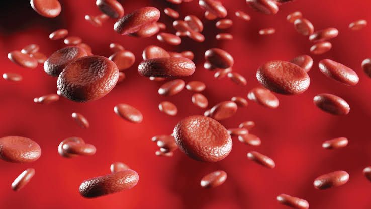
Haemoglobinopathies represent a diverse group of inherited single-gene disorders, making them the most common monogenic diseases globally. These conditions are characterized by abnormalities in the synthesis or structure of the globin chains that constitute haemoglobin, the protein responsible for oxygen transport in red blood cells. Given their significant global burden, understanding their pathogenesis and identifying their morphological signatures on peripheral blood smears is paramount for accurate diagnosis and effective management.
Pathogenesis of Haemoglobinopathies
The pathogenesis of haemoglobinopathies fundamentally stems from genetic mutations affecting the globin genes, leading to either structural variants of haemoglobin (e.g., sickle cell disease) or reduced/absent synthesis of specific globin chains (e.g., thalassemias). The resulting imbalance or dysfunction of haemoglobin subsequently precipitates a cascade of cellular and systemic pathologies.
1. Genetic Basis of Globin Synthesis
Human adult haemoglobin (HbA) is a tetramer composed of two alpha (α) globin chains and two beta (β) globin chains (α2β2). Alpha globin genes (HBA1 and HBA2, totaling four copies) are located on chromosome 16, while beta globin (HBB, two copies), delta globin (HBD), and gamma globin (HBG1 and HBG2) genes are clustered on chromosome 11. Precise regulation of these genes ensures balanced production of globin chains. Disruptions to this delicate balance or modifications to the amino acid sequence of these chains form the foundation of haemoglobinopathies.
2. Pathogenesis of Haemoglobin Structural Variants (e.g., Sickle Cell Disease)
Sickle cell disease (SCD) is the most prevalent haemoglobin structural variant and serves as a prime example of its pathogenesis.
- Genetic Defect: SCD results from a single point mutation in the beta globin gene (HBB gene, located on chromosome 11). Specifically, an adenine-to-thymine transversion at codon 6 (GAG to GTG) leads to the substitution of glutamic acid (a hydrophilic amino acid) by valine (a hydrophobic amino acid) at position 6 of the beta globin chain. This altered beta chain is designated βS. Individuals homozygous for this mutation (HbSS) primarily produce Haemoglobin S (HbS).
- Molecular Consequence (HbS Polymerization): Under deoxygenated conditions (low oxygen tension), the hydrophobic valine residue on the βS chain interacts abnormally with other HbS molecules. This causes the HbS molecules to aggregate and polymerize into long, rigid fibres within the red blood cell cytoplasm. The polymerization is a reversible process initially, but prolonged deoxygenation leads to irreversible sickling.
- Cellular Consequences:
- Red Blood Cell Sickling: The HbS polymers distort the red blood cell membrane, transforming the normally biconcave disc into characteristic, elongated, rigid, crescent-shaped or “sickle” cells. This change impairs the cell’s deformability, its crucial characteristic for navigating microvasculature.
- Haemolysis: Sickle cells are fragile and mechanically unstable. They have reduced lifespans (typically 10-20 days compared to 100-120 days for normal RBCs), leading to chronic extravascular and intravascular haemolysis. Intravascular haemolysis releases free haemoglobin, which scavenges nitric oxide (NO), contributing to endothelial dysfunction and increasing oxidative stress.
- Vaso-occlusion: The rigid, non-deformable sickle cells clump together and obstruct small blood vessels (capillaries and post-capillary venules). This vaso-occlusion is the hallmark of SCD pathogenesis and underlies most acute and chronic complications.
- Inflammation and Endothelial Dysfunction: Chronic inflammation, activation of endothelial cells, increased adhesion molecule expression, and enhanced leukocyte adhesion contribute significantly to the pathophysiology of vaso-occlusion and organ damage.
- Systemic Manifestations: The recurrent episodes of vaso-occlusion and chronic haemolysis lead to a myriad of clinical complications:
- Pain Crises: The most common manifestation, resulting from ischaemia in various tissues.
- Acute Chest Syndrome: Vaso-occlusion in pulmonary vasculature, often triggered by infection.
- Stroke: Cerebral vaso-occlusion.
- Splenic Sequestration and Autoinfarction: Leading to functional asplenia by early childhood, increasing susceptibility to encapsulated bacterial infections.
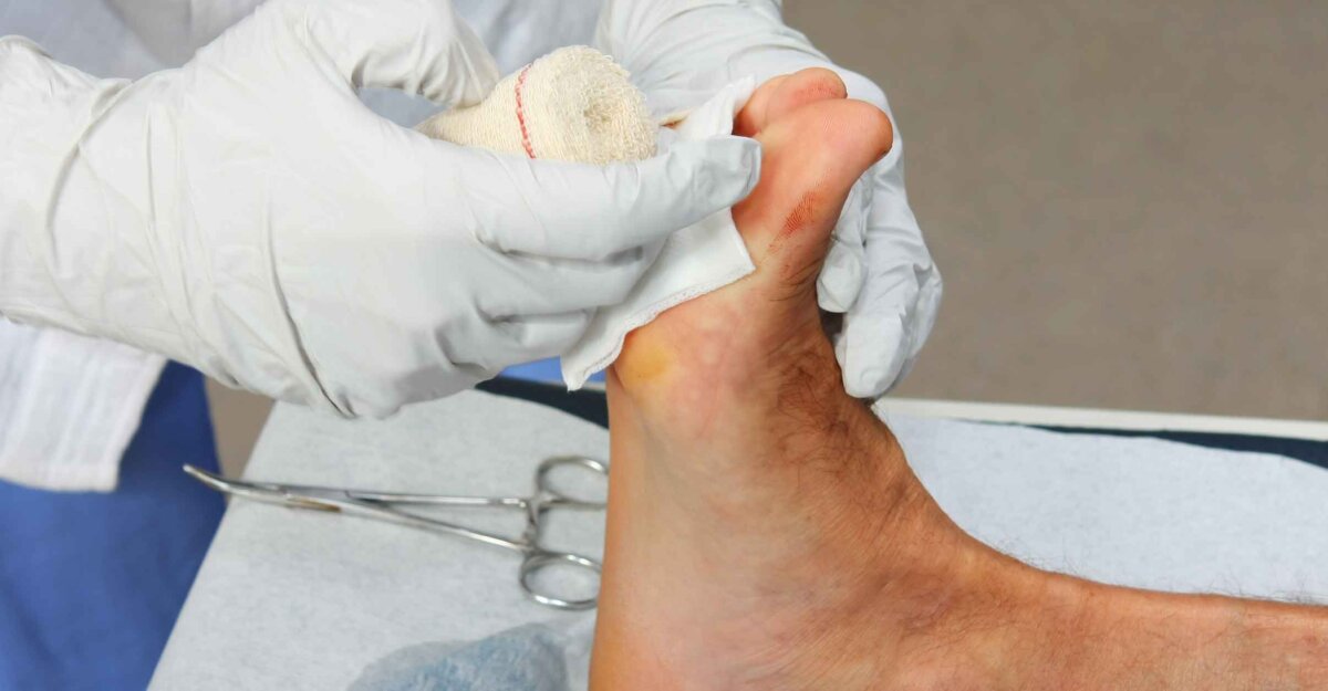
- Renal Failure, Bone Infarction, Leg Ulcers, Priapism, Pulmonary Hypertension, Cholelithiasis (due to chronic haemolysis).
3. Pathogenesis of Thalassemias
Thalassemias are characterized by reduced or absent synthesis of one or more globin chains. They are classified into alpha (α) and beta (β) thalassemias, depending on which globin chain’s production is affected.
- Genetic Defect:
- Alpha Thalassemia: Caused by deletions or, less commonly, point mutations in the alpha globin genes on chromosome 16. Since there are four alpha globin genes, the clinical severity depends on the number of genes affected:
- One gene deletion: Silent carrier (αα/α-).
- Two gene deletion: Alpha thalassemia trait (αα/– or α-/α-).
- Three gene deletion: Hb H disease (–/-α), characterized by excess beta chains forming beta-4 tetramers (Hb H).
- Four gene deletion: Hydrops fetalis (–/–), resulting in excess gamma chains forming gamma-4 tetramers (Hb Barts), incompatible with life.
- Beta Thalassemia: Caused primarily by point mutations or small deletions/insertions in the beta globin gene (HBB) on chromosome 11. These mutations can affect transcription, mRNA processing, or translation, leading to reduced (β+) or absent (β0) beta globin production.
- Heterozygous (β+/β or β0/β): Beta thalassemia trait/minor.
- Homozygous or compound heterozygous (β0/β0, β+/β+, β0/β+): Beta thalassemia major or intermedia.
- Alpha Thalassemia: Caused by deletions or, less commonly, point mutations in the alpha globin genes on chromosome 16. Since there are four alpha globin genes, the clinical severity depends on the number of genes affected:
- Molecular and Cellular Consequences (The Globin Chain Imbalance):
- Imbalanced Globin Chain Synthesis: The core pathogenic mechanism in thalassemia is the disproportionate production of normal globin chains relative to the deficient chain.
- In beta thalassemia, there is an excess of alpha globin chains.
- In alpha thalassemia, there is an excess of beta (or gamma in fetal life) globin chains.
- Precipitation of Unpaired Globin Chains: The surplus, unpaired globin chains are unstable and precipitate within the red blood cell precursors in the bone marrow and mature red blood cells in the circulation.
- Ineffective Erythropoiesis: The precipitate damages the red blood cell membrane and causes oxidative stress. This leads to premature destruction of erythroid precursors in the bone marrow (intramedullary haemolysis), a process known as ineffective erythropoiesis.
- Haemolysis: Unstable red blood cells that escape the marrow are rapidly destroyed in the peripheral circulation, primarily in the spleen (extravascular haemolysis), further contributing to anaemia.
- Iron Overload: Chronic blood transfusions, increased gastrointestinal iron absorption (due to ineffective erythropoiesis), and haemolysis lead to severe iron overload. Iron deposition in vital organs (heart, liver, endocrine glands) causes significant morbidity and mortality.
- Imbalanced Globin Chain Synthesis: The core pathogenic mechanism in thalassemia is the disproportionate production of normal globin chains relative to the deficient chain.
- Systemic Manifestations:
- Severe Anaemia: Primarily due to ineffective erythropoiesis and chronic haemolysis, requiring lifelong blood transfusions in major forms.
- Extramedullary Haematopoiesis: To compensate for severe anaemia, the body attempts to produce red blood cells in sites outside the bone marrow (spleen, liver, paraspinal areas), leading to hepatosplenomegaly and skeletal deformities (e.g., “crew-cut” appearance on skull X-rays).
- Iron Overload Complications: Cardiac failure, liver cirrhosis, diabetes, hypogonadism, hypothyroidism, hypoparathyroidism.
- Growth Retardation and Bone Changes: Due to anaemia and bone marrow expansion.
Morphological Features on Peripheral Blood Smear
Examination of the peripheral blood smear (PBS) is a crucial, initial diagnostic step in evaluating patients suspected of having haemoglobinopathies. The characteristic morphological changes provide valuable clues, distinguishing these conditions from other anaemias and guiding further specific laboratory testing (e.g., haemoglobin electrophoresis, HPLC, genetic studies).
1. General Approach to PBS Examination
When evaluating a PBS for haemoglobinopathies, a systematic approach is essential:
- Assess Red Blood Cell (RBC) Morphology: Size (MCV), shape (poikilocytosis), colour (MCH/MCHC), and inclusions are key.
- Evaluate White Blood Cell (WBC) Morphology: Usually normal unless infection or other specific complications are present.
- Examine Platelet Morphology and Count: Often normal, but can be altered in certain conditions (e.g., thrombocytopenia in hypersplenism).
2. Morphological Features in Sickle Cell Disease (HbSS)
The PBS in SCD reflects both chronic haemolysis and the distinctive red cell sickling phenomenon.
- Sickle Cells (Drepanocytes): These are the most distinctive and pathognomonic feature. They are elongated, crescent-shaped, or oat-shaped red blood cells with pointed ends. Their presence confirms the diagnosis of sickling haemoglobinopathy; however, their number varies and may be scarce during steady-state, becoming abundant during crises or after deoxygenation challenges.
- Target Cells (Codocytes): Found in varying numbers, these cells have a central ‘bullseye’ of haemoglobin surrounded by an area of pallor, then an outer ring of haemoglobin. Target cells are non-specific but common in conditions with increased red cell surface area to volume ratio (e.g., liver disease, post-splenectomy, thalassemias).
- Polychromasia: Indicates reticulocytosis, a compensatory response of the bone marrow to chronic haemolysis. These are larger, bluish-grey red cells representing immature red cells.
- Nucleated Red Blood Cells (NRBCs) / Erythroblasts: Often present, especially in severe anaemia or during aplastic crises, reflecting increased erythropoietic drive and stress erythropoiesis.
- Howell-Jolly Bodies: Small, round, dense basophilic inclusions (remnants of nuclear DNA) typically removed by the spleen. Their presence is highly indicative of functional asplenia or hyposplenism, a common long-term complication of SCD due to recurrent splenic infarctions.
- Basophilic Stippling: Fine, punctate basophilic granules within red cells, representing aggregated ribosomes. Can be seen in various conditions of dyserythropoiesis and chronic anaemia.
- Anisocytosis and Poikilocytosis: Significant variation in red cell size (anisocytosis) and shape (poikilocytosis) are prominent, reflecting the diversity of red cell forms (sickle cells, target cells, fragmented cells).
- Normochromic, Normocytic to Mildly Microcytic Anaemia: Although haemolysis leads to anaemia, the MCV may not be severely microcytic, especially if there is significant reticulocytosis (reticulocytes are larger).
3. Morphological Features in Thalassemias
The PBS findings in thalassemia reflect ineffective erythropoiesis, chronic haemolysis, and the underlying genetic defect in globin chain synthesis. The severity of changes correlates with the clinical phenotype (trait vs. intermedia vs. major).
(a) Beta Thalassemia Major and Intermedia:
- Profound Microcytic, Hypochromic Anaemia: The most striking feature. Red cells are significantly smaller (low MCV) and paler (low MCH/MCHC) than normal, reflecting the impaired haemoglobin synthesis.
- Marked Anisocytosis and Poikilocytosis: Extreme variation in red cell size and shape.
- Target Cells (Codocytes): Very numerous and prominent, due to the increased surface area-to-volume ratio resulting from hypochromia and membrane changes.
- Tear-Drop Cells (Dacryocytes): Pear-shaped or tear-drop-shaped cells, often seen in conditions with ineffective erythropoiesis and extramedullary haematopoiesis (which can lead to marrow fibrosis).
- Elliptocytes/Ovalocytes: Elongated, oval-shaped cells.
- Irregularly Contracted Cells: Red cells with irregular outlines, often representing damaged cells.
- Basophilic Stippling: Coarse and prominent, reflecting ribosomal remnants in severely stressed erythroid precursors and dyserythropoiesis.
- Numerous Nucleated Red Blood Cells (NRBCs) / Erythroblasts: A hallmark feature, often seen in large numbers (sometimes hundreds per 100 WBCs). This signifies intense, compensatory erythropoiesis and severe stress on the bone marrow, often accompanied by extramedullary haematopoiesis.
- Polychromasia: While present due to haemolysis, it is less pronounced relative to the severe anaemia compared to SCD, due to the significant ineffective erythropoiesis.
- Occasional Fragmented Cells (Schistocytes): Can be seen due to the mechanical fragility of the abnormal red cells.
- Pappenheimer Bodies: Small, dark blue granules composed of iron, often visible with Romanowsky stains and confirmed with Prussian blue stain. Result from impaired iron utilization.
- Heinz Bodies (not visible with Wright stain): These are denatured haemoglobin precipitates (e.g., excess alpha chains in beta thalassemia, or Hb H in alpha thalassemia). They are not visible on routine Wright-Giemsa stained smears but can be identified with supravital stains like crystal violet.
(b) Alpha Thalassemia (Hb H Disease):
- Moderate to Severe Microcytic, Hypochromic Anaemia: Similar to beta thalassemia but often less severe than major.
- Marked Anisocytosis and Poikilocytosis: Including many target cells and tear-drop cells.
- Basophilic Stippling and NRBCs: Present, but usually less numerous than in severe beta thalassemia major.
- Cells containing Hb H inclusions: After incubation with a supravital stain (e.g., brilliant cresyl blue), characteristic “golf ball” like inclusions (precipitated Hb H) are visible within red cells in Hb H disease.
(c) Thalassemia Trait (Minor):
- Mild Microcytic, Hypochromic Anaemia or Normal Haemoglobin Levels: Often asymptomatic.
- Mild Anisocytosis and Poikilocytosis:
- Target Cells: Often present, but few in number.
- Elliptocytes/Ovalocytes: Can be seen.
- Occasional Basophilic Stippling: May be present.
- Normal Reticulocyte Count and No NRBCs: Unless there is a co-existing stress.
Conclusion
Haemoglobinopathies represent a fascinating and clinically challenging group of genetic disorders. Their pathogenesis, rooted in intricate molecular defects of globin chain synthesis or structure, leads to profound cellular and systemic consequences, primarily involving chronic anaemia, haemolysis, and end-organ damage. The peripheral blood smear provides invaluable initial insights, with characteristic morphological features such as sickled cells, target cells, microcytic hypochromic red cells, increased nucleated red blood cells, and pathognomonic inclusions acting as critical diagnostic indicators. A thorough understanding of both the pathogenetic mechanisms and the cytological manifestations is crucial for haematologists and clinicians to accurately diagnose, monitor, and manage these conditions, ultimately improving patient outcomes.
References
- Hoffman, R., Benz, E. J., Silberstein, L. E., Heslop, S., Weitz, J., & Anastasi, J. (2018). Hematology: Basic Principles and Practice (7th ed.). Elsevier.
- Rodak, B. F., Carr, J. H., & Smith, L. J. (2016). Rodak’s Hematology: Clinical Principles and Applications (5th ed.). Elsevier.
- Nathan, D. G., & Oski, F. A. (2009). Nathan and Oski’s Hematology of Infancy and Childhood (7th ed.). Saunders Elsevier.
- Weatherall, D. J., & Clegg, J. B. (2001). The Thalassaemia Syndromes (4th ed.). Blackwell Science.
- Steinberg, M. H., Forget, B. G., Higgs, D. R., & Nagel, R. L. (2009). Disorders of Hemoglobin: Genetics, Pathophysiology, and Clinical Management (2nd ed.). Cambridge University Press.
- Kaur, J., & Singh, J. (2018). Peripheral blood smear in hemoglobinopathies: The forgotten tool. Journal of Clinical and Diagnostic Research, 12(1), EC01–EC04.
- Harrison’s Principles of Internal Medicine. (Latest Edition). McGraw-Hill Education.


