Immunoassay techniques represent a cornerstone of modern diagnostics and research, leveraging the exquisite specificity of antigen-antibody reactions to detect and quantify a vast array of biological molecules. From diagnosing infectious diseases and monitoring hormone levels to detecting illicit drugs and environmental contaminants, immunoassays offer powerful tools for highly sensitive and specific analysis.
Introduction to Immunoassays
At its core, an immunoassay is an analytical method that relies on the specific binding between an antibody and its target antigen to detect or quantify a substance (analyte) in a biological sample. This highly specific interaction allows for the isolation and measurement of particular molecules even within complex mixtures. The development of immunoassays revolutionized clinical diagnostics and biomedical research by providing sensitive, rapid, and often cost-effective means of analysis, significantly surpassing previous methods in many applications.
Core Components and Principles
Understanding immunoassay techniques necessitates familiarity with their fundamental building blocks:
A. Antigens: An antigen is any substance that can elicit an immune response, leading to the production of antibodies, and can specifically bind to these antibodies. In immunoassays, the analyte of interest (e.g., a hormone, a pathogen component, a drug) often serves as the antigen to be detected, or an antigen is used to detect specific antibodies in a patient sample.
B. Antibodies: Antibodies are Y-shaped proteins produced by the immune system in response to specific antigens. Their unique ability to bind to an epitope (a specific region on an antigen) with high affinity and specificity is the central mechanism of all immunoassays.
- Polyclonal Antibodies: Produced by multiple B cell clones, they recognize various epitopes on a single antigen. They are heterogeneous and can offer higher signal but may cross-react.
- Monoclonal Antibodies: Produced by a single B cell clone, they recognize only one specific epitope. They are highly specific, consistent, and reproducible, making them ideal for diagnostic kits.
- Recombinant Antibodies: Genetically engineered antibodies offering enhanced control and reproducibility.
C. Labels (Reporters): To enable detection of the antigen-antibody complex, one of the reactants is ‘labeled’ with a detectable marker. The choice of label dictates the detection method and the type of immunoassay. Common labels include:
- Enzymes: Such as horseradish peroxidase (HRP) or alkaline phosphatase (AP), which convert a colorless substrate into a colored, fluorescent, or luminescent product (e.g., in ELISA).
- Fluorophores: Molecules that absorb light at one wavelength and emit it at a longer wavelength (e.g., fluorescein, rhodamine, used in FIA).
- Chemiluminescent molecules: Emit light as a result of a chemical reaction (e.g., luminol, acridinium esters, used in CLIA).
- Radioisotopes: Radioactive atoms that emit radiation (e.g., 125I, used in RIA).
- Biotin/Streptavidin: Biotinylated molecules bind strongly to streptavidin, which can be conjugated to an enzyme or fluorophore, amplifying the signal.
D. Solid Phases: Most immunoassays require immobilizing one of the reactants (antibody or antigen) onto a solid support to facilitate washing steps and separate bound from unbound components. Common solid phases include microtiter plates (96-well or 384-well), magnetic beads, polystyrene beads, nitrocellulose membranes, or glass slides.
E. Key Binding Principles:
- Specificity: The ability of an antibody to bind exclusively to its target antigen and not to other structurally similar molecules.
- Affinity: The strength of the binding between a single antigen epitope and a single antibody binding site.
- Avidity: The overall strength of the interaction between multivalent antibodies and multivalent antigens, taking into account multiple binding sites.
Classification of Immunoassays
Immunoassays can be broadly classified based on several criteria:
A. Based on Label Type:
- Radioimmunoassay (RIA): Uses radioisotopes.
- Enzyme-Linked Immunosorbent Assay (ELISA): Uses enzymes.
- Fluorescent Immunoassay (FIA): Uses fluorophores.
- Chemiluminescent Immunoassay (CLIA): Uses chemiluminescent molecules.
B. Based on Format (Competitive vs. Non-Competitive):
- Competitive Immunoassays: Labeled and unlabeled antigens complete for a limited number of antibody binding sites. The signal is inversely proportional to the analyte concentration.
- Non-Competitive Immunoassays (Sandwich): The analyte (antigen) is “sandwiched” between two antibodies, typically one capture antibody and one detection antibody. The signal is directly proportional to the analyte concentration.
C. Based on Separation (Heterogeneous vs. Homogeneous):
- Heterogeneous Immunoassays: Require physical separation of bound and unbound labeled reagents (e.g., through washing steps). Most ELISAs fall into this category.
- Homogeneous Immunoassays: No physical separation step is needed. The binding event itself causes a change in the signal, often by altering the activity of an enzyme or the fluorescence characteristics of a label (e.g., FPIA, EMIT).
Detailed Discussion of Key Immunoassay Techniques
(i) Enzyme-Linked Immunosorbent Assay (ELISA)
ELISA is one of the most widely used immunoassay formats due to its versatility, sensitivity, and lack of radioactive reagents. It involves immobilizing an antigen or antibody onto a solid phase, followed by a series of binding and washing steps, and finally, enzymatic detection.
A. Direct ELISA (Antigen Detection):
- Coating: Antigen is directly adsorbed onto the wells of a microtiter plate.
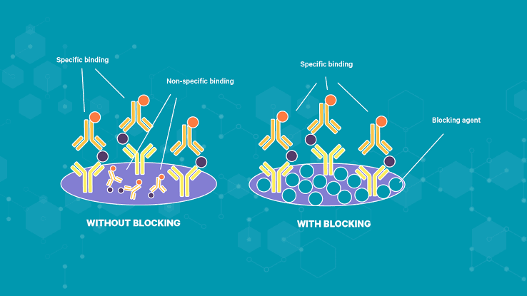
- Blocking: Unoccupied sites on the plate are blocked with an inert protein (e.g., BSA) to prevent non-specific binding.
- Detection: An enzyme-conjugated primary antibody, specific to the antigen, is added and binds to the immobilized antigen.
- Washing: Unbound antibody is washed away.
- Substrate Addition: A chromogenic substrate for the enzyme is added, and the enzyme converts it into a detectable product (color change).
- Measurement: Absorbance is measured spectrophotometrically.
- Application: Primarily for direct antigen quantification.
- Pros: Simple, fast.
- Cons: Less sensitive, potential for high background if primary antibody isn’t pure.
B. Indirect ELISA (Antibody Detection):
- Coating: Antigen (specific to the antibody of interest) is coated onto the plate.
- Blocking: As above.
- Sample Addition: Patient serum (containing primary antibody) is added. If specific antibodies are present, they bind to the coated antigen.
- Washing: Unbound antibodies are washed away.
- Secondary Antibody: An enzyme-conjugated secondary antibody (e.g., anti-human IgG) is added. This secondary antibody binds to the primary antibody.
- Washing: Unbound secondary antibody is washed away.
- Substrate Addition & Measurement: As above.
- Application: Detection of specific antibodies in patient samples (e.g., for infectious diseases like HIV, Lyme disease).
- Pros: Higher sensitivity due to signal amplification by multiple secondary antibodies binding to a single primary antibody, allows use of a single labeled secondary antibody for various primary antibodies.
- Cons: Potential for cross-reactivity with the secondary antibody.
C. Sandwich ELISA (Antigen Detection):
- Coating: A “capture” antibody (specific to the antigen) is coated onto the plate.
- Blocking: As above.
- Sample Addition: The sample containing the antigen is added. The antigen binds to the capture antibody.
- Washing: Unbound components are washed away.
- Detection Antibody: A second, enzyme-conjugated “detection” antibody (recognizing a different epitope on the same antigen) is added, forming a “sandwich” with the captured antigen.
- Washing: Unbound detection antibody is washed away.
- Substrate Addition & Measurement: As above.
- Application: Highly sensitive and specific for antigen detection and quantification (e.g., cytokines, hormones, tumor markers).
- Pros: High specificity (two antibodies binding to different epitopes), high sensitivity, ideal for complex samples as the antigen is “captured.”
- Cons: Requires two antibodies that recognize distinct epitopes on the same antigen, can be affected by “hook effect” at very high antigen concentrations.
D. Competitive ELISA:
- Coating: Either the antigen (labeled or unlabeled) or the capture antibody is coated onto the plate. For instance, if coated with antigen:
- Sample & Labeled Antigen Incubation: The sample (containing unlabeled antigen) and a known amount of enzyme-conjugated antigen are simultaneously added to the well coated with specific antibody (or pre-incubated together).
- Competition: The unlabeled antigen from the sample competes with the labeled antigen for binding to the limited number of antibody sites.
- Washing: Unbound antigens (both labeled and unlabeled) are washed away.
- Substrate Addition & Measurement: As above.
- Application: Detects small molecules that are difficult to “sandwich” (e.g., hormones, drugs, pesticides).
- Pros: Useful for small analytes below 10 kDa which lack multiple binding sites for sandwich assay, high sensitivity.
- Cons: More complex standard curve calculation, lower dynamic range than sandwich ELISAs.
(ii) Radioimmunoassay (RIA)
RIA was the first immunoassay developed (by Yalow and Berson in 1959) and set the standard for sensitivity. It is a competitive assay format where a radioactively labeled antigen competes with unlabeled antigen (from the sample) for binding to a specific antibody.
Procedure Outline:
- A fixed amount of antibody and a fixed amount of radiolabeled antigen are mixed with the sample containing varying amounts of unlabeled antigen.
- The labeled and unlabeled antigens compete for the antibody binding sites.
- After incubation, the antibody-bound antigen is separated from the free antigen (e.g., by precipitation, solid-phase binding).
- The radioactivity in either the bound or free fraction is measured using a gamma counter.
- A standard curve is generated, and the concentration of the unlabeled antigen in the sample is determined by interpolation. The signal is inversely proportional to the analyte concentration.
- Application: Historically used for hormone levels (e.g., insulin, thyroid hormones), therapeutic drug monitoring.
- Pros: Extremely high sensitivity.
- Cons: Health hazards associated with radioactive isotopes, limited shelf-life of labeled reagents, issues with radioactive waste disposal. Largely replaced by non-isotopic methods.
(iii) Chemiluminescent Immunoassay (CLIA)
CLIA harnesses the principle of chemiluminescence, where a chemical reaction generates light without heat. It offers high sensitivity and a wide dynamic range, making it a popular choice in modern automated analyzers.
Principle: Similar to ELISA, CLIA can be competitive or non-competitive (sandwich). The key difference lies in the detection label. Instead of an enzyme producing a colored product, an enzyme (e.g., alkaline phosphatase) acts on a chemiluminescent substrate (e.g., dioxetane derivatives), or a direct chemiluminescent label (e.g., luminol, acridinium esters) is used. This reaction emits light, which is detected by a luminometer.
- Application: Wide range of applications including infectious disease diagnostics, tumor markers, hormones, cardiac markers.
- Pros: Extremely high sensitivity (often surpassing ELISA), wide dynamic range, rapid reaction kinetics, no radioactive waste, suitable for automation.
- Cons: Specialized equipment (luminometer) required, some reagents can be sensitive to light and temperature.
(iv) Fluorescent Immunoassay (FIA)
FIA uses fluorescent molecules as labels. When excited by light of a specific wavelength, these fluorophores emit light at a longer wavelength, which is then detected.
Principle: FIAs can also adopt direct, indirect, sandwich, or competitive formats. The antibody or antigen is labeled with a fluorophore (e.g., fluorescein isothiocyanate, phycoerythrin). After binding and washing steps (for heterogeneous assays), the complex is illuminated, and the emitted fluorescence is measured by a fluorometer. Time-resolved fluorescence immunoassays (TRFIA) use lanthanide chelates (e.g., europium, terbium) with long-lived fluorescence, which helps to separate specific signal from short-lived background fluorescence, enhancing sensitivity.
- Application: Flow cytometry, immunofluorescence microscopy, point-of-care testing (e.g., lateral flow assays with fluorescent reporters), detection of autoantibodies.
- Pros: High sensitivity, rapid detection, no radioactive hazards. TRFIA offers exceptionally high sensitivity and reduced background interference.
- Cons: Susceptible to background fluorescence from biological samples, photobleaching of fluorophores, requires specialized light sources and detectors.
(v) Other Notable Immunoassay Techniques
- Immunochromatographic Assays (Lateral Flow Tests): Rapid, qualitative or semi-quantitative point-of-care tests (e.g., pregnancy tests, COVID-19 antigen tests). They involve a porous membrane strip through which a sample flows by capillary action, encountering labeled reagents and capture lines.
- Immunoblotting (Western Blot): Used to detect specific proteins in a complex mixture. Proteins are separated by gel electrophoresis, transferred to a membrane, and then probed with specific antibodies.
- Immunohistochemistry (IHC) and Immunofluorescence (IF): Used to visualize and localize specific antigens within tissue sections or cells, providing spatial information crucial for pathology and research.
- Flow Cytometry: A cell-based immunoassay that uses fluorescently labeled antibodies to identify and quantify specific cell populations based on surface or intracellular markers.
Key Considerations in Immunoassay Development and Performance
The reliability of immunoassay results hinges on several critical factors:
- Sensitivity: The lowest concentration of an analyte that can be reliably detected.
- Specificity: The ability of an assay to correctly identify only the target analyte without interference from other substances.
- Accuracy: How close the measured value is to the true value.
- Precision (Reproducibility): The degree to which repeated measurements under unchanged conditions show the same results.
- Matrix Effects: Components in the sample (e.g., proteins, lipids) that can interfere with the antigen-antibody reaction or detection system.
- Interferences: Presence of heterophilic antibodies, rheumatoid factors, or human anti-animal antibodies (HAMA) in patient samples can cause false-positive or false-negative results.
- Standardization and Quality Control: Use of certified reference materials, proper calibration, and routine quality control measures are essential for consistent and reliable results.
Applications of Immunoassays
Immunoassays permeate nearly every aspect of modern biology and medicine:
- Clinical Diagnostics: Diagnosis of infectious diseases (e.g., HIV, hepatitis, COVID-19), autoimmune disorders, allergies, cancer (tumor markers), cardiac markers, therapeutic drug monitoring, and endocrine disorders (hormone levels).
- Biomedical Research: Quantification of proteins, cytokines, growth factors; biomarker discovery and validation; drug development; and fundamental studies of protein-protein interactions.
- Food Safety: Detection of allergens, pathogens (e.g., Salmonella, Listeria), toxins, and antibiotic residues.
- Environmental Monitoring: Detection of pollutants, pesticides, and other contaminants in water and soil samples.
Conclusion
Immunoassay techniques stand as a testament to the power of harnessing biological specificity for analytical purposes. From the pioneering days of RIA to the sophisticated automation of modern CLIAs and rapid point-of-care lateral flow tests, these methods have continuously evolved, offering ever-increasing sensitivity, specificity, and throughput. Their continued development, integrating advancements in nanotechnology, microfluidics, and multiplexing, promises even more powerful and diverse applications in diagnostics, research, and beyond, solidifying their role as indispensable tools for understanding and improving health.
References
- Wild, D. (Ed.). (2013). The Immunoassay Handbook. Elsevier Science. (This is a comprehensive reference often cited for immunoassay principles and applications).
- Diamandis, E. P., & Christopoulos, T. K. (1996). Immunoassay. Academic Press. (A classic text covering fundamental aspects and various techniques).
- Liu, X., Lee, J., & Ma, H. (2019). Rapid immunochromatographic detection technologies for food safety: Recent advances and future trends. TrAC Trends in Analytical Chemistry, 114, 24-37.
- Engvall, E., & Perlmann, P. (1971). Enzyme-linked immunosorbent assay (ELISA). Quantitative assay of immunoglobulin G. Immunochemistry, 8(9), 871-874. (Original paper on ELISA).
- Yalow, R. S., & Berson, S. A. (1960). Immunoassay of endogenous plasma insulin in man. Journal of Clinical Investigation, 39(7), 1157-1175. (Original paper on RIA).


