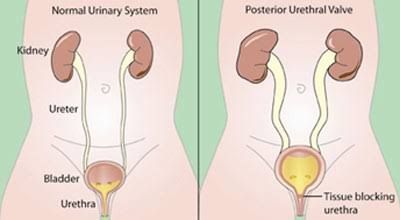
Posterior Urethral Valves (PUV) represent the most common cause of congenital urethral obstruction in male infants and are a significant contributor to end-stage renal disease (ESRD) in children. This condition is characterized by the presence of obstructive folds of tissue in the posterior urethra, leading to varying degrees of bladder outlet obstruction. The resultant high-pressure voiding damages the bladder, ureters, and kidneys, often leading to a spectrum of debilitating urological and renal consequences. Understanding the clinical presentation and adopting a structured management approach is crucial for optimizing outcomes in affected individuals.
Pathophysiology and Embryology
PUV are thought to arise from an anomalous insertion of the Wolffian duct into the cloaca during early embryological development, leading to the formation of sail-like membranes within the posterior urethra. Young’s classification describes three types: Type I (most common) consists of folds extending from the verumontanum distally to the membranous urethra; Type II involves folds extending proximally from the verumontanum towards the bladder neck; and Type III comprises a diaphragm-like membrane distal to the verumontanum. The anatomical obstruction impedes the flow of urine from the bladder, creating suprapubic pressure that progressively dilates and hypertrophies the bladder wall. This chronic obstruction leads to vesicoureteral reflux (VUR) in a significant proportion of cases and obstructive uropathy, causing hydroureteronephrosis and ultimately renal parenchymal damage, often manifesting as dysplasia or progressive scarring (Glassberg et al., 2000).
Clinical Presentation
The clinical presentation of PUV is highly variable, largely depending on the degree of obstruction, the timing of its onset, and the presence of associated complications. Presentation can range from antenatal detection to symptomatic manifestation in later childhood.
1. Antenatal Presentation
With widespread use of prenatal ultrasonography, PUV are increasingly diagnosed in utero. Key sonographic findings suggestive of PUV include:
- Bilateral hydronephrosis and hydroureter: Dilatation of the renal collecting system and ureters due to back pressure.
- Thick-walled, distended bladder: A hallmark sign, often described as a “keyhole” sign due to the dilated posterior urethra.
- Oligohydramnios: Reduced amniotic fluid volume resulting from decreased fetal urine output, indicating significant renal impairment. This is a severe prognostic indicator.
- Ascites or urinoma: Rupture of the collecting system leading to urine leakage into the fetal abdomen.
- Persistent bladder distension after voiding: Suggestive of incomplete emptying.
Antenatal detection allows for early counseling and planning for postnatal management, though intervention in utero (e.g., vesicoamniotic shunt) remains controversial and is reserved for select cases with severe oligohydramnios (Morris et al., 2017).
2. Neonatal Presentation (Birth to 1 month)
Neonates with undiagnosed PUV often present with signs of severe renal or urinary tract dysfunction:
- Poor urinary stream or dribbling: A classic, though sometimes subtle, sign of obstruction.
- Abdominal mass: Palpable distended bladder or hydronephrotic kidneys.
- Respiratory distress: Due to pulmonary hypoplasia secondary to severe oligohydramnios and Potter’s sequence.
- Urosepsis: Fever, lethargy, poor feeding, irritable, often related to urinary tract infection (UTI) in the obstructed system.
- Renal failure: Presenting with electrolyte imbalances (hyponatremia, hyperkalemia, metabolic acidosis), elevated serum creatinine and urea.
- Failure to thrive: General poor growth and development.
3. Infancy and Childhood Presentation
Children who present later often have a less severe obstruction or have compensated initially, but progressive damage eventually becomes symptomatic:
- Recurrent urinary tract infections (UTIs): Due to stasis and incomplete bladder emptying.
- Voiding dysfunction: Straining to void, dribbling, incontinence (daytime or nocturnal enuresis), frequency, urgency.
- Failure to thrive or growth retardation: A hallmark of chronic kidney disease.
- Enlarged bladder and palpable kidneys: On physical examination.
- Hypertension: Due to renovascular changes or chronic kidney disease.
- Hematuria: May occur from bladder wall changes or infection.
- Symptoms of chronic kidney disease: Anemia, bone demineralization, poor appetite.
4. Associated Anomalies and Complications
PUV are frequently associated with other conditions, which can complicate presentation and management:
- Vesicoureteral Reflux (VUR): Present in 30-50% of cases, often unilateral and ipsilateral to a poorly functioning kidney (Duckett & Bellinger, 1989).
- Bladder Dysfunction (PUV bladder): Characterized by detrusor hypertrophy, decreased compliance, and abnormal voiding patterns, leading to urgency, frequency, and incontinence, even after valve ablation.
- Renal dysplasia/hypoplasia: Can coexist, contributing to early renal failure.
- Pulmonary hypoplasia: Resulting from severe oligohydramnios, affecting respiratory function in neonates.
Diagnosis
The diagnosis of PUV relies on a combination of clinical suspicion and specific imaging modalities.
1. Antenatal Ultrasound
As discussed, antenatal ultrasound is pivotal for early detection, identifying the classic “keyhole” sign, hydronephrosis, and oligohydramnios (Lowe & Kaplan, 1999).
2. Postnatal Evaluation
- Clinical Suspicion: Based on presented symptoms (poor stream, distended bladder, signs of renal failure, recurrent UTIs).
- Renal and Bladder Ultrasound (US): Performed postnatally to confirm hydronephrosis, hydroureter, and bladder wall thickening. It also assesses renal parenchymal thickness and echogenicity, offering insights into renal damage.
- Voiding Cystourethrogram (VCUG): This is the definitive diagnostic test for PUV. It demonstrates a dilated and elongated posterior urethra, often with a clear delineation of the valve leaflets at the distal end of the posterior urethra. VCUG also assesses for VUR and bladder morphology (e.g., trabeculation, diverticula).
- Serum Creatinine/Urea and Electrolytes: Essential to assess renal function and detect metabolic derangements. A rising creatinine in the first few days of life is indicative of renal insufficiency.
- Urine Analysis and Culture: To identify infection and assess urine concentration ability.
- Diuretic Renogram (MAG3 scan): May be performed later to assess differential renal function and identify sites of obstruction, particularly in cases of associated VUR or severe hydronephrosis.
Management
The management of PUV involves a multi-pronged approach, focusing initially on stabilization, followed by definitive surgical correction, and comprehensive long-term care to mitigate complications.
1. Initial Stabilization and Resuscitation
For neonates presenting acutely, the immediate priorities are to stabilize their physiological status:
- Urinary Drainage:
- Transurethral catheterization: Placement of a small feeding tube (e.g., 5-8 Fr) into the bladder is the initial step to relieve obstruction, decompress the bladder, and prevent further renal damage. Catheterization also allows for an accurate assessment of urine output and collection for analysis.
- Suprapubic cystostomy or vesicostomy: In cases where transurethral catheterization is difficult or unsuccessful, or in very premature infants, a temporary surgical diversion (e.g., vesicostomy) may be performed to ensure adequate drainage and allow for renal recovery before definitive valve ablation.
- Fluid and Electrolyte Management: Correcting dehydration, acidosis, hyponatremia, and hyperkalemia. These infants can have significant “salt-wasting” or “free-water wasting” nephropathy, requiring careful fluid and electrolyte adjustments.
- Antibiotics: Prophylactic broad-spectrum antibiotics are typically initiated to prevent urosepsis, especially when a catheter is in place or a UTI is suspected.
- Nutritional Support: Adequate nutrition is crucial for growth and renal recovery.
2. Definitive Surgical Management
The primary definitive treatment for PUV is endoscopic ablation.
- Endoscopic Valve Ablation: Performed once the infant is stable and infection-free, typically between 1-2 weeks of age. A small cystoscope is inserted through the urethra, and the valve leaflets are fulgurated (ablated) using a hook electrode or laser. The aim is to create a wide opening for urine flow without damaging the external sphincter. This procedure directly relieves the obstruction and is curative for the valve itself (Smith, 1989).
- Staged Procedures (Less Common): In very ill or extremely premature infants, or those with severe bladder dysfunction, a temporary urinary diversion (e.g., vesicostomy or pyelostomy/ureterostomy) might be performed first to allow for greater renal recovery and bladder decompression, with valve ablation being performed at a later stage. However, the trend is towards primary valve ablation due to potential complications associated with diversions (e.g., stomal stenosis, difficult closure).
3. Long-term Management and Follow-up
Management of PUV extends far beyond valve ablation, focusing on monitoring for and treating the sequelae of chronic obstruction. This requires a multidisciplinary team approach involving pediatric urologists, nephrologists, and specialized nurses.
- Monitoring Renal Function: Regular assessment of serum creatinine, blood urea nitrogen (BUN), electrolytes, and glomerular filtration rate (GFR) is essential to track kidney function and detect progression of renal disease. Urine protein-to-creatinine ratio is also monitored for proteinuria, an indicator of renal damage.
- Managing Bladder Dysfunction (PUV Bladder): Even after valve ablation, many patients experience persistent bladder dysfunction due to irreversible changes in bladder musculature. Management may include:
- Anticholinergic medications: To reduce detrusor overactivity and improve bladder capacity (e.g., oxybutynin, solifenacin).
- Alpha-blockers: To relax the bladder neck (less common).
- Clean Intermittent Catheterization (CIC): For patients with high post-void residuals or significant voiding dysfunction, to ensure complete bladder emptying and prevent UTIs.
- Bladder augmentation: In severe cases of small, non-compliant bladders unresponsive to medical therapy, surgical augmentation using a segment of bowel may be necessary (Kropp et al., 2001).
- Managing Vesicoureteral Reflux (VUR): VUR often resolves spontaneously after valve ablation as bladder pressures decrease. Persistent grade IV-V VUR, especially with recurrent febrile UTIs or progressive renal scarring, may require surgical correction (e.g., ureteral reimplantation or endoscopic bulking agents), though the primary focus is on managing bladder dysfunction and preventing UTIs.
- Preventing and Treating UTIs: Prophylactic antibiotics are often continued for several months post-ablation, especially in the presence of VUR or significant bladder dysfunction. Prompt treatment of any febrile UTI is critical to prevent further renal scarring.
- Addressing Renal Failure: For patients who progress to chronic kidney disease (CKD) or ESRD, management includes:
- Dietary modifications: Low protein, low salt diet.
- Blood pressure control: Using ACE inhibitors or ARBs.
- Anemia management: Erythropoietin supplementation.
- Renal osteodystrophy management: Phosphate binders, vitamin D.
- Dialysis or Renal Transplantation: For ESRD, transplantation offers the best long-term outcome, though bladder dysfunction must be well-managed prior to transplantation (Mattioli et al., 2002).
- Psychosocial Support: Chronic illness can significantly impact the child and family. Support groups, psychological counseling, and education are vital.
Prognosis
Despite advances in management, PUV remain a significant cause of morbidity. Approximately one-third of children with PUV will progress to end-stage renal disease, often requiring dialysis or transplantation. Factors associated with poor renal prognosis include early presentation, severe oligohydramnios, high nadir creatinine in the first year of life, and the presence of severe renal dysplasia or VUR (Churchill et al., 1990). However, timely diagnosis and aggressive, comprehensive management significantly improve outcomes, allowing many children to lead healthy lives.
Conclusion
Posterior Urethral Valves represent a complex congenital anomaly requiring a sophisticated and integrated approach to care. From antenatal detection to definitive treatment and long-term follow-up, a multidisciplinary team is essential to navigate the diverse clinical presentations and potential sequelae. Early recognition, prompt relief of obstruction, diligent management of bladder dysfunction, and close monitoring of renal function are paramount in minimizing the burden of disease and improving the quality of life for these patients, ultimately aiming to preserve renal function and optimize their overall development.
References
- Churchill, B. M., Glassberg, K. I., Packer, M. G., & Belman, A. B. (1990). The management of posterior urethral valves. Journal of Urology, 144(1), 120-123.
- Duckett, J. W., & Bellinger, M. F. (1989). Posterior urethral valves. Urologic Clinics of North America, 16(2), 295-303.
- Glassberg, K. I., Cruz, C., & Noronha, R. F. (2000). The young-PUV syndrome: A proposed classification scheme and review of the literature. Journal of Urology, 163(6), 1894-1899.
- Kropp, B. P., Cheng, E. Y., & Pope, J. C. (2001). Evaluation and management of patients with posterior urethral valves and bladder dysfunction. Pediatric Urology, 58(2), 241-255.
- Lowe, F. C., & Kaplan, G. W. (1999). Posterior urethral valves: An update. Journal of Urology, 161(3), 963-971.
- Mattioli, G., Pini Prato, A., Barabino, A., Montin, D., & Jasonni, V. (2002). Renal transplantation in children with posterior urethral valves. Pediatric Nephrology, 17(11), 934-938.
- Morris, R. K., Kilby, M. D., & Davies, K. E. (2017). Fetoscopic laser ablation of posterior urethral valves: A systematic review and meta-analysis. Prenatal Diagnosis, 37(6), 565-571.
- Smith, G. H. (1989). Posterior urethral valves: A long-term follow-up study. Journal of Pediatric Surgery, 24(7), 652-656.


