Pott’s disease, more formally known as spinal tuberculosis (TB) or tuberculous spondylitis, is an ancient and potentially devastating form of extrapulmonary tuberculosis. It represents approximately 50% of all musculoskeletal tuberculosis cases and continues to be a significant cause of morbidity, particularly in developing nations and immunocompromised populations globally. The term “caries spine” originates from the Latin word caries, meaning “rottenness,” describing the destructive nature of the infection on the vertebral bones.
Understanding the Etiology and Pathogenesis
The primary causative agent of Pott’s disease is Mycobacterium tuberculosis. The infection typically begins elsewhere in the body, most commonly in the lungs (primary focus), and reaches the spine via a hematogenous (blood-borne) spread. The bacteria seed the highly vascularized cancellous bone of the vertebral body, preferring the anterior subchondral region. The lower thoracic and upper lumbar vertebrae are the most frequently involved sites, though any part of the spine can be affected.
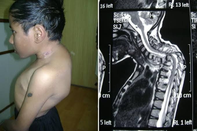
The pathogenesis involves the formation of a granulomatous inflammatory reaction. This leads to caseous necrosis (cheese-like destruction of tissue), osteolysis (bone dissolution), and progressive destruction of the vertebral body. As the anterior vertebral body collapses, a characteristic angular deformity known as kyphosis or “gibbus deformity” develops. The infection can spread beneath the longitudinal ligaments to involve adjacent vertebrae and, critically, can lead to the formation of paravertebral, psoas, or even epidural abscesses. The collapse of bone and extension of abscesses into the spinal canal are the primary mechanisms leading to the most severe complication: neurological deficit from spinal cord compression.
Recognizing the Clinical Presentation
The clinical presentation of Pott’s disease is often insidious and nonspecific, leading to frequent delays in diagnosis. Symptoms can be categorized into three groups:
- Constitutional Symptoms: Patients often present with classic symptoms of chronic infection, including low-grade fever (especially evening pyrexia), night sweats, unintentional weight loss, anorexia, and general malaise.
- Spinal Symptoms: The hallmark is persistent, dull, aching back pain localized to the affected vertebral level. The pain is typically mechanical, worsening with movement, weight-bearing, and at night (often disturbing sleep). The pain becomes progressively more severe as the disease advances. Localized spinal tenderness and palpable spasm of the paraspinal muscles are common findings on physical examination.
- Neurological Symptoms: These are ominous signs and constitute a neurosurgical emergency. They occur in up to 50% of patients and range from radicular pain (pain radiating along a nerve pathway) to motor weakness, sensory deficits, bowel and bladder dysfunction, and ultimately, paraplegia or quadriplegia depending on the level of involvement.
Conducting Diagnostic Investigations
A high index of suspicion is required for diagnosis. The investigative approach is multimodal:
- Imaging:
- Plain Radiographs (X-rays): The initial investigation of choice. Early signs include osteopenia and blurring of the vertebral endplates. Classic late signs include anterior wedging and collapse of the vertebral body, loss of disc height, and the development of a kyphotic deformity. The “vertebra plana” appearance is a severe manifestation.
- Computed Tomography (CT Scan): Excellent for delineating the precise bony anatomy, extent of destruction, fragmentation, and the presence of calcifications within abscesses, which is a characteristic feature of TB.
- Magnetic Resonance Imaging (MRI): The gold standard for imaging. MRI is superior for visualizing soft tissue involvement, including epidural and paravertebral abscesses, spinal cord compression, and meningeal inflammation. It shows characteristic findings like subligamentous spread and prevertebral collections.
- Bone Scan: Can be useful to identify multifocal disease but lacks specificity.
- Laboratory Tests:
- Erythrocyte Sedimentation Rate (ESR) and C-Reactive Protein (CRP): These are invariably elevated and are useful for monitoring response to treatment.
- Tuberculin Skin Test (Mantoux) or Interferon-Gamma Release Assays (IGRAs): These indicate exposure to TB but cannot distinguish between latent and active disease.
- Microbiological and Histopathological Confirmation: This is the definitive diagnostic step. CT or fluoroscopic-guided biopsy of the vertebral lesion or aspiration of an abscess is performed. The sample is sent for acid-fast bacilli (AFB) smear, mycobacterial culture (the gold standard for confirmation, though it takes 4-8 weeks), and polymerase chain reaction (PCR) testing for rapid genetic identification. Histopathology typically shows caseating granulomas.
Implementing Medical Management
Antitubercular chemotherapy is the cornerstone of treatment for Pott’s disease and is effective for the vast majority of patients. The World Health Organization (WHO) recommends a standardized regimen for spinal TB, which typically involves a 6- to 12-month course of four first-line drugs:
- Initial Intensive Phase (2 months): Isoniazid (H), Rifampicin (R), Pyrazinamide (Z), and Ethambutol (E).
- Continuation Phase (4-10 months): Isoniazid (H) and Rifampicin (R).
Treatment must be administered under Directly Observed Therapy (DOT) to ensure adherence and prevent the development of drug-resistant strains. Adjunct therapies include adequate nutrition, pain management, and immobilization of the spine using a brace or cast for a period to rest the spine, prevent deformity, and promote healing.
Determining the Need for Surgical Management
While medical therapy is primary, surgery plays a crucial role in specific scenarios. The common indications for surgery include:
- Neurological Deficit: The development of or progression of paralysis is an absolute indication for surgical decompression of the spinal cord.
- Spinal Instability: Significant vertebral destruction leading to mechanical instability or progressive deformity.
- Failed Medical Therapy: Persistence of symptoms, progression of deformity, or non-resolution of abscesses despite adequate chemotherapy.
- Diagnostic Uncertainty: When a percutaneous biopsy is inconclusive or not feasible, an open biopsy may be required.
- Large Abscesses: Significant paravertebral or psoas abscesses causing symptoms may require surgical drainage.
The goals of surgery are to decompress the neural elements, debride infected tissue, correct the deformity, and achieve spinal stability. Common procedures include:
- Anterior Decompression and Fusion: The traditional and most direct approach to address the pathology, which is typically anterior.
- Posterior Instrumented Fusion: Often used to provide immediate stability and correct kyphotic deformity, sometimes in combination with anterior procedures.
- Debridement and Drainage: For abscesses.
Post-operatively, patients must complete their full course of antitubercular drugs.
Conclusion
Pott’s disease remains a significant clinical challenge. Its diagnosis requires a high index of suspicion and a methodical approach involving advanced imaging and microbiological confirmation. The foundation of treatment is prompt initiation of multi-drug antitubercular therapy. Surgical intervention is reserved for cases with neurological compromise, spinal instability, or complications. With early diagnosis and a comprehensive medical-surgical approach, the prognosis is generally good, aiming to eradicate the infection, preserve neurological function, and prevent crippling spinal deformities.
References:
- Jain, A. K., & Kumar, J. (2013). Tuberculosis of spine: neurological deficit. European Spine Journal, 22(Suppl 4), 624–633.
- Garg, R. K., & Somvanshi, D. S. (2011). Spinal tuberculosis: a review. The Journal of Spinal Cord Medicine, 34(5), 440–454.
- World Health Organization. (2022). WHO consolidated guidelines on tuberculosis: Module 4: Treatment – Drug-resistant tuberculosis treatment. World Health Organization.
- Dunn, R. N., & Ben Husien, M. (2018). Spinal tuberculosis: review of current management. Bone & Joint Journal, 100-B(4), 425-431.
- Nussbaum, E. S., Rockswold, G. L., Bergman, T. A., Erickson, D. L., & Seljeskog, E. L. (1995). Spinal tuberculosis: a diagnostic and management challenge. Journal of Neurosurgery, 83(2), 243–247.


