The circulatory system is a vast and intricate network responsible for transporting life-sustaining substances throughout the body. While arteries and veins serve as the major highways for blood transport, the true work of the circulatory system—the exchange of gases, nutrients, and waste products—occurs at the microscopic level within the capillary beds. These delicate vessels form the crucial interface between the blood and the body’s tissues, governed by a precise interplay of structural design, physiological pressures, and regulatory mechanisms.
Structural Features, Innervation, and Blood Flow of the Capillary System
The structure of a capillary is perfectly tailored to its function of exchange. Understanding its components, control systems, and flow dynamics is fundamental to appreciating its physiological importance.
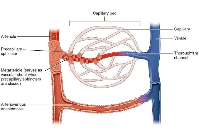
Structural Features: Capillaries are the smallest blood vessels in the body, with a diameter ranging from 5 to 10 micrometers (µm), often just wide enough for red blood cells to pass through in single file. This intimate contact maximizes the surface area and minimizes the diffusion distance for exchange. The capillary wall, or tunica intima, is exceptionally thin, consisting of a single layer of endothelial cells resting on a delicate basement membrane, a non-cellular layer of protein and collagen. Surrounding some capillaries are specialized cells called pericytes, which have contractile properties and play roles in stabilizing the capillary wall, regulating blood flow, and contributing to angiogenesis (the formation of new blood vessels).
There are three primary types of capillaries, distinguished by their permeability:
- Continuous Capillaries: The most common type, found in muscle, skin, lungs, and the central nervous system. The endothelial cells are joined by tight junctions, forming a continuous, uninterrupted lining. Small gaps called intercellular clefts allow for the passage of water and small solutes, but not large molecules. In the brain, these tight junctions are exceptionally robust, forming the basis of the blood-brain barrier, which strictly regulates substance exchange to protect neural tissue.
- Fenestrated Capillaries: These capillaries contain pores, or fenestrae (from the Latin for “windows”), that penetrate the endothelial cells. These pores, often covered by a thin diaphragm, dramatically increase permeability to water and small solutes. Fenestrated capillaries are located in organs where rapid filtration or absorption is required, such as the glomeruli of the kidneys, the villi of the small intestine, and endocrine glands.
- Sinusoidal Capillaries (or Discontinuous Capillaries): The most permeable type, characterized by large intercellular gaps and an incomplete basement membrane. Their wide, irregular shape allows for the passage of large molecules and even blood cells. Sinusoidal capillaries are found in the liver, spleen, bone marrow, and certain endocrine organs, where functions like blood cell formation, removal of old red blood cells, and processing of plasma proteins occur.
Innervation: Direct innervation of the capillary vessels themselves is minimal to non-existent. Instead, the regulation of blood flow into the capillary beds is controlled upstream at the level of the arterioles and precapillary sphincters. These structures are composed of smooth muscle and are richly innervated by the sympathetic nervous system. Sympathetic stimulation causes vasoconstriction of the arterioles, reducing blood flow into the downstream capillary bed. Conversely, a decrease in sympathetic tone leads to vasodilation and increased flow.
More importantly, blood flow at the local level is dominated by autoregulation, where the tissues regulate their own blood supply based on metabolic needs. When a tissue is metabolically active, it produces vasodilator substances like carbon dioxide, adenosine, potassium ions, and hydrogen ions. These local chemical signals cause the smooth muscle of the precapillary sphincters to relax, opening the capillary bed and increasing blood flow to meet the heightened metabolic demand.
Blood Flow: Blood flow through capillaries is characteristically slow and intermittent. The slowness is a direct consequence of the vast total cross-sectional area of the capillary network. While individual capillaries are tiny, their combined area is hundreds of times greater than that of the aorta. According to the principle of continuity in fluid dynamics, flow velocity is inversely proportional to the total cross-sectional area. This slow transit time, typically 1-3 seconds, is critical as it provides ample opportunity for the exchange of substances between the blood and interstitial fluid.
The intermittent nature of the flow is known as vasomotion. It results from the cyclical contraction and relaxation of the precapillary sphincters, which open and close in response to local metabolic signals. This process ensures that blood is directed to areas of the tissue with the greatest need at any given moment, optimizing perfusion efficiency.
The Role of Capillaries as Exchange Vessels
The primary and indispensable role of capillaries is to serve as the site of exchange between the blood and the surrounding interstitial fluid, which bathes the body’s cells. This exchange occurs via three principal mechanisms:
- Diffusion: This is the most important mechanism for the exchange of individual solutes and gases. Substances move down their respective concentration gradients. Oxygen and nutrients, which are in high concentration in the blood, diffuse into the interstitial fluid and then into the cells. Conversely, carbon dioxide and metabolic wastes, which are in high concentration in the tissues, diffuse into the blood to be carried away. Lipid-soluble substances (like O₂ and CO₂) can diffuse directly across the endothelial cell membranes, while water-soluble substances (like ions and glucose) must pass through intercellular clefts or fenestrae.
- Transcytosis: Larger, lipid-insoluble molecules like certain hormones (e.g., insulin) and antibodies are transported across the capillary wall via transcytosis. In this process, the substance is enclosed within a small vesicle (endocytosis) at the luminal side of the endothelial cell, transported across the cell, and released into the interstitial fluid on the other side (exocytosis).
- Bulk Flow: This mechanism involves the movement of a large volume of fluid and its dissolved solutes together across the capillary wall. This movement is not driven by concentration gradients but by pressure gradients. The process of fluid moving from the capillary into the interstitium is called filtration, while the reverse process is called reabsorption. Bulk flow is responsible for regulating the relative volumes of blood plasma and interstitial fluid and is governed by a balance of hydrostatic and osmotic pressures, collectively known as Starling’s forces.
Naming and Approximate Values of Starling’s Forces
The direction and magnitude of bulk flow across the capillary wall are determined by the net balance of four pressures, known as Starling’s forces. These forces can be categorized into two types: hydrostatic pressures (which “push” fluid) and colloid osmotic (or oncotic) pressures (which “pull” fluid).
- Capillary Hydrostatic Pressure (Pc): This is the blood pressure within the capillary, pushing fluid out of the capillary and into the interstitial space. It is the primary force driving filtration. It is higher at the arteriolar end of the capillary and lower at the venous end due to resistance along the vessel.
- Approximate value at arteriolar end: 35 mmHg
- Approximate value at venous end: 15 mmHg
- Interstitial Fluid Hydrostatic Pressure (Pif): This is the pressure of the fluid in the interstitial space, pushing fluid into the capillary. Its value is generally very low and can even be slightly negative in some tissues. For simplicity, it is often considered to be zero.
- Approximate value: 0 mmHg
- Capillary Colloid Osmotic Pressure (πc): Also known as oncotic pressure, this is the osmotic force generated by plasma proteins (primarily albumin) that are too large to easily pass through the capillary wall. These trapped proteins create an osmotic gradient that pulls water into the capillary, favoring reabsorption.
- Approximate value (consistent along the capillary): 25 mmHg
- Interstitial Fluid Colloid Osmotic Pressure (πif): This is the osmotic force created by the small amount of protein present in the interstitial fluid. It tends to pull fluid out of the capillary, favoring filtration. Its value is normally low because very little protein leaks from the capillaries.
- Approximate value: 1 mmHg
The State of Near Equilibrium at the Arteriolar and Venous Ends
The classic Starling principle describes a state of near equilibrium where filtration predominantly occurs at the arteriolar end of the capillary and reabsorption occurs at the venous end. This dynamic can be understood by calculating the Net Filtration Pressure (NFP) at both ends.
NFP = (Forces Favoring Filtration) – (Forces Favoring Reabsorption) NFP = (Pc + πif) – (Pif + πc)
At the Arteriolar End: Here, the capillary hydrostatic pressure (Pc) is high. Using the approximate values:
- Forces Favoring Filtration = Pc (35 mmHg) + πif (1 mmHg) = 36 mmHg
- Forces Favoring Reabsorption = Pif (0 mmHg) + πc (25 mmHg) = 25 mmHg
- NFP = 36 mmHg – 25 mmHg = +11 mmHg
A positive NFP indicates that the net pressure is directed outward, driving the filtration of fluid from the capillary into the interstitial space.
At the Venous End: As blood flows through the capillary, resistance causes the hydrostatic pressure (Pc) to drop significantly, while the oncotic pressure (πc) remains relatively constant.
- Forces Favoring Filtration = Pc (15 mmHg) + πif (1 mmHg) = 16 mmHg
- Forces Favoring Reabsorption = Pif (0 mmHg) + πc (25 mmHg) = 25 mmHg
- NFP = 16 mmHg – 25 mmHg = -9 mmHg
A negative NFP indicates that the net pressure is directed inward, driving the reabsorption of fluid from the interstitial space back into the capillary.
Overall Balance and the Role of the Lymphatic System: Over the entire length of the capillary, the amount of fluid filtered slightly exceeds the amount reabsorbed. This results in a small net loss of fluid from the blood into the interstitial space, amounting to about 2-4 liters per day for the entire body. This excess interstitial fluid, along with any leaked proteins, is collected by the lymphatic system. Lymphatic vessels return this fluid to the circulatory system, thereby preventing its accumulation in the tissues (a condition known as edema) and maintaining the delicate balance of fluid volumes between the blood and the interstitium. This continuous, near-equilibrium state ensures that tissues are adequately perfused and cleared of waste, highlighting the elegant efficiency of the microcirculation.
References:
- Hall, J. E., & Guyton, A. C. (2020). Guyton and Hall Textbook of Medical Physiology (14th ed.). Elsevier.
- Boron, W. F., & Boulpaep, E. L. (2016). Medical Physiology (3rd ed.). Elsevier.
- Pocock, G., Richards, C. D., & Richards, D. A. (2018). Human Physiology (5th ed.). Oxford University Press.
- Levick, J. R., & Michel, C. C. (2010). Microvascular fluid exchange and the revised Starling principle. Cardiovascular Research, 87(2), 198–210. https://doi.org/10.1093/cvr/cvq062


