The intricate web of human metabolism involves a myriad of chemical reactions that sustain life. Among the most crucial are the pathways governing amino acids, the building blocks of proteins. Methionine, cysteine, and cystine constitute a fascinating trio, essential for various physiological processes, from protein synthesis and detoxification to methylation reactions vital for gene expression and neurotransmitter synthesis. Understanding their metabolic pathways is fundamental to comprehending both normal physiological function and the genesis of several significant metabolic disorders.
Metabolic Pathway of Methionine, Cysteine, and Cystine
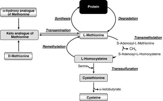
The metabolism of methionine, cysteine, and cystine is deeply interconnected, often referred to as the “sulfur amino acid metabolism” due to the sulfur atoms they contain. Methionine is an essential amino acid, meaning it must be obtained from the diet, while cysteine is conditionally essential, as it can be synthesized from methionine via the transsulfuration pathway. Cystine is simply the disulfide-bonded dimer of two cysteine molecules.
A. The Methionine Cycle (S-Adenosylmethionine (SAM) Cycle)
The methionine cycle is central to the metabolism of all three sulfur amino acids and is critical for numerous biochemical reactions, primarily as a donor of methyl groups.
- Formation of S-Adenosylmethionine (SAM): The cycle begins with methionine reacting with ATP, catalyzed by methionine adenosyltransferase (MAT). This reaction forms S-Adenosylmethionine (SAM), often dubbed “active methionine.” SAM is the primary biological methyl donor, utilized in over 100 methylation reactions involving DNA, RNA, proteins, lipids, and neurotransmitters (e.g., methylation of norepinephrine to epinephrine).
- Methylation and S-Adenosylhomocysteine (SAH) Formation: Once SAM donates its methyl group, it is converted into S-Adenosylhomocysteine (SAH). This step is irreversible under physiological conditions and links the methionine cycle directly to methylation processes.
- Hydrolysis of SAH to Homocysteine: SAH is then hydrolyzed by S-adenosylhomocysteine hydrolase (SAHH) into homocysteine and adenosine. This reaction is reversible, and the accumulation of homocysteine or adenosine can drive the reaction backward, leading to SAH accumulation, which is a potent inhibitor of many SAM-dependent methyltransferases. Therefore, efficient removal of homocysteine is crucial.
B. Homocysteine Disposal and Cysteine Synthesis
Homocysteine, formed from SAH, stands at a critical metabolic crossroads. It can either be remethylated back to methionine or channeled into the transsulfuration pathway to produce cysteine. The balance between these two pathways is vital for maintaining homocysteine levels and ensuring adequate cysteine supply.
- Remethylation Pathway (Homocysteine to Methionine):
- Methionine Synthase Pathway: This is the primary pathway for remethylating homocysteine back to methionine. It requires methionine synthase (MTR), which uses methyl-tetrahydrofolate (methyl-THF) as the methyl donor and vitamin B12 (cobalamin) as a cofactor.
- Betaine-Homocysteine Methyltransferase (BHMT) Pathway: In the liver and kidney, homocysteine can also be remethylated to methionine by betaine-homocysteine methyltransferase (BHMT), using betaine (a derivative of choline) as the methyl donor. This pathway serves as an alternative, particularly when the folate/B12-dependent pathway is compromised or when methionine levels are high.
- Transsulfuration Pathway (Homocysteine to Cysteine): This pathway is the only route for endogenous cysteine synthesis in humans and is irreversible. It requires two key enzymes and vitamin B6 (pyridoxal phosphate) as a cofactor.
- Cystathionine Beta-Synthase (CBS): Homocysteine condenses with serine to form cystathionine, catalyzed by cystathionine beta-synthase (CBS). This is generally considered the rate-limiting step of the transsulfuration pathway.
- Cystathionine Gamma-Lyase (CTH/CSE): Cystathionine is then hydrolyzed by cystathionine gamma-lyase (CTH or CSE) into cysteine, alpha-ketobutyrate, and ammonia. This reaction effectively converts the sulfur of homocysteine into cysteine.
C. Cysteine and Cystine Interconversion and Metabolism
Cysteine, once synthesized or obtained from the diet, serves numerous functions and undergoes further metabolism.
- Cystine Formation: Cysteine contains a sulfhydryl (-SH) group, which can readily undergo oxidation to form a disulfide bond with another cysteine molecule, creating cystine. This reversible dimerization is crucial for protein structure (e.g., disulfide bridges in antibodies) and stabilization. Conversely, cystine reductase enzymes can reduce cystine back to two cysteine molecules.
- Cysteine Catabolism: Cysteine can be catabolized through several oxidative pathways.
- Sulfate Excretion: The main catabolic fate is oxidation of its sulfur to inorganic sulfate, which is then excreted in urine. This pathway yields pyruvate, which can enter the tricarboxylic acid (TCA) cycle for energy production.
- Taurine Synthesis: Cysteine is a precursor for the synthesis of taurine, an important amino acid-like compound involved in bile acid conjugation, osmoregulation, and neurotransmission.
- Glutathione Synthesis: Cysteine is the rate-limiting substrate for the synthesis of glutathione (GSH), a tripeptide (gamma-L-glutamyl-L-cysteinylglycine) that is the body’s primary endogenous antioxidant. GSH plays a critical role in detoxification, redox homeostasis, and immune function.
- Cystine Transport: Cystine, like other amino acids, requires specific transporters for uptake into cells and reabsorption in the kidneys. The primary transporter for cystine (and ornithine, lysine, and arginine – COLA) is the heterodimeric amino acid transporter system b(0,+), composed of SLC7A9 and SLC3A1 subunits.
Metabolic Disorders of Methionine, Cysteine, and Cystine
Disruptions in these intricately linked metabolic pathways can lead to a variety of inherited metabolic disorders, often with severe clinical consequences due to the accumulation of toxic metabolites or deficiencies of essential products.
A. Disorders of Methionine Metabolism
These disorders primarily involve defects in the methionine cycle or related pathways, leading to elevated homocysteine levels (hyperhomocysteinemia) and imbalances in other sulfur amino acids.
- Homocystinuria (Classic Type – Cystathionine Beta-Synthase Deficiency):
- Description: This is the most common inherited disorder of methionine metabolism, caused by a deficiency in the enzyme cystathionine beta-synthase (CBS). This deficiency impairs the transsulfuration pathway, leading to the accumulation of homocysteine and methionine in plasma and urine, while cysteine levels are typically low.
- Pathophysiology: High homocysteine is thought to be toxic to various tissues. The exact mechanisms are complex but involve oxidative stress, endothelial damage, increased thrombogenicity, and interference with collagen cross-linking.
- Clinical Manifestations: Symptoms typically appear in early childhood and include:
- Skeletal abnormalities: Marfanoid habitus (long limbs, scoliosis), osteoporosis, joint laxity.
- Ocular abnormalities: Ectopia lentis (dislocation of the lens), myopia.
- Vascular complications: Thromboembolism (blood clots) in both arteries and veins, a major cause of morbidity and mortality.
- Neurological impairments: Developmental delay, intellectual disability, psychiatric disturbances (e.g., depression, schizophrenia-like illness), seizures.
- Diagnosis: Elevated plasma methionine and homocysteine (total homocysteine >100 µmol/L) and low plasma cysteine are characteristic. Confirmed by CBS enzyme activity assay in fibroblasts or liver, or by genetic testing (CBS gene mutations).
- Treatment:
- Vitamin B6 (Pyridoxine) responsiveness: Approximately 50% of patients respond to high doses of vitamin B6, which acts as a cofactor for CBS, increasing residual enzyme activity.
- Dietary Methionine Restriction: For B6-non-responsive patients, a low-methionine, high-cysteine diet is essential to reduce homocysteine production.
- Betaine Supplementation: Betaine (trimethylglycine) acts as an alternative methyl donor in the BHMT pathway, promoting the remethylation of homocysteine to methionine, thereby lowering homocysteine levels.
- Folate and B12 supplementation: To support the remethylation pathway.
- Disorders of Homocysteine Remethylation: These conditions result from defects in the enzymes and cofactors required to convert homocysteine back to methionine. They are characterized by elevated plasma homocysteine but low or normal plasma methionine (unlike CBS deficiency where methionine is high).
- Methylenetetrahydrofolate Reductase (MTHFR) Deficiency:
- Description: Deficiency in MTHFR prevents the synthesis of 5-methyl-THF, the methyl donor for methionine synthase.
- Clinical Manifestations: Variable, ranging from severe (early-onset neurological dysfunction, developmental delay, seizures, microcephaly) to milder forms depending on residual enzyme activity. Elevated homocysteine and low/normal methionine.
- Treatment: High-dose folate (methyl-THF) and betaine supplementation, often with B12.
- Methionine Synthase (MTR) Deficiency and Cobalamin (Vitamin B12) Metabolism Defects:
- Description: Defects in MTR itself or in the enzymes responsible for reducing dietary B12 to its active forms (methylcobalamin and adenosylcobalamin) lead to impaired MTR activity. This includes disorders like methylmalonic aciduria with homocystinuria (e.g., cblC, cblD, cblF defects), which impair both methylmalonyl-CoA mutase and methionine synthase.
- Clinical Manifestations: Neurological symptoms (developmental delay, hypotonia, seizures, megaloblastic anemia) are prominent, along with hyperhomocysteinemia and hypomethioninemia.
- Treatment: Parenteral vitamin B12 supplementation (often hydroxocobalamin), betaine, and folate.
- Methylenetetrahydrofolate Reductase (MTHFR) Deficiency:
B. Disorders of Cysteine and Cystine Metabolism/Transport
These disorders typically involve issues with the direct handling, transport, or lysosomal processing of cysteine and its dimer, cystine.
- Cystinuria:
- Description: An autosomal recessive disorder characterized by defective transport of dibasic amino acids (cystine, ornithine, lysine, and arginine – COLA) in the renal tubules and intestinal tract. The defect is in the system b(0,+), a heteromeric amino acid transporter (SLC3A1 and SLC7A9 genes).
- Pathophysiology: Due to impaired reabsorption in the renal tubules, high concentrations of these amino acids, particularly cystine (which is poorly soluble), accumulate in the urine. This supersaturation leads to the formation of hexagonal-shaped cystine stones in the kidneys, ureters, and bladder.
- Clinical Manifestations: The primary clinical manifestation is recurrent nephrolithiasis (kidney stones), which can cause pain, urinary tract infections, hematuria, and progressive kidney damage, potentially leading to renal failure. Usually presents in childhood or young adulthood.
- Diagnosis: Detection of characteristic hexagonal cystine crystals in urine sediment, positive urinary cyanide-nitroprusside test, and elevated urinary excretion of cystine and dibasic amino acids.
- Treatment:
- High Fluid Intake: To dilute urine and reduce cystine concentration.
- Urinary Alkalinization: Medications like potassium citrate raise urinary pH, increasing cystine solubility.
- Thiol-binding drugs: For severe cases, drugs like D-penicillamine or tiopronin can react with cystine to form more soluble disulfide complexes, preventing stone formation.
- Dietary modifications: Limiting methionine intake may be considered, as it is a precursor to cystine.
- Cystinosis:
- Description: A rare, autosomal recessive lysosomal storage disorder caused by mutations in the CTNS gene, encoding cystinosin. Cystinosin is a lysosomal membrane protein responsible for transporting cystine out of lysosomes into the cytoplasm.
- Pathophysiology: Defective cystine transport leads to the accumulation of cystine crystals within lysosomes in various cells throughout the body. This accumulation is toxic, causing cellular dysfunction and premature cell death.
- Clinical Manifestations: Three main forms, varying in severity:
- Infantile Nephropathic Cystinosis (most common and severe): Presents in the first year of life with Fanconi syndrome (renal tubular dysfunction leading to excessive urinary loss of electrolytes, glucose, amino acids, and phosphate), growth retardation, rickets, and renal failure by age 10. Systemic involvement includes ocular manifestations (corneal crystals, photophobia), thyroid dysfunction, diabetes mellitus, and muscle weakness.
- Intermediate (Juvenile) Cystinosis: Later onset of renal Fanconi syndrome and slower progression to renal failure.
- Ocular Non-Nephropathic Cystinosis (Adult Cystinosis): Primarily affects the eyes with corneal cystine crystals and photophobia, with minimal to no renal involvement.
- Diagnosis: Measurement of elevated cystine levels in white blood cells (leukocytes) is the gold standard. Slit-lamp examination reveals pathognomonic corneal cystine crystals. Genetic testing confirms CTNS mutations.
- Treatment:
- Cysteamine (Cystagon®): The cornerstone of therapy. Cysteamine reacts with lysosomal cystine to form cysteine and a mixed disulfide, which can then exit the lysosome via another transporter, effectively depleting lysosomal cystine. Oral cysteamine significantly slows the progression of kidney disease and other systemic complications.
- Topical Cysteamine Eye Drops: Used to dissolve corneal crystals and alleviate photophobia.
- Supportive therapy: Management of Fanconi syndrome (fluid, electrolyte, and nutrient supplementation), growth hormone therapy, thyroid hormone replacement, and eventually renal dialysis or transplantation for end-stage renal disease.
While other rare disorders affect specific aspects of sulfur amino acid metabolism (e.g., alpha-ketobutyric aciduria due to CTH deficiency, sulfocysteinuria due to molybdenum cofactor deficiency), the ones detailed above represent the most clinically significant and prevalent conditions.
Conclusion
The metabolic pathways of methionine, cysteine, and cystine are fundamental to human health, underpinning essential processes like methylation, antioxidant defense, and protein structure. The interconnectedness of the methionine cycle, remethylation, and transsulfuration pathways highlights the delicate balance required for proper cellular function. When this balance is disrupted by genetic defects, the consequences can be profound, leading to severe multisystem disorders such as homocystinuria, cystinuria, and cystinosis. Early diagnosis through newborn screening programs and targeted biochemical testing, combined with specific therapeutic interventions—ranging from dietary modifications and vitamin supplementation to enzyme replacement and targeted drug therapies—are crucial for mitigating the devastating effects of these inherited metabolic diseases and improving patient outcomes.
References:
- Scriver, C. R., Beaudet, A. L., Sly, W. S., Valle, D., Kinzler, K. W., & Vogelstein, B. (Eds.). (2001). The Metabolic and Molecular Bases of Inherited Disease (8th ed.). McGraw-Hill. (Chapter on Disorders of Sulfur Amino Acid Metabolism, and Specific Chapters on Homocystinuria, Cystinuria, Cystinosis).
- Nelson, D. L., Cox, M. M., & Lehninger, A. L. (2021). Lehninger Principles of Biochemistry (8th ed.). W. H. Freeman. (Chapters on Amino Acid Metabolism, One-Carbon Metabolism).
- Applegarth, D. A., & Toone, J. R. (2001). The metabolism of methionine, homocysteine and cysteine in humans. Clinical Biochemistry, 34(6), 503-509.
- Graham, I. M., Daly, L. E., Refsum, H. M., Robinson, L., Brattstrom, G. L., Palma-Reis, P., … & Nygard, O. (1997). Plasma homocysteine as a risk factor for cardiovascular disease. The European Concerted Action Project. JAMA, 277(22), 1775-1781. (Relevant for context on homocysteine toxicity).
- Gahl, W. A., Thoene, J. G., & Schneider, J. A. (2002). Cystinosis: A disorder of lysosomal membrane transport. New England Journal of Medicine, 347(16), 111-121.
- Emma, F., & Montini, G. (2007). Cystinosis: a disease of the twenty-first century. Pediatric Nephrology, 22(8), 1115-1123.
- D’Angelo, R., & Capasso, G. (2021). Cystinuria: Pathophysiology and advances in treatment. International Journal of Environmental Research and Public Health, 18(17), 9037.
- Kraus, J. P., & Kozich, V. (2018). Homocystinuria and other disorders of homocysteine metabolism. In Online Metabolic and Molecular Bases of Inherited Disease (OMMBID). McGraw-Hill.
- Online Mendelian Inheritance in Man (OMIM): A comprehensive, authoritative compendium of human genes and genetic phenotypes. (Access via https://www.omim.org/ for specific gene and disorder information like CBS, MTHFR, CTNS, SLC3A1, SLC7A9).


