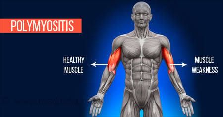
Polymyositis (PM) and Dermatomyositis (DM) are two of the major idiopathic inflammatory myopathies (IIMs), a group of rare, acquired autoimmune disorders. Their primary characteristic is chronic, progressive, and typically symmetrical inflammation of skeletal muscles, leading to significant weakness. While they share the core feature of myositis, their underlying pathology, clinical presentation—most notably the presence of a characteristic rash in DM—and associated risks are distinct. A thorough understanding of these differences is crucial for accurate diagnosis and effective management.
Understanding Pathogenesis – The Underlying Mechanisms
Although both conditions involve an autoimmune attack on muscle tissue, the specific cellular and molecular pathways differ significantly.
1. Polymyositis (PM): A Cell-Mediated Attack
The pathogenesis of PM is primarily considered a T-cell-mediated process. The current understanding is that cytotoxic CD8+ T-lymphocytes directly attack muscle fibers (myofibers).
- Mechanism: In a genetically predisposed individual, an unknown trigger (potentially a viral infection or other environmental factor) causes muscle cells to aberrantly express Major Histocompatibility Complex (MHC) class I molecules on their surface.
- Immune Response: This MHC class I expression presents endogenous muscle antigens to the immune system. CD8+ cytotoxic T-cells recognize these antigens as foreign, leading them to infiltrate the muscle tissue.
- Muscle Damage: These T-cells surround and invade healthy, non-necrotic myofibers, releasing cytotoxic granules like perforin and granzymes, which induce apoptosis (programmed cell death) of the muscle cell. This inflammation occurs deep within the muscle fascicle, in an area known as the endomysium.
2. Dermatomyositis (DM): A Humoral-Mediated Microangiopathy
In contrast, DM is largely a humorally mediated disease characterized by an attack on the small blood vessels (capillaries and arterioles) that supply the muscles and skin. This is often described as a complement-mediated microangiopathy.
- Mechanism: The initial trigger leads to the production of various autoantibodies. These antibodies, along with other immune components, form immune complexes that activate the complement cascade.
- Vascular Damage: The deposition of complement, particularly the C5b-9 membrane attack complex (MAC), within the walls of small blood vessels in the muscle (perimysium) and skin causes endothelial cell damage and necrosis.
- Ischemia: This leads to a reduction in the number of capillaries, resulting in microinfarctions and ischemic damage to the muscle fibers. The muscle damage is most pronounced at the periphery of the muscle fascicles, leading to a hallmark finding on biopsy known as perifascicular atrophy. The skin manifestations are also a direct result of this underlying vasculopathy.
Recognizing the Clinical Features
Both conditions present with the insidious onset of progressive, symmetrical proximal muscle weakness. Patients often report difficulty with tasks like rising from a chair, climbing stairs, lifting objects, or combing their hair. Distal muscle weakness is less common but can occur. In addition to muscle weakness, systemic symptoms such as fatigue, low-grade fever, and weight loss may be present.
Distinguishing Features of Dermatomyositis (DM): The key differentiator for DM is the presence of characteristic cutaneous manifestations, which may precede or occur concurrently with the muscle weakness.
- Gottron’s Papules: Erythematous to violaceous papules located over the extensor surfaces of the metacarpophalangeal (MCP) and interphalangeal (IP) joints. This is considered pathognomonic for DM.
- Heliotrope Rash: A violaceous (lilac-colored) rash on the upper eyelids, often accompanied by periorbital edema.
- Shawl Sign and V-Sign: A widespread, photosensitive, erythematous rash over the upper back and shoulders (Shawl Sign) or the anterior neck and upper chest (V-Sign).
- Mechanic’s Hands: Roughened, cracked, hyperkeratotic skin on the palmar and lateral aspects of the fingers.
Systemic Involvement and Major Associations:
- Interstitial Lung Disease (ILD): Can occur in both PM and DM and is a major cause of morbidity and mortality. It is particularly common in patients with the anti-Jo-1 antibody (Antisynthetase Syndrome).
- Dysphagia: Weakness of oropharyngeal and esophageal muscles can lead to difficulty swallowing and an increased risk of aspiration pneumonia.
- Malignancy: Adult-onset DM is strongly associated with an increased risk of underlying cancer. This paraneoplastic association is less established for PM. The most common malignancies are ovarian, lung, pancreatic, stomach, and colorectal cancers.
The Diagnostic Investigation Pathway
Diagnosis is based on a combination of clinical features, laboratory tests, electrophysiological studies, and muscle biopsy.
A. Laboratory Tests:
- Muscle Enzymes: Creatine kinase (CK) is the most sensitive marker of muscle damage and is often elevated 10 to 50 times the upper limit of normal. Aldolase, lactate dehydrogenase (LDH), aspartate aminotransferase (AST), and alanine aminotransferase (ALT) may also be elevated.
- Autoantibodies: Myositis-Specific Antibodies (MSAs) are highly specific for IIMs and can provide prognostic information.
- Anti-Jo-1: The most common MSA, associated with Antisynthetase Syndrome (myositis, ILD, “mechanic’s hands,” arthritis, and Raynaud’s phenomenon).
- Anti-Mi-2: Highly specific for DM, often associated with the classic skin rashes and a good response to therapy.
- Anti-TIF1-γ and Anti-NXP2: Strongly associated with cancer-associated dermatomyositis in adults.
B. Electromyography (EMG): EMG helps confirm a myopathic process and rule out a neurogenic cause. Typical findings include:
- Increased insertional activity and spontaneous fibrillations.
- Short-duration, low-amplitude, polyphasic motor unit potentials.
C. Muscle Biopsy (The Gold Standard): A muscle biopsy provides the definitive diagnosis by revealing the distinct histopathological patterns.
- Polymyositis Biopsy: Shows endomysial inflammation with CD8+ T-cells surrounding and invading non-necrotic muscle fibers.
- Dermatomyositis Biopsy: The hallmark finding is perifascicular atrophy, with inflammation that is predominantly perivascular (around blood vessels) and located in the interfascicular septa, composed mainly of B-cells and CD4+ T-cells.
D. Additional Screening:
- Malignancy Screening (in DM): Given the strong association, all adults with a new diagnosis of DM should undergo age-appropriate and risk-stratified cancer screening. This may include CT scans of the chest, abdomen, and pelvis, mammography, and colonoscopy.
- Interstitial Lung Disease (ILD) Screening: A baseline high-resolution computed tomography (HRCT) of the chest and pulmonary function tests are recommended.
Formulating a Differential Diagnosis
It is essential to exclude other conditions that can mimic PM and DM.
- Inclusion Body Myositis (IBM): Typically affects older individuals (>50 years), presents with both proximal and distal weakness (especially of finger flexors and quadriceps), is often asymmetrical, and is largely refractory to immunosuppressive therapy.
- Drug-Induced Myopathy: Statins are a common cause. Other culprits include corticosteroids, colchicine, and antimalarials.
- Endocrine Myopathies: Hypothyroidism, hyperthyroidism, and Cushing’s syndrome can all cause proximal muscle weakness.
- Muscular Dystrophies: These are genetic disorders that present with progressive muscle weakness.
- Systemic Lupus Erythematosus (SLE): Can present with a rash and myalgias, but true inflammatory myositis is less common, and its rash (malar rash) typically spares the nasolabial folds.
Implementing a Management Strategy
Management is multidisciplinary, aiming to improve muscle strength, prevent complications, and enhance quality of life.
A. First-Line Pharmacological Therapy:
- High-Dose Corticosteroids: Oral prednisone (or prednisolone) at a dose of 1 mg/kg/day is the cornerstone of initial treatment. This is continued until CK levels normalize and strength improves, followed by a very slow taper over many months to minimize side effects.
B. Steroid-Sparing Agents: To reduce the cumulative dose and toxicity of long-term steroid use, a second agent is typically introduced early.
- Methotrexate or Azathioprine are the most common first-choice steroid-sparing agents.
C. Refractory Disease: For patients who do not respond to the above or who have severe organ involvement (e.g., severe ILD), other options include:
- Mycophenolate Mofetil
- Rituximab (a B-cell depleting antibody)
- Intravenous Immunoglobulin (IVIg)
D. Non-Pharmacological Management:
- Physical and Occupational Therapy: A structured, progressive exercise program is crucial to rebuild muscle strength and improve function.
- Sun Protection: For DM patients, rigorous use of sunscreen and protective clothing is essential to manage the photosensitive rash.
- Speech Therapy: For patients with dysphagia to assess swallowing function and recommend diet modifications.
E. Monitoring: Patients require long-term follow-up to monitor muscle strength, CK levels, and screen for treatment-related side effects and disease-related complications like ILD and malignancy.
Conclusion
Polymyositis and Dermatomyositis are complex systemic autoimmune diseases that demand a systematic approach to diagnosis and a comprehensive, multidisciplinary management plan. While both are characterized by debilitating muscle weakness, understanding their distinct pathogenic pathways—a T-cell-mediated attack in PM versus a complement-mediated microangiopathy in DM—is key to interpreting their unique clinical and histopathological findings. A timely diagnosis, aggressive initial therapy, and vigilant screening for associated conditions like interstitial lung disease and malignancy are paramount to improving patient outcomes and quality of life.
References:
- Dalakas, M. C. (2015). Inflammatory Muscle Diseases. New England Journal of Medicine, 372(18), 1734–1747. DOI: 10.1056/NEJMra1402225
- Lundberg, I. E., Tjärnlund, A., & Bottai, M., et al. (2021). 2017 European League Against Rheumatism/American College of Rheumatology classification criteria for adult and juvenile idiopathic inflammatory myopathies and their major subgroups. Annals of the Rheumatic Diseases, 76(12), 1955-1964.
- Oldroyd, A. G., Lilleker, J. B., & Chinoy, H. (2017). Idiopathic inflammatory myopathies – a guide to subtypes, classification and management. Clinical Medicine, 17(4), 322–328. DOI: 10.7861/clinmedicine.17-4-322
- DeWane, M. E., Waldman, R., & Lu, J. (2020). Dermatomyositis: Clinical features and pathogenesis. Journal of the American Academy of Dermatology, 82(2), 267-281.
- Mammen, A. L. (2016). Autoantibodies in the idiopathic inflammatory myopathies: clinical and pathogenic importance. Rheumatic Disease Clinics of North America, 42(2), 205-215.


