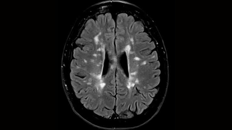
Computed Tomography (CT) scans of the brain are indispensable diagnostic tools in modern medicine, particularly in emergency settings and for initial evaluations of neurological conditions. They provide rapid, non-invasive imaging that can detect a wide range of intracranial pathologies. A fundamental skill for healthcare professionals involved in patient care is the ability to accurately identify common brain lesions on CT scans.
Understanding CT Scan Basics for Lesion Identification
Before delving into specific lesions, it’s crucial to understand the basic principles of CT imaging. CT scans utilize X-rays to create cross-sectional images of the body. Different tissues attenuate X-rays to varying degrees, which is then translated into a grayscale image. This attenuation is quantified in Hounsfield Units (HU):
- Hyperdense (Bright White): High attenuation, typically bone (1000+ HU), acute blood (50-90 HU), or calcifications.
- Isodense (Gray): Similar attenuation to normal brain parenchyma (20-40 HU), often seen with subacute hematomas or tumors.
- Hypodense (Dark Gray/Black): Low attenuation, typically cerebrospinal fluid (CSF) (0-15 HU), fat (-50 to -100 HU), air (-1000 HU), or areas of edema/infarction.
The ability to recognize these density differences is paramount for identifying pathologies.
General Principles of Brain Lesion Identification on CT
A systematic approach is crucial when reviewing brain CT scans to avoid missing critical findings:
- Assess Symmetry: The brain is largely symmetrical. Any asymmetry in density, sulcal pattern, or ventricular size should prompt further investigation.
- Evaluate Grey-White Matter Differentiation: Normal brain shows a clear distinction between the darker grey matter (cortex, deep nuclei) and the lighter white matter. Loss of this differentiation can indicate pathology like edema or ischemia.
- Examine Sulcal and Gyral Patterns: The sulci (grooves) and gyri (folds) of the brain cortex are normally prominent. Effacement (flattening or disappearance) of sulci suggests underlying swelling or mass effect.
- Review Ventricular System: Assess the size, shape, and symmetry of the lateral, third, and fourth ventricles. Abnormalities can indicate hydrocephalus or mass effect.
- Check for Mass Effect: This refers to the displacement or compression of normal brain structures by an abnormal lesion. Signs include:
- Effacement of sulci over the convexity.
- Compression or effacement of ventricles.
- Midline shift: Displacement of the falx cerebri or other midline structures from the center. This is a critical sign of significant mass effect.
- Analyze Extra-Axial Spaces: These are the spaces surrounding the brain parenchyma, including the epidural and subdural spaces, where hematomas often occur.
- Bone Windows: Always review the images in bone windows to identify skull fractures, which can be associated with intracranial hemorrhage.
1. Cerebral Edema
Definition: Cerebral edema refers to an abnormal accumulation of fluid within the brain parenchyma, leading to brain swelling. This swelling increases intracranial pressure (ICP) and can compromise cerebral blood flow, leading to secondary brain injury.
Types and Mechanisms:
- Vasogenic Edema: Most common type, resulting from disruption of the blood-brain barrier. Plasma constituents leak into the extracellular space, primarily affecting the white matter. Common causes include tumors, abscesses, severe ischemia, and trauma.
- Cytotoxic Edema: Occurs due to cellular dysfunction, where failure of the Na+/K+ pump leads to intracellular swelling of neurons, glial cells, and endothelial cells. It affects both grey and white matter. The most common cause is severe ischemia (e.g., stroke), hypoxia, or metabolic disorders.
- Interstitial Edema: Primarily seen in severe hydrocephalus, where CSF permeates the periventricular white matter through the ventricular ependyma due to elevated intraventricular pressure.
CT Appearance: How to Identify
- Hypodensity (Darkening): The most characteristic CT finding is a regional or diffuse area of decreased attenuation (darker appearance) within the brain parenchyma, indicating fluid accumulation.
- Vasogenic: Typically causes finger-like, patchy hypodensity predominantly in the white matter, often associated with a mass lesion (tumor, abscess).
- Cytotoxic: Results in more uniform hypodensity involving both grey and white matter, often respecting vascular territories in cases of infarction. The loss of the normal grey-white matter interface is a hallmark.
- Interstitial: Characterized by bilateral, periventricular hypodensity, appearing as a “halo” around the ventricles.
- Loss of Grey-White Matter Differentiation: The normal distinct boundary between grey matter (cortex and deep nuclei) and white matter becomes blurred or indistinct in areas of edema, particularly in cytotoxic edema.
- Sulcal Effacement: The normal sulci (grooves) over the affected brain surface appear narrowed or completely flattened due to outward expansion of the swollen brain tissue.
- Ventricular Compression/Effacement: As the brain swells, the adjacent ventricle(s) may be compressed, narrowed, or even completely effaced (obliterated).
- Mass Effect: Significant edema can lead to mass effect, causing midline shift (displacement of the falx cerebri), transtentorial herniation (displacement of brain tissue across the tentorium cerebelli), or uncal herniation.
Distinguishing Features: The pattern of hypodensity, its distribution (grey vs. white matter predominance), and the presence of underlying lesions (e.g., stroke for cytotoxic, tumor for vasogenic) help differentiate types of edema.
2. Ventricular Enlargement (Hydrocephalus)
Definition: Ventricular enlargement, clinically termed hydrocephalus, refers to the abnormal accumulation of cerebrospinal fluid (CSF) within the cerebral ventricles, leading to their dilation. While “hypertrophy” implies cellular enlargement, it is an inaccurate term for the ventricles; “enlargement” or “dilation” is correct. This buildup occurs due to an imbalance between CSF production and absorption, or an obstruction in its flow.
Types and Mechanisms:
- Communicating (Non-obstructive) Hydrocephalus: CSF flow within the ventricular system is unobstructed, but there is impaired reabsorption of CSF into the venous system (e.g., post-meningitis, subarachnoid hemorrhage, or idiopathic normal pressure hydrocephalus). All ventricles are typically enlarged.
- Non-communicating (Obstructive) Hydrocephalus: CSF flow is blocked within the ventricular system (e.g., by a tumor, congenital aqueductal stenosis, or intraventricular hemorrhage). The ventricles proximal to the obstruction will enlarge, while those distal to it may be normal or collapsed.
CT Appearance: How to Identify
- Enlarged Ventricles: The most obvious sign is the clear dilation of one or more of the cerebral ventricles (lateral, third, and/or fourth ventricles).
- General Enlargement: The lateral ventricles appear rounded and prominent, especially the frontal and temporal horns. The third and fourth ventricles may also be enlarged.
- Specific Patterns for Obstructive: If the obstruction is at the cerebral aqueduct, the lateral and third ventricles will be enlarged, but the fourth ventricle will be normal or small. If the obstruction is at the outlet foramina of the fourth ventricle, all four ventricles will enlarge.
- Periventricular Hypodensity (Transependymal Edema): In acute or severe hydrocephalus, the increased intraventricular pressure forces CSF into the surrounding white matter. This appears as areas of periventricular hypodensity (darker halo) around the ventricles, representing interstitial edema.
- Sulcal Effacement (Variable): In acute hydrocephalus, the enlarged ventricles can exert pressure on the surrounding brain parenchyma, leading to effacement of the cortical sulci. In chronic hydrocephalus, particularly Normal Pressure Hydrocephalus (NPH), the sulci may appear prominent despite ventricular enlargement, as the brain parenchyma has atrophied.
- Specific Angles/Measurements (for NPH): In Normal Pressure Hydrocephalus, the callosal angle (the angle formed by the roofs of the lateral ventricles on a coronal view) may be acutely angled (<90 degrees), distinguishing it from hydrocephalus ex vacuo (ventricular enlargement due to brain atrophy, where the angle is preserved).
Distinguishing Features: The pattern of ventricular enlargement (symmetrical vs. asymmetrical, which ventricles are affected), the presence of periventricular hypodensity, and the status of the sulci help differentiate types of hydrocephalus and distinguish it from hydrocephalus ex vacuo (brain atrophy presenting with enlarged ventricles but prominent sulci and no periventricular edema).
3. Epidural Hematoma (EDH)
Definition: An epidural hematoma is a collection of blood that forms in the potential space between the dura mater (the outermost meningeal layer) and the inner table of the skull. It is typically arterial in origin, commonly from a tear in the middle meningeal artery after a skull fracture.
CT Appearance: How to Identify
- Biconvex (Lenticular/Lens-shaped) Hyperdense Collection: This is the classic appearance. The hematoma has a characteristic convex shape towards the brain and a convex shape towards the skull, resembling a lens. It is hyperdense (bright white) if acute (within hours to days of injury) due to fresh blood.
- Does NOT Cross Suture Lines: This is a crucial distinguishing feature. The dura mater is firmly attached to the skull at the cranial sutures. Therefore, an epidural hematoma is typically limited by suture lines and does not cross them, leading to its characteristic focal, well-demarcated appearance. However, it can cross the midline only if it extends across the superior sagittal sinus.
- Location: Most commonly found over the temporoparietal region due to the vulnerability of the middle meningeal artery, but can occur anywhere.
- Significant Mass Effect: Due to its arterial origin and rapid expansion, EDHs typically exert significant mass effect for their size, causing:
- Compression of the underlying brain parenchyma.
- Effacement of adjacent sulci.
- Compression or effacement of the ipsilateral ventricle.
- Midline shift, which can be severe and rapid, leading to herniation.
- Associated Skull Fracture: A skull fracture in the overlying bone is present in 80-95% of EDH cases. Always check bone windows.
Distinguishing Features: The biconvex shape and its inability to cross suture lines are key differentiators from a subdural hematoma. The rapid onset of symptoms and significant mass effect are also characteristic.
4. Subdural Hematoma (SDH)
Definition: A subdural hematoma is a collection of blood located in the potential space between the dura mater and the arachnoid mater. It typically results from tears in bridging veins that traverse the subdural space, connecting the cerebral cortex to the dural sinuses. SDHs are often associated with falls, minor head trauma (especially in the elderly, alcoholics, or those on anticoagulants), or rapid acceleration-deceleration injuries.
CT Appearance: How to Identify
- Crescent-shaped (Concavoconvex) Collection: This is the hallmark. The hematoma conforms to the curved surface of the brain, appearing like a crescent moon or banana. The inner margin is concave towards the brain, and the outer margin is convex towards the skull.
- Crosses Suture Lines but NOT the Falx/Tentorium: Unlike EDHs, SDHs are not limited by suture lines because the subdural space is continuous. They can spread widely over the cerebral hemisphere. However, they are typically limited by the falx cerebri (midline dural fold) and the tentorium cerebelli (dural fold separating cerebrum from cerebellum), meaning they usually don’t cross the midline over the convexities.
- Variable Density Based on Age:
- Acute SDH (0-3 days): Hyperdense (bright white, 50-90 HU) due to clotted blood.
- Subacute SDH (3 days – 3 weeks): Isodense (similar density to normal brain parenchyma). This is the most challenging phase to diagnose as the hematoma blends in with the brain. Look for subtle signs of mass effect, effacement of sulci, or subtle asymmetry.
- Chronic SDH (>3 weeks): Hypodense (darker than brain parenchyma, similar to CSF) as the blood degrades and lyses. A fluid-fluid level may be seen if there’s ongoing bleeding or rebleeding.
- Conforms to Brain Surface: The collection spreads along the cerebral convexity, reflecting the natural contours of the brain.
- Mass Effect (Variable): Mass effect depends on the size and rapidity of accumulation.
- Acute SDHs can cause significant mass effect, leading to sulcal effacement, ventricular compression, and midline shift.
- Chronic SDHs may cause less acute mass effect but can still compress the brain and lead to neurological deficits.
- Location: Most common over the cerebral convexities, but can occur along the falx (interhemispheric SDH), tentorium, or in the posterior fossa.
Distinguishing Features: The crescent shape, ability to cross suture lines (but not midline over convexities), and variable density based on age are critical for differentiating SDHs from EDHs and other lesions. Bilateral SDHs can be difficult to detect if they are symmetrical and isodense, requiring careful assessment of sulcal effacement and ventricular size.
Step-by-Step Approach to CT Scan Review for Lesions
- Patient Information & Clinical Context: Start by reviewing patient demographics, mechanism of injury (if traumatic), and presenting symptoms. This helps narrow the differential diagnosis.
- Initial Overview (“Scout” or “Topogram”): Get a general sense of the scan quality and patient positioning.
- Bone Window Review: Systematically scroll through the bone window images to identify any skull fractures, which are highly relevant for hematomas.
- Brain Parenchyma (Brain Window Review – Systemic Scroll):
- Start at the Vertex: Begin from the top of the head and scroll down slice by slice.
- Symmetry and Density: Look for any asymmetry in brain tissue density or sulcal patterns. Are there areas of hypodensity (edema, stroke, chronic SDH) or hyperdensity (acute blood, calcifications)?
- Grey-White Matter Differentiation: Assess whether the normal distinction between grey and white matter is preserved, or if there’s effacement indicating edema.
- Sulcal Effacement: Note if the normal cortical sulci appear narrowed or flattened, suggesting swelling or mass effect.
- Ventricles: Examine the size, shape, and symmetry of the lateral, third, and fourth ventricles. Look for enlargement (hydrocephalus) or compression/effacement (mass effect). Check for periventricular hypodensity.
- Basal Cisterns: Ensure the basal cisterns (e.g., suprasellar, ambient) are open and not effaced, which is a sign of severe mass effect or herniation.
- Midline Shift: Crucially, check if the falx cerebri, septum pellucidum, and third ventricle are shifted from the true midline. Measure the shift if present.
- Extra-axial Spaces: Look for collections in the epidural (biconvex, respects sutures) and subdural (crescent, crosses sutures, variable density) spaces.
- Posterior Fossa: Evaluate the brainstem and cerebellum for any lesions, effacement of the fourth ventricle, or hydrocephalus.
- Systematic Comparison: Compare one side of the brain to the other. Use a mental checklist for each region.
Conclusion
Proficiently identifying common brain lesions on CT scans is a critical skill for any healthcare professional involved in neurological care. By understanding the fundamental principles of CT imaging, the distinct appearances of cerebral edema, ventricular enlargement, epidural hematomas, and subdural hematomas, and by employing a systematic approach to scan review, clinicians can make timely and accurate diagnoses, directly impacting patient management and outcomes. Always integrate CT findings with the patient’s clinical presentation for the most comprehensive assessment.
References
- Osborn, A. G. (2012). Osborn’s Brain: Imaging, Pathology, and Anatomy. Elsevier Mosby.
- Rubin, G. D. (2016). Computed Tomography: Principles, Design, Applications. CRC Press.
- Weishaupt, D., Koechli, V. F., & Marincek, B. (2012). How to Review a Brain CT. Springer.
- Barkhof, F. (2011). Clinical Neuroimaging: A Case-Based Approach. Cambridge University Press.
- Lee, S. H., & Rao, K. C. V. G. (2006). Cranial Computed Tomography. McGraw-Hill Education.


