Leishmaniasis constitutes a complex spectrum of diseases caused by parasitic protozoa of the genus Leishmania. Transmitted to humans through the bite of infected female phlebotomine sandflies, this neglected tropical disease presents in various clinical forms, ranging from self-healing skin lesions to fatal systemic infections. Its endemic presence in approximately 90 countries, coupled with significant morbidity and mortality, underscores the critical need for understanding its multifaceted nature, epidemiological patterns, and the strategic measures required for its prevention and control.
1. Defining Leishmaniasis and Its Clinical Expressions
Leishmaniasis is fundamentally a vector-borne parasitic disease that manifests with diverse clinical outcomes primarily dependent on the Leishmania species involved, the geographic region, and the host’s immune response. The parasites exist in two main morphological forms: promastigotes (flagellated, found in the sandfly gut) and amastigotes (non-flagellated, found within host macrophages).
1.1. Core Definition
Leishmaniasis is a parasitic infection caused by protozoa belonging to the genus Leishmania. These microscopic parasites are transmitted to humans and other mammals (reservoir hosts) through the bite of infected female sandflies, which are tiny, blood-feeding insects. Once inside the host, Leishmania parasites primarily infect macrophages, leading to chronic infections that can affect the skin, mucous membranes, or internal organs.
1.2. Types of Leishmaniasis
The clinical presentation of Leishmaniasis is broadly categorized into three main forms, each with distinct pathological features and associated Leishmania species:
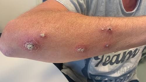
a) Cutaneous Leishmaniasis (CL): This is the most common form of Leishmaniasis, primarily affecting the skin. CL typically presents as localized skin lesions, which can begin as papules, nodules, or ulcers. The lesions often have raised borders and a central crater, sometimes resembling volcanic craters. While many CL lesions heal spontaneously within months to years, they often leave disfiguring scars, which can lead to social stigma and psychological distress, especially when lesions are on the face.
- Causative Species: Diverse species cause CL, including L. major, L. tropica, and L. aethiopica in the Old World, and L. mexicana, L. amazonensis, and various species of the L. braziliensis complex in the New World.
- Variations:
- Diffuse Cutaneous Leishmaniasis (DCL): A rare anergic form characterized by widespread non-ulcerating nodules, often resistant to treatment.
- Post-Kala-azar Dermal Leishmaniasis (PKDL): A dermal manifestation that can appear months or years after successful treatment for Visceral Leishmaniasis, characterized by macules, papules, or nodules, primarily on the face and trunk. It can serve as a reservoir for infection.
b) Mucocutaneous Leishmaniasis (MCL): Also known as “espundia,” this severe and destructive form of Leishmaniasis typically follows an initial, often untreated, cutaneous lesion. It involves the metastatic spread of parasites from the skin to the mucous membranes of the nose, mouth, and throat. MCL can lead to extensive tissue destruction, causing severe disfigurement, difficulty in breathing, speaking, and eating, and significant morbidity. Secondary bacterial infections are common, further complicating management.
- Causative Species: Predominantly associated with species of the L. braziliensis complex in the New World (e.g., L. braziliensis, L. guyanensis, L. panamensis).
c) Visceral Leishmaniasis (VL) / Kala-azar: This is the most severe form of Leishmaniasis and, if left untreated, is almost always fatal. VL targets internal organs, primarily the spleen, liver, and bone marrow. It is characterized by irregular bouts of fever, weight loss, enlargement of the spleen (splenomegaly) and liver (hepatomegaly), pancytopenia (reduction in all blood cell types leading to anemia, leukopenia, and thrombocytopenia), and generalized weakness. Over time, individuals become highly susceptible to other infections due to a severely compromised immune system.
- Causative Species: Primarily caused by L. donovani in the Old World (Indian subcontinent, East Africa) and L. infantum (syn. L. chagasi in the Americas) in the Mediterranean basin, Middle East, and Latin America.
- Co-infection: VL is a significant opportunistic infection in individuals with HIV, where the co-infection often exacerbates the disease and complicates treatment.
2. The Epidemiology of Leishmaniasis
The epidemiology of Leishmaniasis is remarkably diverse and complex, influenced by a dynamic interplay of biological, ecological, environmental, and socioeconomic factors. It is a disease of poverty, disproportionately affecting vulnerable populations.
2.1. Global Distribution and Burden Leishmaniasis is endemic in approximately 90 countries across tropical, subtropical, and temperate regions. The World Health Organization (WHO) estimates that 700,000 to 1 million new cases of Cutaneous Leishmaniasis and 50,000 to 90,000 new cases of Visceral Leishmaniasis occur annually.
- VL hotspots: Over 90% of global VL cases occur in six countries: Brazil, Ethiopia, India, Kenya, Somalia, and Sudan.
- CL hotspots: The majority of CL cases are reported from Afghanistan, Algeria, Brazil, Colombia, Costa Rica, Ethiopia, Iran, Iraq, Libya, Morocco, Nicaragua, Pakistan, Peru, Saudi Arabia, Syria, Tunisia, and Venezuela.
- Disease Burden: Leishmaniasis contributes significantly to the global burden of disease, measured in Disability-Adjusted Life Years (DALYs), reflecting both morbidity from CL and MCL, and mortality from VL.
2.2. Transmission Cycle The life cycle of Leishmania involves two hosts: a vertebrate host (human or animal) and a sandfly vector.
- Infection of Sandfly: An uninfected female sandfly bites an infected human or animal, ingesting macrophages containing amastigotes.
- Transformation in Sandfly: Amastigotes transform into flagellated promastigotes in the sandfly’s midgut, multiply, and migrate to the proboscis.
- Infection of Vertebrate Host: The infected sandfly bites a new human or animal host, regurgitating promastigotes into the skin.
- Transformation in Vertebrate Host: Promastigotes are phagocytized by macrophages and other phagocytic cells, where they transform into amastigotes, multiply, and rupture the host cell, infecting new cells.
2.3. Key Epidemiological Factors
a) Vector Factors:
- Species Diversity: Over 90 species of sandflies (genus Phlebotomus in the Old World and Lutzomyia in the New World) are known or suspected vectors. Each Leishmania species typically has specific sandfly vectors.
- Habitat: Sandflies thrive in warm, humid environments, often in cracks in walls, animal burrows, and organic debris.
- Biting Behavior: Sandflies are typically active from dusk to dawn, primarily biting outdoors. Their small size (2-3 mm) allows them to pass through standard mosquito nets.
b) Reservoir Hosts:
- Zoonotic Leishmaniasis: Many forms are zoonotic, meaning animals serve as primary reservoirs. Dogs are the most important reservoir for L. infantum (causing VL and CL) in the Mediterranean, Middle East, and Latin America. Rodents (e.g., gerbils, hyraxes) are common reservoirs for L. major and L. tropica.
- Anthroponotic Leishmaniasis: In some forms, humans are the sole reservoir (e.g., L. donovani in the Indian subcontinent, L. tropica in urban areas).
c) Environmental Factors:
- Deforestation and Urbanization: Ecological changes, such as deforestation for agriculture or human settlement into previously wild areas, can bring humans into closer contact with enzootic cycles. Rapid, unplanned urbanization can create favorable breeding sites for sandflies.
- Climate Change: Changes in temperature, rainfall, and humidity patterns can alter the geographical distribution and seasonality of sandflies and Leishmania parasites.
- Housing Conditions: Poor housing quality, inadequate sanitation, and proximity to animal shelters can increase exposure to sandflies.
d) Socioeconomic Factors:
- Poverty: Malnutrition, poor housing, lack of access to healthcare, and illiteracy are significant risk factors. Malnutrition weakens the immune system, increasing susceptibility to severe disease.
- Population Displacement: Conflict, migration, and natural disasters force populations into new, often endemic, areas with limited shelter and sanitation, increasing exposure risk.
- Occupational Exposure: Agricultural workers, soldiers, and ecotourists may be at higher risk due to outdoor exposure in endemic areas.
e) Host Factors:
- Immunity: The immune status of an individual profoundly influences disease progression. Malnutrition and co-infections (e.g., HIV) severely weaken the immune system, predisposing individuals to VL or more severe CL.
- Genetic Predisposition: Genetic factors in humans are also thought to play a role in susceptibility and disease outcome.
3. Preventive and Control Measures for Leishmaniasis
Controlling Leishmaniasis requires a comprehensive and integrated approach, recognizing the complexity of the parasite, vector, reservoir, and human hosts. Effective strategies prioritize reducing exposure to sandflies, managing infected cases, and controlling reservoir hosts.
3.1. Vector Control
Reducing human-sandfly contact is a cornerstone of prevention.
- Insecticide-Treated Nets (ITNs) and Long-Lasting Insecticidal Nets (LLINs): While sandflies are smaller than mosquitoes, ITNs/LLINs can offer protection, especially where people sleep outdoors or in poorly screened dwellings. However, effectiveness varies depending on sandfly biting habits.
- Indoor Residual Spraying (IRS): Spraying the internal surfaces of houses with insecticides can reduce sandfly populations by targeting their resting sites. This is particularly effective for anthroponotic VL where sandflies are endophilic (prefer to rest indoors).
- Personal Protection Measures:
- Insect Repellents: Applying repellents containing DEET, picaridin, or IR3535 to exposed skin.
- Protective Clothing: Wearing long sleeves and trousers, especially during peak sandfly activity (dusk to dawn).
- Avoiding Outdoor Exposure: Limiting outdoor activities during peak biting hours in endemic areas.
- Environmental Management:
- Improved Housing: Sealing cracks in walls, screening windows and doors, and maintaining clean peridomestic environments to reduce sandfly breeding and resting sites.
- Waste Management: Proper disposal of organic waste and rubbish near homes, as these can serve as sandfly breeding sites.
3.2. Reservoir Host Control
Controlling animal reservoirs is crucial for zoonotic forms of Leishmaniasis.
- Canine Leishmaniasis (for L. infantum):
- Insecticide-Impregnated Collars: Dog collars treated with insecticides like deltamethrin reduce sandfly bites and parasite transmission from dogs.
- Canine Vaccination: Vaccines for dogs are available in some regions (e.g., Europe, Brazil), aimed at reducing infection rates and transmission to humans, though their impact on human disease burden is still being evaluated.
- Treatment and Culling of Infected Dogs: Treatment of infected dogs can reduce parasite load, but culling is a controversial and often impractical measure.
- Rodent Control: In certain foci of zoonotic cutaneous leishmaniasis (e.g., L. major), rodent control measures (e.g., trapping, burrow destruction) may be implemented.
3.3. Case Management and Surveillance
Early diagnosis and effective treatment are essential for reducing morbidity, preventing progression to severe forms, and interrupting transmission, especially in anthroponotic Leishmaniasis.
- Early Diagnosis and Treatment: Prompt access to diagnostic tools (e.g., microscopy, rapid diagnostic tests, PCR) and effective anti-leishmanial drugs (e.g., amphotericin B, miltefosine, paromomycin, sodium stibogluconate) is critical.
- Drug Resistance Monitoring: Continuous surveillance for drug resistance is vital to inform treatment guidelines.
- Active Case Finding: In highly endemic areas, active surveillance and case finding are important to detect and treat cases that might otherwise go undiagnosed.
- Disease Surveillance Systems: Robust surveillance systems are necessary to monitor disease trends, identify outbreaks, map risk areas, and evaluate the effectiveness of control programs. Integrated surveillance that includes vector and reservoir monitoring is ideal.
3.4. Health Education and Community Engagement
Empowering communities with knowledge about Leishmaniasis is fundamental for effective control.
- Awareness Campaigns: Educating the public about the disease, its symptoms, transmission, available treatments, and preventive measures.
- Behavior Change Communication: Promoting practices such as using ITNs, seeking early treatment, and personal protection.
- Community Participation: Engaging local communities in planning and implementing control activities to ensure their sustainability and acceptance.
3.5. Research and Development
Continued investment in research is vital for developing new and improved tools.
- Diagnostics: Development of highly sensitive, specific, and user-friendly point-of-care diagnostic tests.
- Drugs: Discovery and development of new, safer, more effective, and orally available drugs with shorter treatment regimens, particularly to combat drug resistance and improve adherence.
- Vaccines: Research into effective human vaccines is ongoing, but none are currently widely available. Continued development of canine vaccines is also important.
- Vector Control Tools: Development of novel insecticides, traps, or biological control methods for sandflies.
3.6. Policy and Partnerships
Effective Leishmaniasis control requires strong political commitment and collaborative efforts.
- National Control Programs: Development and implementation of well-funded national Leishmaniasis control programs, often integrated within broader neglected tropical disease (NTD) initiatives.
- International Collaboration: Partnerships between endemic countries, international organizations (e.g., WHO), research institutions, NGOs, and pharmaceutical companies for resource mobilization, technical support, and knowledge sharing.
In conclusion, Leishmaniasis remains a significant public health challenge with a complex epidemiology influenced by environmental, socioeconomic, and biological factors. A multi-pronged approach encompassing robust vector control, effective reservoir management, timely case detection and treatment, community engagement, and sustained research and development efforts is essential for reducing its burden and moving towards its eventual elimination in endemic regions.
References:
- World Health Organization (WHO). Leishmaniasis Fact Sheet. (Regularly updated online. Accessed via searching “WHO Leishmaniasis Fact Sheet”).
- Centers for Disease Control and Prevention (CDC). Parasites – Leishmaniasis. (Accessed via searching “CDC Leishmaniasis”).
- Peters, W., & Killick-Kendrick, R. (Eds.). (1987). The Leishmaniases in Biology and Medicine. Academic Press. (A foundational text, though older, principles remain relevant).
- Desjeux, P. (2004). Leishmaniasis: current situation and new perspectives. Comparative Immunology, Microbiology and Infectious Diseases, 27(5-6), 305-312.
- Reithinger, R., Dujardin, J. C., Louzir, H., Pirmez, C., Alexander, B., & Brooker, S. (2007). Cutaneous leishmaniasis. The Lancet Infectious Diseases, 7(9), 581-592.
- Alvar, J., Vélez, I. D., Bern, C., Herrero, M., Desjeux, P., Cano, J., … & WHO Leishmaniasis Control Team. (2012). Leishmaniasis Worldwide and Global Estimates of Its Incidence. PLoS One, 7(5), e35671.
- Murray, H. W., Berman, J. D., Davies, C. R., & Saravia, N. G. (2005). Advances in Leishmaniasis. The Lancet, 366(9496), 1537-1547.


