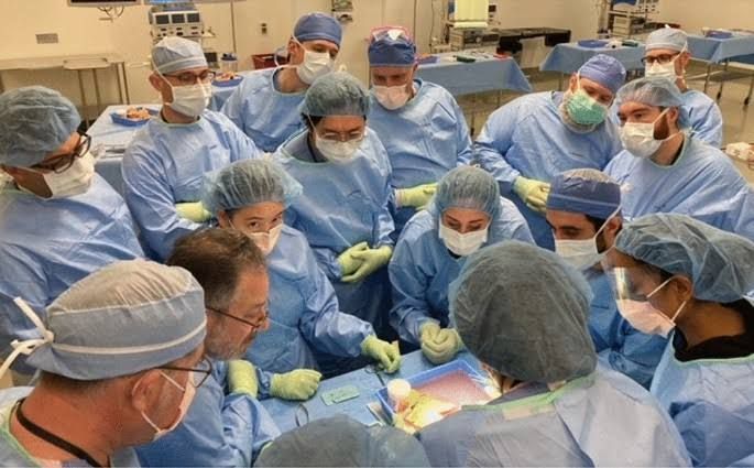
The success of pancreas transplantation hinges not only on precise surgical technique during recipient implantation but also critically on the meticulous back table preparation of the donor organ. This ex-vivo stage, performed under sterile conditions, is paramount for ensuring graft viability, optimizing vascular inflow and outflow, and minimizing recipient operative time and complications.
Introduction to Back Table Preparation
Upon retrieval from the donor, the pancreas, often procured en-bloc with the duodenum and sometimes segments of the aorta and iliac vessels, is immediately immersed in cold preservation solution. The back table preparation is then initiated in a sterile environment, typically on a dedicated instrument table. The primary objectives are to trim excess tissue, identify and prepare all vascular structures for anastomosis, manage the duodenal segment for exocrine drainage, and ensure the graft is optimized for implantation. This careful preparation directly impacts perfusions, reduces anastomotic complications, and is fundamental to long-term graft function.
1. Removal of the Spleen
The spleen is typically procured en-bloc with the pancreas to ensure an intact splenic artery and vein supply to the pancreatic body and tail. However, for pancreas transplantation, the spleen is almost universally removed during back table preparation.
- Rationale for Splenectomy:
- Immunogenicity: The spleen is a highly lymphoid organ. Its removal reduces the total immunological burden from the donor tissue, potentially decreasing the risk of recipient sensitization to donor antigens, although its direct impact on long-term outcomes is debated.
- Space and Handling: Removing the bulky spleen simplifies the graft’s geometry, making it easier to handle, position, and implant into the recipient’s abdomen, especially in cases of limited space.
- Complication Prevention: Retaining the spleen could theoretically introduce risks such as splenic infarction, rupture, or abscess formation post-transplant. While these are rare, their prevention is a consideration.
- Vascular Dissection: Splenectomy provides better exposure and easier dissection of the splenic artery and vein, which are crucial for the arterial reconstruction and portal vein formation.
- Technique:
- The dissection begins by identifying the splenic artery and vein as they emerge from the pancreatic hilum or near the tail.
- These vessels are carefully ligated and divided as close to the splenic hilum as safely possible, preserving their length and integrity for their course within the pancreatic tissue.
- The spleen is then carefully dissected away from the greater curvature of the stomach and the tail of the pancreas. Care must be taken to avoid injuring the pancreatic capsule or parenchyma during this separation.
- Any small, short gastric vessels or gastrosplenic ligaments are ligated and divided.
- Once detached, the spleen is removed from the sterile field. The splenic artery and vein stumps are inspected for hemostasis.
2. Role for Shortening of the Duodenum
The pancreas is procured with a segment of the duodenum because the pancreatic head is intimately associated with the C-loop of the duodenum, sharing a common blood supply via the pancreaticoduodenal arcades. This duodenal segment serves as the conduit for the exocrine secretions of the pancreas into the recipient’s gastrointestinal tract.
- Rationale for Shortening:
- Anastomotic Simplicity: A shorter, manageable duodenal segment simplifies the Roux-en-Y duodenojejunostomy, which is the standard technique for exocrine drainage. A long segment can be cumbersome and predispose to kinking or obstruction.
- Reduced Complication Risk: A lengthy duodenal segment can increase the surface area for potential bacterial contamination from the recipient’s gut, theoretically leading to a higher risk of anastomotic leaks, infections, or fistulas. Shortening minimizes this exposure.
- Fit and Position: A compact duodenal patch is easier to position within the recipient’s abdominal cavity, especially when space is at a premium (e.g., in simultaneous pancreas-kidney transplants).
- Blood Supply Optimization: The transection lines of the duodenum must be carefully chosen to ensure the segment retains robust blood flow from the pancreaticoduodenal arcade, which originates from the superior mesenteric artery.
- Technique:
- The duodenal segment is carefully inspected to identify the pancreatic head and the duodenal C-loop.
- The optimal length for the duodenal segment is typically 2-4 cm, preserving the ampulla of Vater and ensuring adequate surrounding tissue for anastomosis. This creates a “duodenal patch.”
- The duodenum is transected proximally (often superior to the first part of the duodenum) and distally (inferior to the second or third part, often at the level of the ligament of Treitz, if the full C-loop is procured).
- Transection is performed with a linear stapler or sharp dissection, ensuring that the blood supply from the pancreaticoduodenal arteries (originating from the superior mesenteric artery) remains intact to the entire duodenal patch and, critically, to the head of the pancreas.
- The transected ends are then typically oversewn with a continuous non-absorbable suture to reinforce the staple line and create a closed patch ready for side-to-side anastomosis to the recipient’s jejunum.
3. Control of Mesenteric Vessels
The proper identification, dissection, and preparation of the superior mesenteric artery (SMA) and superior mesenteric vein (SMV) are paramount, as these vessels are primary components of the pancreatic graft’s vascular supply and drainage.
- Superior Mesenteric Artery (SMA):
- Function: Provides the main arterial inflow to the pancreatic head and the duodenal segment via the pancreaticoduodenal arcades.
- Preparation: The SMA is meticulously dissected free from surrounding lymphatic tissue and fat. It is often procured with a segment of the donor aorta (a “Carrel patch”) which encompasses its ostium and sometimes also the ostium of the celiac axis. This aortic patch facilitates a wide, robust anastomosis to the recipient’s arterial system. Any small, non-essential branches originating directly from the SMA that do not supply the graft are ligated and divided to prevent bleeding or steal phenomena. The integrity of the pancreaticoduodenal arteries must be confirmed.
- Superior Mesenteric Vein (SMV) and Portal Vein:
- Function: The SMV constitutes the primary venous drainage of the duodenum and joins the splenic vein to form the main portal vein, which drains the entire pancreas. This portal vein is the primary venous outflow for the graft.
- Preparation: The SMV is carefully dissected. All jejunal and ileal branches are individually ligated and divided to isolate the main trunk of the SMV. Similarly, the splenic vein is dissected to its confluence with the SMV, forming the portal vein. Any small collateral veins or lymphatic channels encountered around the portal vein are ligated. The goal is to present a clean, adequately long, and patent portal vein for anastomosis to the recipient’s venous system (either the SMV for portal venous drainage or the common iliac vein for systemic venous drainage).
4. Y-Graft or Use of Alternative Arterial Reconstruction
The pancreas receives arterial blood from two main sources: the splenic artery (supplying the body and tail) and the superior mesenteric artery (supplying the head and duodenum). These two arteries originate separately from the celiac axis and aorta, respectively. To simplify the arterial inflow to the graft in the recipient, a single arterial anastomosis is typically created on the back table.
- Y-Graft Reconstruction:
- Concept: A segment of a donor artery, commonly a portion of the donor common iliac artery bifurcation, is used as a “Y” conduit.
- Technique: The splenic artery and the superior mesenteric artery (or their respective origins on an aortic patch) are anastomosed end-to-side to the two branches of the donor iliac bifurcation. For instance, the splenic artery (usually smaller) might be anastomosed to the internal iliac branch, and the SMA (larger and higher flow) to the external iliac branch. The common trunk of the donor iliac artery then serves as the single inflow vessel for the entire pancreatic graft, ready for anastomosis to the recipient’s iliac artery (common, external, or internal) or aorta. This creates a robust, single point of inflow.
- Aortic Patch Reconstruction (Alternative):
- Concept: If the donor pancreas is procured with a large aortic segment that includes the ostia of both the celiac axis (from which the splenic artery originates) and the SMA, a single Carrel patch containing both origins can be created.
- Technique: The redundant aortic tissue is trimmed to form a smooth, elliptical patch encompassing both vessel ostia. This single patch is then anastomosed to the recipient’s arterial system. This is often the preferred method when anatomically feasible and when sufficient donor aortic tissue is available.
- Other Arterial Reconstructions: Less common alternatives might include direct end-to-end anastomoses if the recipient’s vessels allow for it, or using other donor arterial segments (e.g., saphenous vein graft from another donor) if the primary options are unavailable or unsuitable. The objective remains a tension-free, wide, and single arterial inflow.
5. Role for Portal Vein Extension Graft
The length of the native donor portal vein, formed by the confluence of the splenic and superior mesenteric veins, may be inadequate for tension-free anastomosis to the recipient’s chosen venous drainage site. This is particularly relevant in systemic venous drainage or in recipients with challenging anatomy (e.g., obesity, prior surgery).
- Rationale for Extension:
- Tension-Free Anastomosis: A primary goal in vascular surgery is to avoid tension on anastomoses, as tension can lead to kinking, thrombosis, or disruption. An extension graft ensures sufficient length for an optimal connection.
- Optimal Flow: Adequate length prevents kinking or compression of the vein, which could impede venous outflow, leading to congestion, thrombosis, and potential graft loss.
- Anatomical Variations: Recipient anatomical variations or surgical approach may dictate the need for a longer venous conduit.
- Technique:
- A suitable donor vein segment is harvested, typically from the same donor (e.g., a segment of the internal or external iliac vein, or sometimes a segment of saphenous vein from a different donor if necessary).
- One end of the chosen extension graft is anastomosed end-to-end to the resected end of the donor portal vein on the back table. This anastomosis is usually performed with a fine, continuous, non-absorbable monofilament suture.
- Once completed, the portal vein graft, now with its extension, is ready for anastomosis to the recipient’s superior mesenteric vein (for portal venous drainage) or common iliac vein (for systemic venous drainage), ensuring ample length and a wide lumen for optimal outflow.
Conclusion
The back table preparation of the pancreas graft is a sophisticated and intricate process requiring expert anatomical knowledge and meticulous surgical skill. Each step, from splenectomy and duodenal shortening to the intricate arterial and venous reconstructions, plays a vital role in ensuring the viability and long-term function of the transplanted organ. A well-prepared graft minimizes the operative time required in the recipient, reduces the risk of post-transplant complications, and ultimately contributes significantly to successful patient outcomes. This ex-vivo stage is not merely a preparatory step but a critical component of the overall pancreas transplantation procedure.


