Hearing is a cornerstone of human interaction and quality of life. Impairment in this vital sense can significantly impact an individual’s communication, social engagement, and overall well-being.
Sensorineural Hearing Loss (SNHL)
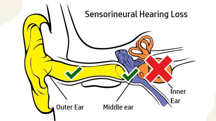
Sensorineural Hearing Loss (SNHL) represents a form of hearing impairment resulting from damage to the inner ear (cochlea) or the auditory nerve pathways that transmit sound signals to the brain. Unlike conductive hearing loss, which involves issues with sound conduction to the inner ear, SNHL often leads to permanent hearing reduction and challenges with speech clarity.
1. Pathophysiology: The inner ear houses delicate hair cells within the cochlea that convert sound vibrations into electrical signals. These signals are then transmitted via the auditory nerve (cranial nerve VIII) to the brain for interpretation. SNHL occurs when these hair cells are damaged or lost, or when the auditory nerve itself is compromised, leading to a diminished or distorted electrical signal reaching the brain. This damage can reduce the perception of sound intensity and lead to difficulties distinguishing between different frequencies, particularly in noisy environments.
2. Etiology and Differential Diagnosis: SNHL is a complex condition with a wide array of potential causes, necessitating a thorough differential diagnosis to pinpoint the underlying etiology:
- Presbycusis (Age-Related Hearing Loss): The most common cause of SNHL, it is a progressive, symmetrical high-frequency hearing loss that occurs naturally with aging due to the degeneration of cochlear hair cells and nerve fibers.
- Noise-Induced Hearing Loss (NIHL): Prolonged or intense exposure to loud noise (e.g., occupational noise, recreational activities like shooting or loud music) damages the hair cells in the cochlea, leading to a characteristic “noise notch” on audiograms, typically at 4000 Hz.
- Genetic Factors: SNHL can be congenital (present at birth) or develop later in life. It can be syndromic (associated with other medical conditions, e.g., Usher syndrome, Waardenburg syndrome) or non-syndromic, involving specific genetic mutations affecting inner ear development or function.
- Infections:
- Viral: Mumps, measles, rubella, cytomegalovirus (CMV, especially congenital), herpes zoster oticus (Ramsay Hunt syndrome), and HIV can cause SNHL by directly infecting inner ear structures.
- Bacterial: Bacterial meningitis, syphilis, and Lyme disease can lead to significant SNHL if left untreated.
- Autoimmune Inner Ear Disease (AIED): A rare condition where the body’s immune system attacks the inner ear, causing bilateral, progressive SNHL, often fluctuating and rapidly progressing over weeks to months.
- Meniere’s Disease: Characterized by episodic vertigo, fluctuating low-frequency SNHL, tinnitus, and aural fullness, caused by an excess of endolymphatic fluid in the inner ear.
- Ototoxicity: Damage to the inner ear caused by certain medications or chemicals. This will be discussed in detail in Section III.
- Trauma: Head injuries (e.g., temporal bone fractures), barotrauma (sudden pressure changes, e.g., diving, flying), or acoustic trauma (sudden, very loud sounds) can cause SNHL.
- Vascular Causes: Conditions affecting blood supply to the cochlea, such as stroke, transient ischemic attacks, or disorders like hypercoagulability, can lead to sudden or progressive SNHL.
- Neoplastic Conditions: Acoustic neuroma (vestibular schwannoma), a benign tumor on the vestibular nerve, can cause unilateral progressive SNHL, tinnitus, and balance issues. Other rare tumors can also affect auditory pathways.
- Sudden Sensorineural Hearing Loss (SSNHL): Defined as a rapid onset (within 72 hours) of SNHL of at least 30 dB in three contiguous frequencies. While many cases are idiopathic, potential causes include viral infections, vascular compromise, or autoimmune disorders. Prompt medical attention is crucial.
3. Diagnosis of SNHL: Diagnosis involves a multi-faceted approach:
- Comprehensive Audiological Evaluation:
- Pure-Tone Audiometry: Determines the quietest sounds (thresholds) a person can hear at different frequencies, identifying the degree and configuration of hearing loss.
- Speech Audiometry: Assesses speech understanding abilities, including speech reception thresholds (SRT) and word recognition scores (WRS).
- Tympanometry: Measures middle ear function, helping differentiate SNHL from conductive components.
- Otoacoustic Emissions (OAEs): Tests the function of outer hair cells in the cochlea. Absent OAEs suggest cochlear damage.
- Auditory Brainstem Response (ABR): Measures the electrical activity in the auditory nerve and brainstem in response to sound, useful for diagnosing retrocochlear lesions or for assessing infant hearing.
- Medical History and Physical Examination: Detailed history of onset, associated symptoms, medical conditions, medication use, and family history. Otoscopic examination to rule out external or middle ear pathologies.
- Imaging: Magnetic Resonance Imaging (MRI) of the internal auditory canal and brain is often performed to rule out retrocochlear pathology (e.g., acoustic neuroma), especially in unilateral or asymmetrical SNHL.
- Laboratory Tests: Blood tests may be ordered to investigate infectious, autoimmune, or metabolic causes.
4. Management of SNHL: The management of SNHL is primarily focused on rehabilitation and addressing the underlying cause where possible:
- Hearing Aids: Electronic devices that amplify sound, highly effective for most forms of SNHL, customized to the individual’s hearing loss profile.
- Cochlear Implants: For individuals with severe to profound SNHL who receive limited benefit from hearing aids, these devices bypass damaged hair cells and directly stimulate the auditory nerve.
- Assistive Listening Devices (ALDs): FM systems, amplified telephones, and alerting devices can complement hearing aids or cochlear implants.
- Auditory Rehabilitation: Includes auditory training, speech-language therapy, and communication strategies to help individuals adapt to and optimize their residual hearing.
- Medical Management: For specific etiologies, such as corticosteroids for sudden SNHL, immunosuppressants for AIED, or antiviral therapy for viral infections.
- Surgical Intervention: For conditions like acoustic neuroma or Meniere’s disease, surgery may be considered.
- Prevention: Noise protection (earplugs, earmuffs) is crucial for preventing NIHL.
Tinnitus
Tinnitus is the perception of sound in one or both ears or in the head without any external acoustic stimulus. It is a symptom, not a disease, and can significantly impact an individual’s quality of life, leading to distress, sleep disturbances, and impaired concentration.
1. Types of Tinnitus:
- Subjective Tinnitus: The most common type, audible only to the individual experiencing it. It is often described as ringing, buzzing, hissing, clicking, or roaring. It is typically associated with damage to the auditory system.
- Objective Tinnitus: A rare form where the sound can be heard by an examiner using a stethoscope or by simply listening closely. It is usually caused by a somatic source near the ear, such as vascular abnormalities or muscular spasms.
2. Causes and Associations of Tinnitus: Tinnitus is frequently a symptom accompanying SNHL, but it can also arise from various other conditions:
- Hearing Loss: Over 90% of individuals with tinnitus have some degree of hearing loss, particularly SNHL. The brain’s response to reduced auditory input (e.g., from damaged hair cells) is thought to contribute to the generation of tinnitus.
- Ototoxicity: Many ototoxic medications (discussed in Section III) can induce or exacerbate tinnitus, often as an early warning sign of inner ear damage.
- Meniere’s Disease: Tinnitus is a characteristic symptom, often fluctuating in intensity with episodes of vertigo and hearing loss.
- Vascular Conditions (Objective Tinnitus):
- Arteriovenous Malformations (AVMs): Abnormal connections between arteries and veins.
- Carotid Artery Stenosis: Narrowing of the carotid artery.
- Venous Hum: Turbulent blood flow in the jugular vein, often positional.
- Glomus Tumors: Vascular tumors in the middle ear or jugular bulb.
- These conditions can cause pulsatile tinnitus, synchronized with the heartbeat.
- Muscular Spasms (Objective Tinnitus): Spasms of the middle ear muscles (tensor tympani, stapedius) or Eustachian tube muscles can produce clicking sounds.
- Temporomandibular Joint (TMJ) Disorders: Dysfunction of the jaw joint can sometimes lead to tinnitus, possibly due to proximity to the ear or shared nerve pathways.
- Head and Neck Trauma: Whiplash injuries or direct trauma to the head can disrupt auditory pathways or cause vascular changes leading to tinnitus.
- Neurological Conditions: Rarely, tinnitus can be associated with conditions like multiple sclerosis, migraines, or acoustic neuroma.
- Metabolic Disorders: Thyroid dysfunction, vitamin B12 deficiency (less common).
- Stress and Psychological Factors: While not a direct cause, stress, anxiety, depression, and fatigue can significantly worsen the perception and impact of tinnitus.
- Cerumen Impaction or Foreign Body: Blockage of the ear canal can cause temporary tinnitus.
3. Diagnosis of Tinnitus: A thorough evaluation is essential to identify treatable causes and determine the most appropriate management strategy:
- Detailed History: Character (pitch, quality, pulsatile/non-pulsatile), laterality, duration, severity, aggravating and alleviating factors, and impact on life (sleep, concentration, mood).
- Audiological Evaluation: Essential to determine if hearing loss is present and to characterize it. Tinnitus matching (pitch and loudness matching) may be performed.
- Physical Examination: Including otoscopy, auscultation of the head and neck for vascular bruits (especially for pulsatile tinnitus), and examination of the TMJ.
- Imaging Studies: MRI (to rule out acoustic neuroma or other retrocochlear lesions) or CT angiography/venography (for vascular causes) may be indicated, especially for unilateral, pulsatile, or rapidly progressive tinnitus.
- Laboratory Tests: Blood tests for metabolic or autoimmune conditions sparingly.
4. Management of Tinnitus: As tinnitus is often a symptom, management focuses on alleviating distress, improving coping mechanisms, and addressing any underlying treatable conditions. There is currently no universal cure for subjective tinnitus.
- Counselling and Education: Understanding tinnitus, its commonality, and the fact that it is rarely a sign of serious illness can significantly reduce anxiety and distress.
- Sound Therapy:
- Masking: Using external sounds (e.g., white noise, nature sounds, background music) to cover or reduce the perception of tinnitus.
- Tinnitus Retraining Therapy (TRT): A habituation-based therapy that combines directive counseling with low-level broadband noise generators to help the brain habituate to the tinnitus signal, making it less noticeable and bothersome.
- Neuromonics Tinnitus Treatment: Combines acoustic neural stimulation with counseling to help desensitize the auditory pathways to tinnitus.
- Cognitive Behavioral Therapy (CBT): Helps individuals change their negative thoughts and emotional reactions to tinnitus, improving coping strategies and reducing the impact on quality of life.
- Pharmacological Interventions: No drug is universally effective for tinnitus. Antidepressants (e.g., tricyclic antidepressants, SSRIs) or anxiolytics may be prescribed to manage associated anxiety, depression, or sleep disturbances, rather than directly treating tinnitus. Evidence for supplements like Ginkgo Biloba is mixed.
- Lifestyle Modifications: Stress reduction techniques (mindfulness, meditation, yoga), avoiding known triggers (caffeine, nicotine, excessive alcohol), ensuring adequate sleep, and regular exercise.
- Hearing Aids: If tinnitus is associated with hearing loss, hearing aids can improve audibility, reduce the contrast between external sounds and tinnitus, and often provide some masking benefit.
- Management of Objective Tinnitus: Treatment focuses on addressing the underlying vascular or muscular cause, which may involve medication or surgical intervention.
- Emerging Therapies: Research is ongoing into neuromodulation techniques such as transcranial magnetic stimulation (TMS) and transcranial direct current stimulation (tDCS), but these are still considered experimental.
Ototoxicity
Ototoxicity refers to the deleterious effects of certain therapeutic agents or chemicals on the inner ear (cochlea and/or vestibule) or the auditory nerve, leading to hearing loss, tinnitus, or balance disorders. These effects can be temporary or permanent, and often dose-dependent.
1. Common Ototoxic Agents: Numerous medications and substances have the potential for ototoxicity. Key categories include:
- Aminoglycoside Antibiotics: (e.g., Gentamicin, Amikacin, Tobramycin, Streptomycin) These are potent antibiotics primarily used for severe bacterial infections. They can cause irreversible damage to inner ear hair cells, leading to SNHL and vestibulo-toxicity (balance issues). Damage is often cumulative and can progress even after cessation.
- Loop Diuretics: (e.g., Furosemide, Bumetanide, Ethacrynic Acid) Used to treat fluid retention (edema) and hypertension. Ototoxicity is typically transient and more common with rapid intravenous infusion, very high doses, or in patients with renal impairment. They primarily affect the stria vascularis.
- Chemotherapeutic Agents:
- Cisplatin: A highly ototoxic platinum-based chemotherapy drug commonly used for various cancers. It causes irreversible, progressive, bilateral SNHL, particularly in the high frequencies, and is often co-administered with other ototoxic agents.
- Carboplatin: Less ototoxic than cisplatin but can still cause SNHL, especially in children or at higher doses.
- Other chemotherapeutics: Bleomycin, Vincristine, Vinblastine can have ototoxic potential.
- Salicylates: (e.g., High-dose Aspirin) Ototoxicity is typically reversible and dose-dependent, manifesting as bilateral SNHL (often flat or high-frequency) and tinnitus.
- Non-Steroidal Anti-Inflammatory Drugs (NSAIDs): (e.g., Ibuprofen, Naproxen) Similar to salicylates, can cause reversible SNHL and tinnitus, usually at high doses.
- Quinine and Chloroquine/Hydroxychloroquine: Used for malaria and autoimmune diseases. Can cause reversible SNHL and tinnitus.
- Macrolide Antibiotics: (e.g., Erythromycin) Rare, typically reversible SNHL, especially with high IV doses in patients with renal/hepatic impairment.
- Environmental/Industrial Chemicals: Heavy metals (e.g., lead, mercury), solvents (e.g., toluene, xylene), and carbon monoxide can also be ototoxic.
2. Mechanism of Damage: While specific mechanisms vary among agents, common pathways involve:
- Oxidative Stress: Many ototoxic drugs generate reactive oxygen species (ROS) that damage cellular components, particularly in the metabolically active hair cells.
- Apoptosis: Programmed cell death of hair cells, leading to their irreversible loss.
- Damage to Stria Vascularis: Affects the delicate blood supply and ionic balance within the cochlea.
- Neurotransmitter Disruption: Interference with the function of neurotransmitters in the auditory pathway.
3. Symptoms of Ototoxicity:
- Hearing Loss: Often bilateral, symmetrical, and initially affecting high frequencies, progressing to lower frequencies with continued exposure. It can be sudden or gradual.
- Tinnitus: Often the first symptom, appearing as a high-pitched ringing or buzzing, and can sometimes precede measurable hearing loss.
- Vertigo/Balance Issues: Indicative of vestibulo-toxicity, causing dizziness, unsteadiness, or oscillopsia (sensation that the visual world is jiggling).
- Aural Fullness: A sensation of pressure or fullness in the ears.
4. Diagnosis of Ototoxicity: Early detection is paramount to minimize irreversible damage.
- Baseline Audiological Assessment: Essential before initiating treatment with potentially ototoxic drugs. This includes pure-tone audiometry (especially high-frequency greater than 8 kHz), OAEs.
- Regular Audiological Monitoring: During and after treatment, serial audiograms are crucial to detect changes. High-frequency audiometry (>8 kHz) is particularly sensitive for early detection. OAEs can also be used as they reflect outer hair cell function.
- Vestibular Testing: If balance issues are reported, tests like VEMP (Vestibular Evoked Myogenic Potentials) or vHIT (Video Head Impulse Test) can assess vestibular function.
- Patient Symptom Monitoring: Regular inquiry about hearing changes, tinnitus, or dizziness.
5. Management of Ototoxicity: Management focuses on prevention, early detection, and rehabilitation.
- Prevention:
- Careful Drug Selection and Dosing: Avoiding ototoxic drugs when equally effective, less toxic alternatives are available. Using the lowest effective dose and shortest duration.
- Therapeutic Drug Monitoring (TDM): For agents like aminoglycosides, monitoring serum peak and trough levels can help optimize dosing and reduce toxicity risk, especially in patients with impaired renal function.
- Screening for Risk Factors: Identifying patients at higher risk (renal impairment, pre-existing hearing loss, genetic predispositions, concurrent use of multiple ototoxic drugs).
- Otoprotectants: While research is ongoing, some agents (e.g., sodium thiosulfate for cisplatin) are being investigated or selectively used to mitigate ototoxicity, but their widespread clinical application is limited.
- Early Detection: Strict adherence to audiological monitoring protocols.
- Cessation or Dose Adjustment: If ototoxicity is detected, the medication should be discontinued or the dose reduced if clinically feasible, in consultation with the prescribing physician.
- Rehabilitation: For irreversible hearing loss, hearing aids or cochlear implants may be necessary. Vestibular rehabilitation therapy can help individuals adapt to vestibular deficits.
- Patient Education: Informing patients about the potential for ototoxicity and instructing them to report any new auditory or vestibular symptoms immediately.
Conclusion
Sensorineural Hearing Loss, Tinnitus, and Ototoxicity represent a significant spectrum of auditory health challenges, often interconnected in their presentation and underlying mechanisms. Understanding their diverse etiologies, diagnostic approaches, and management strategies is crucial for healthcare professionals. From the progressive nature of presbycusis and NIHL to the distressing symptom of tinnitus and the iatrogenic effects of ototoxicity, comprehensive and individualized care is paramount. Early diagnosis, diligent monitoring, and tailored rehabilitative interventions are key to mitigating the impact of these conditions, preserving hearing, and improving the quality of life for affected individuals.


