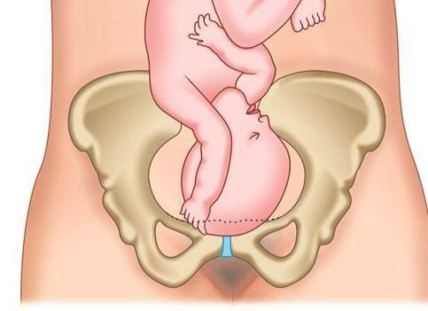
In the intricate field of obstetrics, the process of childbirth is a finely tuned sequence of events designed to safely navigate the fetus through the maternal pelvis. While most deliveries proceed without significant anatomical aberrations, certain deviations can present unique challenges. One such uncommon yet clinically significant obstetric complication is compound presentation, sometimes referred to as ‘complex presentation.’ This condition involves the descent of a fetal extremity alongside the head or breech into the maternal pelvis, potentially causing obstruction and increasing the risk of adverse outcomes for both mother and fetus. Given its potential for complications, a thorough understanding of its definition, causes, diagnosis, and management is paramount for healthcare professionals.
Defining Compound Presentation
Compound presentation occurs when a fetal extremity — typically a hand, arm, or, less commonly, a foot — prolapses alongside the main presenting part (usually the fetal head, or less frequently, the breech) into the maternal pelvis and potentially into the vagina during labor. This coexistence of two fetal parts simultaneously presenting at the pelvic inlet or within the birth canal differentiates it from other malpresentations.
Unlike a simple hand or foot presentation where the extremity is the presenting part, in a compound presentation, the extremity is merely an additional part presenting alongside the primary one. For example, a common scenario is a fetal head with an accompanying hand or arm. This simultaneous descent can mechanically impede the normal progress of labor, leading to dystocia (difficult labor).
While relatively rare, occurring in approximately 1 in 700 to 1 in 1000 deliveries, its unpredictability and potential for rapid deterioration necessitate prompt recognition and intervention. It is crucial to distinguish compound presentation from a simple umbilical cord prolapse, although an umbilical cord prolapse can coexist with a compound presentation, compounding the complexity and urgency of the situation.
Causes of Compound Presentation
The occurrence of compound presentation is often multifactorial, stemming from conditions that prevent the proper engagement of the fetal presenting part, thereby creating space for an extremity to descend alongside it. These predisposing factors can broadly be categorized into maternal, fetal, and iatrogenic causes:
1. Maternal Factors:
- Multiparity: Women who have had multiple previous pregnancies may have lax abdominal muscles and an overly capacious uterus, making it more difficult for the fetal head to engage properly in the pelvis, thus allowing an extremity to descend.
- Pelvic Abnormalities: A contracted pelvis (cephalopelvic disproportion – CPD) or an abnormally shaped pelvis can obstruct the normal descent of the presenting part, leaving space for an accompanying extremity.
- Polyhydramnios: An excessive amount of amniotic fluid provides increased fetal mobility within the uterus. This can prevent the presenting part from settling firmly against the cervix and pelvic inlet, allowing a limb to slip down.
- Abnormal Uterine Shape/Pathology: Uterine anomalies such as a bicornuate uterus, or the presence of large uterine fibroids, can distort the uterine cavity, hindering fetal positioning and engagement.
- Placenta Previa: If the placenta is implanted low in the uterus, potentially covering the cervical os, it can obstruct the descent and engagement of the presenting part, making compound presentation more likely.
2. Fetal Factors:
- Prematurity: Premature fetuses are generally smaller and have less tone, making them more mobile within the uterine cavity. Their smaller size also means there is more space around the presenting part, allowing an extremity to prolapse.
- Multiple Gestation: In twin or higher-order pregnancies, the reduced space and potential for abnormal lie or presentation of one or both fetuses can predispose to compound presentation.
- Abnormal Fetal Lie or Position:
- Transverse or Oblique Lie: Before correction, these lies inherently prevent the head or breech from engaging properly, increasing the chance of an extremity prolapsing.
- Malpositions of the Head: Conditions like deflexed head (brow or face presentation where the head is not fully flexed) or asynclitism (where the sagittal suture is not in the middle of the pelvis) can prevent snug engagement, leaving room for an extremity.
- Fetal Anomalies: Certain fetal anomalies, such as hydrocephalus (excess fluid in the brain, leading to an enlarged head) or anencephaly (absence of a major portion of the brain and skull), can alter the fetal shape and prevent proper engagement.
3. Iatrogenic Factors:
- Artificial Rupture of Membranes (AROM): If AROM is performed when the presenting part is not well-engaged, especially with polyhydramnios, the sudden gush of amniotic fluid can wash down an extremity alongside the presenting part.
- Misguided Vaginal Procedures: Rarely, procedures like internal podalic version (now rarely done) or attempts at manual repositioning of the fetus without proper technique could theoretically contribute.
Complications Associated with Compound Presentation
The simultaneous descent of an extremity alongside the presenting part introduces several risks, significantly increasing the potential for complications for both the mother and the fetus.
1. Maternal Complications:
- Prolonged and Obstructed Labor: The presence of an additional fetal part increases the diameter of the presenting part, leading to mechanical obstruction. This can result in prolonged labor, uterine exhaustion, and ineffective contractions.
- Increased Risk of Operative Delivery: Due to prolonged labor and obstruction, the likelihood of requiring instrumental vaginal delivery (forceps or vacuum extraction) or, more commonly, an emergency Cesarean section significantly increases.
- Maternal Trauma: Obstructed labor can lead to severe cervical, vaginal, and perineal lacerations, rupture of the uterus (though rare, a critical concern with prolonged obstruction), and damage to the bladder or rectum.
- Postpartum Hemorrhage (PPH): Prolonged labor, uterine exhaustion, and operative delivery are major risk factors for uterine atony and subsequent PPH.
- Infection: Prolonged rupture of membranes and frequent vaginal examinations increase the risk of intra-amniotic infection (chorioamnionitis) and postpartum puerperal sepsis.
2. Fetal Complications:
- Fetal Distress and Asphyxia: The most critical fetal complication is umbilical cord compression or prolapse. If the umbilical cord descends alongside the extremity or becomes entrapped, it can lead to acute fetal hypoxia and acidosis, requiring immediate intervention. Fetal compromise can also arise from prolonged labor.
- Fetal Injury to the Prolapsed Extremity: While rare, the prolapsed extremity (e.g., hand or arm) can sustain injuries such as edema, nerve damage (e.g., brachial plexus injury), or even fracture if it becomes entrapped or is mishandled during delivery.
- Increased Perinatal Morbidity and Mortality: All the aforementioned fetal complications contribute to a higher risk of perinatal morbidity (e.g., neonatal intensive care unit admission) and, in severe cases, mortality. This risk is further amplified if the compound presentation is associated with prematurity or underlying fetal anomalies.
How to Diagnose Compound Presentation
The diagnosis of compound presentation is primarily clinical, relying on a high index of suspicion and a thorough vaginal examination.
1. Clinical Suspicion:
- Abnormal Labor Progression: The most common indicator is slow or arrested labor progress despite adequate uterine contractions. The presenting part may remain high, or descent may be minimal.
- Abnormal Fetal Heart Rate (FHR) Patterns: FHR abnormalities, such as decelerations, can suggest umbilical cord compression, which may be an early clue, especially if the cord has prolapsed with the extremity. Continuous FHR monitoring is essential.
- High Presenting Part on Abdominal Palpation: Leopold’s maneuvers may reveal that the fetal presenting part (head or breech) is not well-engaged, or there might be difficulty in outlining it clearly.
2. Vaginal Examination (Definitive Diagnosis):
- Technique: A careful and thorough sterile vaginal examination is the cornerstone of diagnosis. This should be performed whenever labor progress is abnormal, or FHR abnormalities are detected.
- Palpation: The key finding is the simultaneous palpation of a fetal extremity (e.g., hand, arm, or foot) alongside the fetal head or breech.
- Distinguishing Features: The examiner must be able to clearly identify the primary presenting part (e.g., sutures and fontanelles of the head, sacrum and ischial tuberosities of the breech) and independently identify the prolapsed extremity.
- Relationship: Note which extremity it is (left or right hand/foot) and its relationship to the main presenting part (e.g., “head with a left hand”).
- Rule out Cord Prolapse: During the examination, it is imperative to also palpate for the presence of the umbilical cord, which, if prolapsed, constitutes an immediate obstetric emergency.
- Assessment of Labor Progress: Concurrently, assess cervical dilation, effacement, and the station and position of the presenting part, as well as membrane status (intact or ruptured).
3. Ultrasound:
- Confirmatory Role: While usually diagnosed by vaginal examination, ultrasound can be a valuable adjunct. It can confirm the presence of the prolapsed extremity alongside the presenting part, providing a visual confirmation of the diagnosis.
- Additional Information: Ultrasound can also provide crucial information regarding:
- Fetal lie and position.
- Any associated fetal anomalies that might predispose to compound presentation (e.g., hydrocephalus).
- Amniotic fluid volume (polyhydramnios).
- Placental location (placenta previa).
- Fetal size and estimated weight, which can influence management decisions.
- Guidance: In complex cases, ultrasound can guide attempts at repositioning or assist in planning the most appropriate mode of delivery.
Different Ways of Managing Compound Presentation
The management of compound presentation is highly individualized, depending on several critical factors: the specific type of presentation (e.g., head with hand, breech with foot), fetal size, maternal pelvic adequacy, cervical dilation, the presence or absence of umbilical cord prolapse, fetal well-being, and the progress of labor. The primary goals are to ensure maternal and fetal safety.
1. General Principles of Management:
- Immediate Assessment: Prompt and thorough evaluation of both maternal and fetal status is paramount.
- Continuous Fetal Heart Rate Monitoring: Strict continuous FHR monitoring is essential to detect any signs of fetal distress, especially potential cord compression.
- Preparation for Operative Delivery: Given the high likelihood of intervention, preparations for an emergency Cesarean section, including establishing intravenous access, cross-matching blood, and notifying anesthetic and neonatal teams, should be initiated early.
2. Conservative Management:
- Expectant Management (Rarely Successful Alone): In very specific and rare circumstances, if the extremity is small (e.g., a few fingers) and not genuinely impeding labor progress, and if the fetal head is well-engaged with a roomy pelvis, expectant management with close monitoring might be considered. This is often only possible in cases of extreme prematurity where the fetus is very small.
- Repositioning (Manual Reduction): This involves attempting to gently push the prolapsed extremity back above the presenting part.
- Indications: Most successful when membranes have only recently ruptured, or are still intact, and when the cervix is not fully dilated.
- Procedure:
- Place the mother in steep Trendelenburg position or knee-chest position to utilize gravity to move the presenting part out of the pelvis.
- Under sterile conditions and with gentle digital pressure, attempt to push the prolapsed extremity upwards and away from the presenting part during a uterine contraction.
- Once the extremity is retracted, try to facilitate the descent and engagement of the primary presenting part (e.g., by applying fundal pressure if permitted by institutional guidelines, or assisting head flexion).
- Caveats: This maneuver is often difficult to achieve and carries significant risks, including recurrent prolapse, exacerbating cord prolapse, rupture of membranes (if intact), or worsening the fetal lie. It requires an experienced obstetrician. It is often not recommended if the cervix is fully dilated or if the presenting part is deeply engaged.
3. Active Management – Vaginal Delivery (Very Seldom Recommended for Most Cases):
- If the Extremity Does Not Impede Progress: Very rarely, especially with very small, premature fetuses or in multiparous women with exceptionally large pelves, a compound presentation might not cause significant obstruction. In such cases, if the fetal well-being is assured and labor progresses spontaneously, a vaginal delivery might occur. Close supervision is essential.
- Assisted Vaginal Delivery (Forceps/Vacuum): This is generally discouraged due to the high risk of fetal injury and worsening of the situation. Forceps or vacuum extraction might only be considered in extremely select cases where the head is deeply engaged, the extremity is not causing significant obstruction, and a safe application of the instrument is unequivocally possible to complete delivery promptly. The risk of trapping the extremity or extensive maternal trauma makes this option highly unfavorable.
4. Active Management – Cesarean Section (Most Common and Safest Approach):
Cesarean section is the most frequently chosen and often the safest mode of delivery for compound presentations, particularly when conditions are unfavorable for vaginal delivery or when complications arise.
- Indications for Cesarean Section:
- Failed Attempt at Repositioning: If manual reduction of the extremity is unsuccessful or unsustainable.
- Fetal Distress: Any signs of fetal compromise (e.g., non-reassuring FHR patterns) especially if associated with potential cord compression. This is an immediate indication.
- Lack of Labor Progress: Arrest of labor, cervical dilation, or descent despite adequate uterine contractions (failure to progress).
- Presence of Umbilical Cord Prolapse: This is an obstetric emergency that necessitates immediate action to relieve cord compression (e.g., manual elevation of the presenting part, Trendelenburg or knee-chest position for the mother) followed by urgent Cesarean section.
- Associated Complications: Suspected cephalopelvic disproportion, transverse or oblique fetal lie, or any other factor making vaginal delivery unsafe.
- Breech Presentation with Prolapsed Extremity: Management of breech with a foot or hand prolapsing is almost universally by C-section, as it indicates an incomplete or footling breech and mechanical obstruction.
- Unfavorable Conditions for Vaginal Delivery: Cervix not fully dilated, presenting part unengaged, or other maternal/fetal conditions that preclude a safe vaginal birth.
- Procedure: A standard Cesarean section is performed. During extraction, particular care must be taken to avoid injury to the prolapsed fetal extremity.
5. Management of Specific Scenarios:
- Head with Hand/Arm: If the hand/arm is not preventing head engagement and labor is progressing, close observation is possible. However, if progress arrests or fetal distress occurs, C-section is the preferred route. Manual reduction is often difficult but may be attempted in very specific circumstances.
- Breech with Foot/Hand: This situation almost always requires a Cesarean section, as the foot or hand usually indicates an incomplete breech and significantly impedes descent.
Conclusion
Compound presentation represents a significant challenge in obstetrics, demanding prompt recognition and expert management. While relatively rare, its association with increased risks of both maternal and fetal complications, particularly prolonged labor, operative delivery, and fetal distress, underscores the importance of a clear understanding of its pathophysiology and appropriate interventions. The diagnostic cornerstone remains a meticulous vaginal examination, while management strategies range from cautious observation to manual repositioning, with Cesarean section emerging as the most frequent and often the safest definitive solution. By adhering to a systematic, professional approach, healthcare providers can significantly mitigate the risks associated with compound presentation, ensuring the best possible outcomes for both the mother and newborn.


