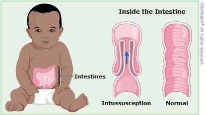
Intussusception is a critical pediatric surgical emergency characterized by the telescoping of one segment of the intestine into an adjacent, more distal segment. This condition can lead to bowel obstruction, ischemia, and potentially life-threatening complications if not promptly diagnosed and managed. Understanding its definition, epidemiology, pathogenesis, clinical presentation, etiology, diagnostic approaches, and management strategies is paramount for healthcare professionals.
Definition and Epidemiology of Intussusception
Definition: Intussusception refers to the invagination of a proximal segment of the bowel (the intussusceptum) into the lumen of an immediately distal segment (the intussuscipiens). This process creates a “telescope” effect, leading to mechanical bowel obstruction. The most common form is ileocolic intussusception, where the ileum invaginates into the colon. Other less frequent types include ileoileal, colocolic, and jejunojejunal intussusception.
Epidemiology:
- Incidence: Intussusception is the most common cause of acute abdominal emergency in infants and young children, excluding appendicitis. Its incidence varies globally but is generally reported as approximately 1 to 4 cases per 1,000 live births.
- Age Distribution: The condition predominantly affects infants and toddlers, with a peak incidence between 5 and 9 months of age. Approximately 60% of cases occur before the first birthday, and 80-90% occur before two years of age. It is rare in neonates and older children, where underlying pathological lead points are more common.
- Gender Predominance: There is a consistent male predominance, with boys affected 1.5 to 2 times more frequently than girls.
- Seasonal Variation: A seasonal pattern is often observed, with an increased incidence during spring and autumn, coinciding with the peak seasons for viral gastrointestinal or respiratory infections. This observation supports the theory of a link between viral infections and lymphoid hyperplasia in the bowel.
Pathogenesis of Intussusception
The pathogenesis of intussusception involves a complex interplay of anatomical and physiological factors that lead to the telescoping of the bowel.
- Mechanism of Invagination: The initial event is typically the invagination of the ileum into the cecum, often initiated by a segment of bowel wall or an enlarged lymphoid tissue (Peyer’s patch) acting as a “leading point.” Peristaltic waves then propel this leading point, drawing the more proximal bowel segment into the distal lumen. The mesenteric attachment, which is shorter proximally and longer distally, contributes to the bowel’s tendency to fold into itself rather than fold back out.
- Consequences of Obstruction: As the intussusception progresses, it results in a series of pathological changes:
- Mechanical Obstruction: The telescoping bowel blocks the passage of intestinal contents, leading to proximal bowel dilation, accumulation of gas and fluid, and vomiting.
- Venous and Lymphatic Obstruction: The mesentery, containing blood vessels and lymphatic channels, is drawn into the intussusception. This compression initially obstructs venous and lymphatic return, leading to edema and hemorrhage within the bowel wall and mesentery.
- Bowel Wall Ischemia: Persistent venous and lymphatic congestion, followed by arterial compression, compromises blood supply to the trapped bowel segment. This causes ischemia, which can progress to infarction (necrosis) and perforation.
- “Currant Jelly” Stools: Ischemia and congestion cause the bowel mucosa to slough off, leading to the characteristic passage of blood and mucus in the stool, resembling “currant jelly.”
- Systemic Manifestations: If untreated, bowel ischemia and necrosis can lead to peritonitis, sepsis, and hypovolemic shock due to fluid shifts and loss of blood into the bowel lumen.
- Role of Leading Points:
- Idiopathic Intussusception: In the vast majority of cases in infants (idiopathic intussusception), no specific anatomical lead point is identified. However, hypertrophied Peyer’s patches (lymphoid tissue in the ileum) are frequently implicated. These can become swollen due to viral infections (e.g., adenovirus, rotavirus), acting as a transient lead point during peristalsis.
- Pathological Lead Points: In older children and adults, or in recurrent cases, an identifiable pathological lead point is more common. These can include Meckel’s diverticulum, intestinal polyps, lymphoma, duplication cysts, appendicitis, or even an inverted appendiceal stump. These fixed points disrupt normal peristalsis and facilitate invagination.
Clinical Presentation of Intussusception
The classic clinical presentation of intussusception is often described by a triad of symptoms, though not all three may be present, especially in early or atypical cases.
- Classic Triad:
- Colicky Abdominal Pain: This is the most consistent and often the first symptom. The pain is typically sudden in onset, severe, intermittent, and crampy. Infants may draw their knees to their chest, cry inconsolably, and appear pale and lethargic between episodes of pain.
- Vomiting: Initially, vomiting is non-bilious, reflecting proximal obstruction. As the obstruction progresses and becomes more distal, or if the bowel becomes ischemic, vomiting may become bilious (greenish).
- “Currant Jelly” Stools: This characteristic finding (stool mixed with blood and mucus) occurs late in the disease process due to mucosal ischemia and sloughing. It is present in approximately 50-60% of cases and indicates significant bowel injury.
- Other Key Clinical Signs:
- Palpable Abdominal Mass: A “sausage-shaped” mass is often palpable in the right upper quadrant or epigastrium. This represents the intussuscepted bowel. The right lower quadrant may feel empty (Dance’s sign) as the cecum is displaced.
- Lethargy and Irritability: Between episodes of pain, infants may appear unusually quiet, lethargic, or irritable. This lethargy can sometimes be the predominant or even sole symptom, especially in very young infants, making diagnosis challenging.
- Dehydration and Shock: As the condition progresses, fluid shifts into the bowel lumen and peritoneal cavity, leading to dehydration. If ischemia and perforation occur, the child can rapidly descend into septic shock with tachycardia, hypotension, and altered mental status.
- Rectal Bleeding: May present as frank blood or occult blood detected on examination.
- Fever: May be present, especially if there is bowel necrosis or perforation.
- Atypical Presentations: It’s crucial to note that not all children present with the classic triad. Infants, in particular, may present predominantly with lethargy, poor feeding, or just subtle changes in behavior without significant pain or vomiting initially. This highlights the importance of a high index of suspicion in any infant presenting with unexplained systemic symptoms.
Etiology (Idiopathic or Due to Underlying Causes)
The etiology of intussusception is broadly categorized into idiopathic and those with an identifiable pathological lead point.
- Idiopathic Intussusception (Approximately 90-95% of cases):
- This is the most common type, primarily affecting infants between 3 months and 2 years of age, with a peak incidence around 6 months.
- No obvious anatomical abnormality is found to cause the intussusception.
- Suspected Mechanism: The prevailing theory involves enlargement of lymphoid tissue (Peyer’s patches) in the terminal ileum. These patches can become hypertrophied due to viral infections (e.g., adenovirus, rotavirus, enteroviruses) or bacterial gastroenteritis. The enlarged lymphoid tissue then acts as a temporary “leading point” that is caught by peristaltic waves and pulled into the distal bowel segment.
- Seasonal Variation: The observed seasonal peaks in incidence often correlate with the prevalence of common viral infections, lending support to the viral etiology hypothesis.
- Rotavirus Vaccine: The introduction of rotavirus vaccines in many countries has shown a complex relationship with intussusception. While early versions of the vaccine (Rotashield) were associated with a small increased risk of intussusception, newer vaccines (Rotarix, RotaTeq) have a much safer profile, with only a very small, transient increase in risk in the first week post-vaccination, which is generally outweighed by the benefits of preventing severe rotavirus gastroenteritis. Overall, routine vaccination has not significantly altered the baseline incidence of intussusception in the general population.
- Intussusception Due to Pathological Lead Point (Approximately 5-10% of cases):
- More common in older children (>2 years) and adults, and in cases of recurrent intussusception.
- An identifiable anatomical lesion serves as the fixed “leading point” that initiates the telescoping.
- Common Pathological Lead Points:
- Meckel’s Diverticulum: The most common pathological lead point in children, a remnant of the vitelline duct that can invert or present as a polypoid mass.
- Intestinal Polyps: Benign or malignant growths within the bowel lumen.
- Lymphoma: Malignant proliferation of lymphoid tissue in the bowel wall (e.g., Burkitt’s lymphoma).
- Duplication Cysts: Congenital anomalies where a segment of the bowel wall is duplicated.
- Appendicitis/Inverted Appendiceal Stump: The inflamed appendix or an inverted stump after appendectomy can act as a lead point.
- Other Rare Causes: Henoch-Schönlein Purpura (HSP) leading to intramural hematoma, celiac disease, cystic fibrosis, hemangiomas, surgical adhesions, or foreign bodies.
- Recognition of a pathological lead point is crucial as these cases typically require surgical intervention for definitive management, as non-operative reduction is unlikely to be successful or curative.
Differential Diagnosis and How to Diagnose Intussusception
Diagnosing intussusception requires a high index of suspicion and is primarily based on clinical history, physical examination, and confirmed by imaging.
- Differential Diagnosis: The symptoms of intussusception can mimic various other pediatric abdominal conditions. It is essential to consider:
- Acute Gastroenteritis: Common cause of vomiting and abdominal pain, though typically without the classic intermittent, severe pain or “currant jelly” stools.
- Acute Appendicitis: More common in older children, presents with localized right lower quadrant pain, fever, and leukocytosis.
- Intestinal Volvulus: Twisting of the bowel, causing severe pain, bilious vomiting, and rapid deterioration, often without blood in stool initially.
- Mesenteric Adenitis: Inflammatory enlargement of mesenteric lymph nodes, often mimicking appendicitis or intussusception, but usually self-limiting.
- Renal Colic/UTI: Abdominal pain, but associated with urinary symptoms.
- Pneumonia: Lower lobe pneumonia can sometimes cause referred abdominal pain.
- Constipation: Can cause abdominal pain and discomfort, but usually without the acute, severe, intermittent pattern.
- Incarcerated Hernia: A bulge in the groin area with pain and vomiting.
- Diagnosis Methods:
- Clinical Assessment: A thorough history focusing on the onset, character, and progression of symptoms, combined with a detailed physical examination to look for abdominal tenderness, distention, palpable mass, and signs of shock, is the first step.
- Imaging Studies (Cornerstone of Diagnosis):
- Abdominal Ultrasound (Gold Standard): This is the diagnostic modality of choice due to its high sensitivity (97-100%) and specificity (97-100%), non-invasiveness, and lack of radiation exposure. Key findings include:
- “Target Sign” or “Doughnut Sign” (Transverse View): Concentric rings of hyperechoic and hypoechoic bowel, representing the layers of intussuscepted bowel and mesentery.
- “Pseudokidney Sign” (Longitudinal View): Layers of bowel wall resembling a kidney.
- Presence of trapped fluid or lymph nodes within the intussusception.
- Plain Abdominal Radiographs (X-ray): While not diagnostic for intussusception itself, X-rays are crucial in the initial evaluation. They can show signs of bowel obstruction (dilated loops of bowel, air-fluid levels), an absence of gas in the right lower quadrant (Dance’s sign equivalent), or a soft tissue mass. Most importantly, they are used to rule out free intraperitoneal air, which indicates bowel perforation and is a contraindication to non-operative reduction.
- Computed Tomography (CT) Scan: Rarely used as the primary diagnostic tool in children due to radiation exposure. However, it can be useful in older children, atypical presentations, or when ultrasound is equivocal, especially if a pathological lead point is suspected. It can clearly delineate the intussusception and any associated masses.
- Abdominal Ultrasound (Gold Standard): This is the diagnostic modality of choice due to its high sensitivity (97-100%) and specificity (97-100%), non-invasiveness, and lack of radiation exposure. Key findings include:
- Laboratory Tests: No specific lab test diagnoses intussusception. However, blood tests (Complete Blood Count, Electrolytes, Blood Urea Nitrogen/Creatinine) are important for assessing hydration, electrolyte imbalances, and ruling out other conditions. Leukocytosis (elevated white blood cell count) may be present, particularly with bowel necrosis or perforation.
Investigations and Management of Intussusception
Prompt and appropriate management is critical to prevent complications and ensure a favorable outcome. The management strategy depends on the child’s clinical condition, duration of symptoms, and the presence of complications.
- Initial Investigations and Stabilization:
- Clinical Assessment: Immediate assessment of airway, breathing, and circulation (ABCs).
- Vital Signs Monitoring: Continuous monitoring for signs of shock (tachycardia, hypotension).
- Intravenous Access and Fluid Resuscitation: Essential to correct dehydration and hypovolemia.
- Nasogastric (NG) Tube Insertion: For gastric decompression to relieve vomiting and prevent aspiration.
- Laboratory Tests: CBC, electrolytes, BUN/creatinine, blood type and cross-match (if surgery is anticipated).
- Imaging: Abdominal X-ray (to rule out perforation) followed by abdominal ultrasound (for confirmation of diagnosis).
- Management Strategies:
- Non-Operative Reduction (Enema Reduction):
- First-line treatment for uncomplicated ileocolic intussusception without signs of peritonitis or perforation.
- Mechanism: Involves injecting air or fluid (saline) into the colon under fluoroscopic or ultrasound guidance, creating hydrostatic or pneumatic pressure that “pushes” the intussuscepted bowel back into its normal position.
- Types of Enema:
- Pneumatic (Air) Enema: Considered the preferred method in many centers. It is cleaner, allows better visualization of potential perforations (free air into the peritoneum), and has a high success rate (70-90%).
- Hydrostatic (Saline) Enema: Performed with saline under ultrasound guidance. Offers the advantage of no radiation exposure during the reduction attempt.
- Barium enema is largely historical due to the disadvantages associated with barium contamination in case of perforation.
- Pre-requisites for Enema Reduction: Patient must be stable, no signs of peritonitis, no evidence of bowel perforation on X-ray, and symptoms should not be excessively prolonged (>24-48 hours), as this increases the risk of bowel necrosis.
- Contraindications: Peritonitis, signs of bowel perforation (free air on X-ray), shock, prolonged symptoms/evidence of gangrenous bowel.
- Success Rate & Complications: High success rates (70-90%). Complications are rare but include bowel perforation (around 0.5-1%). Recurrence after successful non-operative reduction occurs in 5-10% of cases, usually within 24-48 hours but can be later.
- Surgical Reduction:
- Indications for Surgery:
- Failed non-operative reduction attempts (usually after 2-3 attempts).
- Contraindications to non-operative reduction (e.g., signs of peritonitis, perforation, severe shock, prolonged symptoms highly suggestive of bowel necrosis).
- Suspicion of a pathological lead point (e.g., older child, recurrent intussusception).
- Procedure:
- Laparotomy: An open abdominal incision is traditionally used. The surgeon manually reduces the intussusception by “milking” the distal bowel back, avoiding traction on the proximal bowel to minimize risk of tearing.
- Laparoscopy: Minimally invasive approach increasingly used in stable patients, particularly for diagnosis and gentle reduction.
- Bowel Resection: If the bowel is found to be necrotic (non-viable) or perforated, or if a pathological lead point cannot be simply removed, the affected segment of the bowel is resected, and an anastomosis (rejoining of bowel ends) is performed.
- Post-operative Care: Includes IV fluids, NG tube decompression until bowel function returns, pain management, and antibiotics.
- Indications for Surgery:
- Non-Operative Reduction (Enema Reduction):
- Monitoring and Follow-up: Regardless of the reduction method, patients are monitored closely for recurrence (especially in the first 24-48 hours after non-operative reduction) and for any signs of complications. Parents are educated about the signs of recurrence.
In conclusion, intussusception is a time-sensitive emergency in pediatric populations. Its prompt recognition through characteristic clinical features, rapid diagnosis with abdominal ultrasound, and timely intervention—either non-operative enema reduction or surgical intervention—are crucial for minimizing morbidity and mortality. A high index of suspicion, especially in infants with non-specific symptoms, remains key to optimal outcomes.


