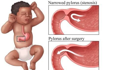
Hypertrophic pyloric stenosis (HPS) is a common cause of gastric outlet obstruction in infants, characterized by the progressive hypertrophy and hyperplasia of the circular and longitudinal muscular layers of the pylorus. This thickening leads to a functional obstruction, preventing the normal passage of gastric contents into the duodenum. Understanding the various facets of HPS, from its incidence patterns to its intricate physiological derangements and effective management strategies, is crucial for healthcare professionals.
Epidemiology of Pyloric Stenosis
Pyloric stenosis is one of the most frequently encountered surgical conditions in young infants, with distinct epidemiological characteristics:
- Incidence: The global incidence varies, but it is generally reported to be between 1 to 3 per 1,000 live births. There are observed geographical and ethnic variations, with higher rates in White populations, particularly those of Scandinavian descent, and lower rates in Black and Asian populations.
- Gender Predominance: HPS exhibits a strong male predominance, with males affected four to five times more frequently than females. The exact reason for this disparity is not fully understood, but it suggests a role for sex-linked or hormonal factors.
- Age of Onset: While present from birth, symptoms typically manifest between 2 to 8 weeks of age, with a peak incidence around 3 to 6 weeks. It is rare for symptoms to appear before 1 week or after 5 months of age.
- Familial Predisposition: There is a well-established genetic component to HPS. Infants with a first-degree relative (parent or sibling) who had HPS have an increased risk. The risk is higher if the mother had HPS compared to the father, suggesting a more complex inheritance pattern, possibly multifactorial with a lower threshold for expression in males. For instance, the risk for male infants of affected mothers is approximately 20%, while for male infants of affected fathers, it is around 5%.
- Environmental Factors:
- Macrolide Exposure: Postnatal exposure to macrolide antibiotics (e.g., erythromycin, azithromycin) in young infants, and even prenatal or postnatal exposure in mothers who are breastfeeding, has been linked to an increased risk of HPS. The mechanism is thought to involve the antibiotic’s motilin-like effect, stimulating excessive pyloric contractility.
- Bottle Feeding: Some studies have suggested a potential association between bottle feeding and HPS, possibly due to differences in gastric emptying patterns or formula composition compared to breast milk. However, this link is less definitively established than the macrolide association.
- Maternal Age and Parity: While not as strong a factor, some studies show a slight increase in incidence with firstborn infants and younger mothers.
Pathophysiology of Pyloric Stenosis
The pathophysiology of HPS revolves around the anatomical obstruction of the gastrointestinal tract and its subsequent systemic consequences:
- Anatomical Anomaly: The hallmark of HPS is the progressive hypertrophy (increase in cell size) and hyperplasia (increase in cell number) of the circular and, to a lesser extent, the longitudinal muscular layers of the pylorus. The pylorus is the muscular valve connecting the stomach to the duodenum. This thickening leads to elongation and narrowing of the pyloric channel, making it difficult for gastric contents to pass into the small intestine.
- Gastric Outlet Obstruction: As the pyloric muscle mass increases, the lumen of the pyloric canal progressively narrows, leading to a functional gastric outlet obstruction. This obstruction prevents food and liquids from leaving the stomach, causing them to accumulate.
- Vomiting: The stomach attempts to push its contents through the obstructed pylorus, leading to forceful peristaltic contractions. When these contractions are insufficient to overcome the obstruction, the accumulated contents are expelled through projectile vomiting. This vomiting is typically non-bilious because the obstruction is proximal to the ampulla of Vater, meaning bile does not enter the stomach.
- Fluid and Electrolyte Imbalance: Prolonged and repeated vomiting of gastric contents, which are rich in hydrochloric acid (HCl), potassium (K+), and chloride (Cl-), leads to significant fluid and electrolyte derangements:
- Dehydration: Loss of gastric fluid results in intravascular volume depletion.
- Hypochloremia: Significant loss of chloride ions, which are integral to HCl, leads to low serum chloride levels.
- Hypokalemia: Vomiting also leads to direct loss of potassium. Furthermore, the ensuing metabolic alkalosis causes an intracellular shift of potassium as hydrogen ions move out of cells to buffer the alkalosis, further lowering serum potassium.
- Metabolic Alkalosis: This is a hallmark of HPS. The continuous loss of gastric HCl results in a net gain of bicarbonate (HCO3-) in the extracellular fluid. The stomach lining secretes HCl and reabsorbs bicarbonate; when vomiting prevents HCl from reaching the duodenum, bicarbonate absorption continues unopposed, leading to a rise in serum bicarbonate and a consequent metabolic alkalosis (elevated pH and HCO3-).
- Compensatory Mechanisms and Renal Response: The body attempts to compensate for these imbalances. The kidneys initially try to excrete excess bicarbonate to correct the alkalosis. However, as dehydration and hypovolemia worsen, the kidneys prioritize volume conservation over acid-base balance. This leads to complex renal responses, including paradoxical aciduria.
Paradoxical Aciduria
Paradoxical aciduria is a critical and often misunderstood consequence of the severe metabolic derangements in HPS. It refers to the presence of an acidic urine pH (low pH) despite systemic metabolic alkalosis (high blood pH and bicarbonate).
- Mechanism:
- Severe Hypovolemia: Prolonged vomiting leads to significant dehydration and marked intravascular volume depletion. This hypovolemia triggers the renin-angiotensin-aldosterone system (RAAS) and antidiuretic hormone (ADH) release, signaling the kidneys to conserve sodium (Na+) and water to restore circulating volume.
- Aldosterone Effects: Aldosterone, released due to hypovolemia, acts on the renal collecting ducts to increase sodium reabsorption. To maintain electroneutrality, sodium reabsorption is coupled with the excretion of potassium (K+) and hydrogen ions (H+).
- Hypokalemia’s Role: The pre-existing hypokalemia (due to direct loss from vomiting and intracellular shift) further exacerbates the situation. When serum potassium is low, the kidneys compensate by shifting potassium out of cells in exchange for hydrogen ions entering cells. This reduces the intracellular hydrogen ion concentration, making more hydrogen ions available for secretion into the urine by renal tubular cells.
- Bicarbonate Retention: Despite the body being in a state of alkalosis, the kidneys are unable to excrete the excess bicarbonate effectively. The need to conserve sodium to maintain fluid volume overrides the need to excrete bicarbonate. Chloride depletion also plays a role; normally, chloride is reabsorbed with sodium, but in hypochloremia, bicarbonate is preferentially reabsorbed with sodium to preserve electroneutrality, thus maintaining the alkalosis.
- Net Result: The combination of intense renal sodium-for-hydrogen exchange (driven by aldosterone and hypokalemia) and the inability to excrete bicarbonate due to volume depletion and chloride deficiency results in the excretion of an acidic urine despite the systemic metabolic alkalosis. The kidneys are effectively sacrificing acid-base balance to preserve vital blood volume.
Understanding paradoxical aciduria is crucial because it highlights the severity of the dehydration and electrolyte imbalances, emphasizing the need for meticulous pre-operative rehydration and electrolyte correction.
Clinical Presentation of Pyloric Stenosis
The clinical presentation of HPS is often characteristic and follows a predictable pattern, typically emerging in infants between 2 and 8 weeks of age:
- Projectile Non-Bilious Vomiting: This is the cardinal symptom.
- Projectile: The vomit is expelled with significant force, often across the room, far beyond the infant’s typical range.
- Non-Bilious: The vomit does not contain bile, as the obstruction is proximal to the ampulla of Vater. It typically consists of formula, breast milk, or gastric contents.
- Progressive: The vomiting starts subtly, often as occasional spitting up, and gradually escalates in frequency and force over several days to weeks.
- Post-Feeding: Vomiting usually occurs shortly after feeding, though it can be delayed.
- Hunger After Vomiting: A distinctive feature is that despite vigorous vomiting, the infant remains hungry and eager to feed again almost immediately. This is because the food has not passed into the intestine for absorption, and the stomach quickly empties.
- Weight Loss and Failure to Thrive: Due to persistent vomiting and inadequate nutrient absorption, infants with HPS typically fail to gain weight or even lose weight. They may appear emaciated or “scrawny.”
- Signs of Dehydration: As fluid and electrolyte losses mount, the infant develops signs of dehydration, including:
- Decreased urine output and dry diapers.
- Dry mucous membranes.
- Sunken fontanelle.
- Reduced skin turgor (tenting of skin).
- Lethargy, irritability, or listlessness.
- Electrolyte Imbalance Symptoms: Symptoms related to hypochloremia, hypokalemia, and metabolic alkalosis can manifest:
- Hypokalemia: Muscle weakness, lethargy, constipation, or even cardiac arrhythmias (though less common in infants).
- Metabolic Alkalosis: Rarely causes overt central nervous system symptoms in infants, but can contribute to lethargy.
- Physical Examination Findings:
- Visible Peristaltic Waves: After a feed, prominent gastric peristaltic waves may be observed moving from the left upper quadrant across the epigastrium to the right side as the stomach attempts to force contents through the narrowed pylorus. This is a highly specific sign.
- Palpable “Olive” Mass: This is the most diagnostic physical finding. During feeding or examination, especially after vomiting or gastric emptying, a firm, mobile, olive-shaped mass (representing the hypertrophied pylorus) can be palpated in the epigastrium or right upper quadrant, typically just to the right of the midline, below the liver edge. Palpation may require patience and a relaxed infant.
- Jaundice: Approximately 2% to 5% of infants with HPS develop unconjugated hyperbilirubinemia (jaundice). The exact mechanism is not fully understood, but it is thought to be related to starvation, dehydration, and increased enterohepatic recirculation of bilirubin due to delayed gastric emptying.
Investigation and Surgical Management of Pyloric Stenosis
Prompt diagnosis and appropriate management are critical for infants with HPS to prevent severe dehydration and electrolyte derangements.
Investigations
- Clinical Assessment: A detailed history focusing on the characteristics of vomiting and feeding patterns, combined with a thorough physical examination (including palpation for the “olive”), is often highly suggestive of HPS.
- Laboratory Tests:
- Arterial Blood Gas (ABG) or Venous Blood Gas (VBG): Essential for confirming metabolic alkalosis (elevated pH, elevated HCO3-) and assessing the degree of compensation.
- Serum Electrolytes: Characteristically reveal hypochloremia, hypokalemia, and normal or slightly low sodium. Elevated blood urea nitrogen (BUN) and creatinine indicate dehydration and pre-renal azotemia.
- Blood Glucose: Hypoglycemia can occur, especially in sick or severely malnourished infants.
- Bilirubin Levels: To assess for concomitant unconjugated hyperbilirubinemia.
- Diagnostic Imaging:
- Abdominal Ultrasonography: This is the diagnostic gold standard for HPS due to its high accuracy (nearly 100%), non-invasiveness, and lack of radiation exposure. Key ultrasound findings include:
- Pyloric Muscle Thickness: Measured greater than 3-4 mm (the most reliable criterion).
- Pyloric Channel Length: Measured greater than 14-17 mm.
- Pyloric Diameter: Measured greater than 10-14 mm.
- “Target Sign” or “Donut Sign”: A cross-sectional view showing the thickened pyloric muscle surrounding the central lumen.
- “Cervix Sign” or “Antral Nipple Sign”: Longitudinal view showing the elongated, narrowed pyloric canal resembling a uterine cervix.
- Failure of gastric contents to pass into the duodenum.
- Upper Gastrointestinal (UGI) Series (Barium Swallow): Less commonly used now due to the reliability of ultrasound and radiation exposure, but can be helpful in ambiguous cases or to rule out other causes of vomiting. Findings include:
- “String Sign”: Elongated, markedly narrowed pyloric channel.
- “Railroad Sign” or “Double-Track Sign”: Mucosal folds compressed within the narrowed pyloric channel.
- Delayed gastric emptying.
- Abdominal Ultrasonography: This is the diagnostic gold standard for HPS due to its high accuracy (nearly 100%), non-invasiveness, and lack of radiation exposure. Key ultrasound findings include:
Surgical Management (Fredet-Ramstedt Pyloromyotomy)
Surgical intervention is the definitive treatment for HPS. However, surgery is never an emergency. Pre-operative stabilization of the infant’s fluid and electrolyte status is paramount and takes precedence over immediate surgical correction.
- Pre-operative Stabilization: This is the most crucial step and can take 24-48 hours, or even longer in severe cases.
- Fluid Resuscitation: Intravenous fluids are initiated to correct dehydration and restore intravascular volume. Initially, an isotonic solution (e.g., 0.9% normal saline with 5% dextrose) is used, ensuring adequate urine output.
- Electrolyte Correction: Once urine output is established, potassium chloride (KCl) is added to the IV fluids (e.g., 0.45% or 0.9% normal saline with D5 and KCl) to correct hypokalemia. Chloride levels are also carefully monitored and corrected.
- Correction of Metabolic Alkalosis: Rehydration and chloride repletion allow the kidneys to excrete excess bicarbonate and correct the alkalosis. Adequately corrected electrolytes (specifically plasma chloride > 100 mEq/L and potassium > 3.5 mEq/L) are indicators of readiness for surgery.
- Gastric Decompression: A nasogastric (NG) tube may be inserted to decompress the stomach and prevent aspiration during stabilization.
- Surgical Procedure: Fredet-Ramstedt Pyloromyotomy: This procedure aims to relieve the obstruction by incising the hypertrophied muscle layer while leaving the mucosa intact.
- Technique: A longitudinal incision is made through the serosa and muscular layers of the pylorus, extending from the stomach to the duodenum, carefully avoiding perforation of the underlying submucosa and mucosa. The muscle fibers are then spread apart, allowing the mucosa to bulge through the incision, effectively widening the pyloric channel.
- Approaches:
- Open Pyloromyotomy: A traditional approach involving a small incision (often right upper quadrant or umbilical).
- Laparoscopic Pyloromyotomy: The preferred approach in many centers. It involves smaller incisions, leads to less post-operative pain, quicker recovery, and improved cosmetic outcomes.
- Post-operative Care:
- Gradual Reintroduction of Feeds: Feeding is typically initiated a few hours after surgery, starting with small, frequent clear fluid feeds, gradually transitioning to full-strength formula or breast milk over 24-48 hours.
- Post-operative Vomiting: Some vomiting is common in the immediate post-operative period due to pyloric spasm and mucosal edema. This usually resolves within a few days.
- Monitoring for Complications: Potential complications, though rare, include pyloric perforation during surgery (requiring repair), incomplete myotomy (leading to persistent obstruction), wound infection, or apnea (particularly in pre-term infants).
- Discharge: Infants are typically discharged once tolerating full feeds, demonstrating adequate weight gain, and showing no signs of significant post-operative complications.
In conclusion, hypertrophic pyloric stenosis is a significant yet treatable condition in infants. A thorough understanding of its epidemiological patterns, the intricate physiological consequences of gastric outlet obstruction, the phenomenon of paradoxical aciduria, its characteristic clinical presentation, and the precise steps involved in its investigation and surgical management are fundamental for timely diagnosis and successful outcomes. With proper pre-operative stabilization and a well-executed pyloromyotomy, infants with HPS typically recover fully with an excellent prognosis.


