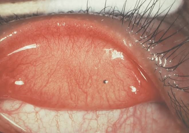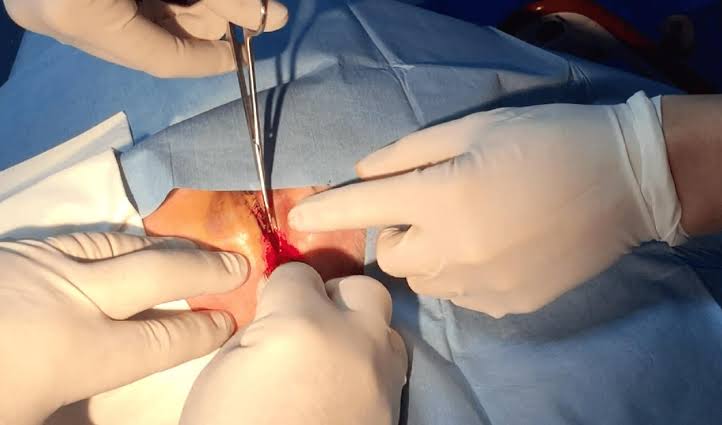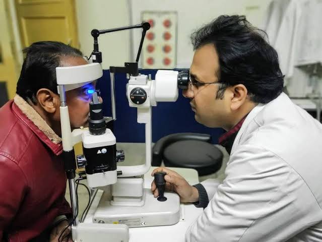
The human eye is an intricate and delicate organ, indispensable for daily life and perception. Its exposed position, however, makes it susceptible to various forms of injury, with foreign bodies and chemical exposures being among the most common and potentially sight-threatening. Understanding the types of ocular foreign bodies, appropriate removal techniques, critical referral criteria, emergency management of chemical trauma, and essential preventive measures through protective eyewear is paramount for healthcare professionals and the general public alike.
Types of Foreign Bodies in the Conjunctiva of the Eye
The conjunctiva is a thin, transparent membrane that lines the inner surface of the eyelids (palpebral conjunctiva) and covers the white part of the eyeball (bulbar conjunctiva), extending up to the edge of the cornea. Its primary function is to lubricate and protect the eye. Due to its exposed nature, it is a common site for foreign body lodging. Foreign bodies in the conjunctiva can vary widely in their nature, size, and potential for harm. They are generally categorized based on their composition and potential for irritation or damage:
- Inert/Non-corrosive Foreign Bodies: These are the most common types and typically cause irritation rather than significant tissue damage unless they abrade the surface. Examples include:
- Dust and Sand Particles: Common in windy or dusty environments.
- Eyelashes and Hair: Often shed from the individual’s own body or from others.
- Fibers: From clothing, tissues, or environmental sources.
- Small Insects: Especially common outdoors.
- Tiny Wood Splinters: From carpentry or gardening activities.
- Irritating/Corrosive Foreign Bodies: These can cause localized inflammation, tearing, and pain due to their chemical properties or sharp edges. Prolonged contact can lead to more severe irritation or even chemical burns.
- Metal Shavings/Filings: From grinding, drilling, or machining processes (e.g., rust rings can form if not removed promptly).
- Chemical Particles: Small fragments from detergents, fertilizers, or other chemicals.
- Organic Foreign Bodies: Originating from living matter, these can carry a higher risk of infection due to the presence of microorganisms.
- Plant Material: Thorns, seeds, or small leaf fragments.
- Insect Parts: Fragments of wings or legs.
- Inorganic Foreign Bodies: Non-living, non-metallic particles.
- Small Glass Shards: From broken objects.
- Stone Fragments: From chipping or breaking rocks.
Symptoms of a conjunctival foreign body typically include acute foreign body sensation (feeling something in the eye), excessive tearing (lacrimation), redness (conjunctival injection), light sensitivity (photophobia), and pain or discomfort.
Procedures for Foreign Body Removal from the Conjunctiva
Safe and effective removal of a foreign body from the conjunctiva requires good lighting, a cooperative patient, and a gentle, sterile approach. Before any procedure, ensure proper hand hygiene (washing hands thoroughly with soap and water or using an alcohol-based hand rub) and explain the steps to the patient to gain their cooperation and minimize anxiety.
A. Removing a Foreign Body from the Conjunctiva Over the Sclera (White of the Eye)
- Patient Positioning and Lighting: Have the patient sit comfortably with their head supported, ideally in a well-lit area. A penlight or examination lamp is often helpful for direct illumination.
- Locate the Foreign Body: Ask the patient to look in various directions (up, down, left, right) while you gently retract their eyelids to expose the entire bulbar conjunctiva.
- Initial Attempt (Rinsing): The least invasive method is often the most effective.
- Sterile Saline or Eye Wash: Gently irrigate the eye with a steady stream of sterile physiological saline or a commercial eye wash solution. Direct the stream from the nasal (inner) corner of the eye towards the temporal (outer) corner. Ask the patient to blink several times during and after irrigation.
- Clean Water (Emergency): If sterile saline is unavailable, clean tap water can be used as an emergency measure, though it may cause some temporary discomfort due to osmolarity differences.
- Assisted Removal (If Rinsing Fails): If the foreign body persists after irrigation:
- Sterile Cotton Swab/Gauze: Moisten the tip of a sterile cotton swab or the corner of a sterile gauze pad with sterile saline. Gently dab or lightly touch the foreign body to lift it away from the conjunctival surface. Avoid vigorous rubbing, which can embed the foreign body further or cause abrasion.
- Sterile Saline Irrigation (Syringe without Needle): A syringe without a needle, filled with sterile saline, can provide a more forceful, directed stream to dislodge the foreign body.
- Post-Removal Assessment: After removal, re-examine the eye to ensure the foreign body is completely gone and to check for any residual irritation or abrasion. If an abrasion is suspected, consider applying a topical antibiotic eye drop or ointment to prevent infection. Advise the patient that they may experience a persistent foreign body sensation for a short period, even after successful removal, due to minor irritation.
B. Removing a Foreign Body Beneath the Lower Eyelid
- Patient Positioning: Have the patient look upwards.
- Expose the Lower Conjunctiva: Gently depress the lower eyelid by placing a finger on the skin below the eyelashes and pulling downwards. This will expose the palpebral conjunctiva of the lower lid and the inferior fornix (the pocket where the conjunctiva folds).
- Locate and Remove: The foreign body is usually visible on the exposed conjunctival surface.
- Rinsing: Irrigate with sterile saline as described above.
- Sterile Cotton Swab/Tissue: Gently wipe the foreign body away using a moistened sterile cotton swab or the clean corner of a tissue.
- Post-Removal: Release the lower eyelid and re-examine.
C. Removing a Foreign Body Beneath the Upper Eyelid
This location is often more challenging because the foreign body may be lodged in the superior fornix or adherent to the palpebral conjunctiva, causing repeated abrasion of the cornea with each blink. Eversion of the upper eyelid is usually necessary.
- Patient Positioning: Have the patient look downwards and keep their eye still.
- Prepare for Eversion:
- Grasp the upper eyelid eyelashes gently between your thumb and forefinger.
- Place a sterile cotton swab or the shank of a cotton applicator horizontally across the upper eyelid, just above the tarsal plate (a firm structure in the lid), approximately 1 cm from the lid margin.
- Evert the Eyelid:
- While holding the eyelashes, gently pull the lid upwards and outwards, simultaneously pressing downwards on the cotton swab. The eyelid should fold back over the swab, exposing the inner surface of the upper lid (palpebral conjunctiva).
- Once everted, maintain the everted position by holding the eyelashes against the brow.
- Locate and Remove:
- Examine the everted conjunctiva carefully. The foreign body may be small and difficult to see.
- Rinsing: Direct a stream of sterile saline onto the everted conjunctiva.
- Sterile Cotton Swab: Gently wipe away the foreign body with a moistened sterile cotton swab.
- Revert the Eyelid: Once the foreign body is removed, ask the patient to look up, or gently pull the eyelashes forward and release. The eyelid should return to its normal position. If it doesn’t, a gentle blink will usually effect it.
- Post-Removal: Re-examine the eye and consider antibiotic drops if there’s any corneal abrasion.
Referral Criteria for Ocular Foreign Bodies: When to Seek Specialized Care
While many conjunctival foreign bodies can be safely removed in a primary care setting, certain situations demand immediate referral to an ophthalmologist or specialized medical facility. The health post in-charge plays a critical role in recognizing these red flags and ensuring prompt, appropriate referral to prevent severe, irreversible vision loss or permanent structural damage to the eye.
A. Foreign Body on the Pupil or Cornea
The cornea is the transparent, outermost layer of the eye that covers the iris, pupil, and anterior chamber. It is highly innervated and crucial for clear vision. A foreign body embedded on the pupil or the cornea carries significant risks:
- Risk of Corneal Abrasion and Ulceration: Even small foreign bodies can cause a scratch (abrasion) on the delicate corneal surface. If left untreated or compounded by infection, this can progress to a corneal ulcer, a potentially vision-threatening condition.
- Visual Impairment: A foreign body on the visual axis (over the pupil) directly interferes with light entry, causing blurred vision. Even after removal, scarring or irregular astigmatism can persist, affecting visual acuity.
- Need for Specialized Equipment: Removal of corneal foreign bodies often requires a slit lamp microscope for magnified visualization and specialized instruments (e.g., sterile spud, fine needle) for precise removal, which are not available at a health post. Attempting removal without proper equipment can cause more harm.
- Risk of Infection: Foreign bodies, especially organic ones or those from contaminated sources, can introduce bacteria or fungi into the cornea, leading to severe infections that are difficult to treat and can result in permanent corneal opacification or even loss of the eye.
- Rust Rings: Metallic foreign bodies can quickly form rust rings on the cornea, which need to be removed by an ophthalmologist using a specialized corneal burr.
Therefore, any foreign body suspected to be on or embedded in the pupil or cornea necessitates immediate referral to an ophthalmologist.
B. Penetrating Eyeball Injury (Globe Penetration)
A penetrating eye injury occurs when an object pierces the outer layers of the eye (sclera or cornea), entering the globe. This is a true ophthalmological emergency with potentially devastating consequences. The health post in-charge should suspect globe penetration if:
- Visible Entry Wound: A clear wound on the sclera or cornea, sometimes with a foreign body still protruding.
- Distorted Pupil: The pupil may appear irregular or “teardrop” shaped, especially if the iris has prolapsed (protruded) through the wound.
- Decreased Vision: Sudden and significant loss of vision in the affected eye.
- Hyphema: Blood visible in the anterior chamber (the space between the cornea and the iris).
- Prolapse of Intraocular Contents: Any internal eye tissue (e.g., iris, vitreous) visible outside the globe.
- Severe Pain: Intense, unrelenting eye pain.
Reasons for Immediate Referral (Emergency):
- Risk of Endophthalmitis: A severe, sight-threatening intraocular infection that can rapidly destroy the eye’s internal structures.
- Risk of Retinal Detachment: The trauma can cause the retina to pull away from its normal position, leading to severe vision loss.
- Risk of Sympathetic Ophthalmia: A rare but devastating inflammatory condition that affects the uninjured eye days to years after injury to the other eye, potentially leading to bilateral blindness.
- Requires Surgical Intervention: Globe penetration almost always requires urgent surgical repair and possibly intraocular foreign body removal by an ophthalmologist to preserve the globe’s integrity and maximize visual outcome.
- Prevention of Further Damage: Any pressure on the eye, including attempting removal or even too much blinking, can cause further prolapse of intraocular contents and worsen the injury.
First Aid for Penetrating Injuries (Prior to Referral):
- Do NOT Attempt Removal: Never try to remove a penetrating foreign body or apply pressure to the eye.
- Cover the Eye: Protect the eye with a rigid shield (e.g., the bottom half of a paper cup taped over the eye) to prevent accidental pressure or rubbing.
- Avoid Pressure: Instruct the patient not to rub the eye, blink forcefully, or strain.
- Keep Patient Calm and Supine: Minimize movement.
- Expedite Transport: Arrange for immediate, safe transport to an ophthalmological center.
How to Rinse the Eye When Chemical Trauma Has Occurred
Chemical burns to the eye are medical emergencies that require immediate and prolonged irrigation to minimize damage and preserve vision. The severity of the burn depends on the chemical’s type (acid or alkali), concentration, volume, duration of contact, and temperature. Alkali burns (e.g., lye, ammonia, lime, cement) are typically more dangerous than acid burns because they cause saponification of cell membranes, leading to deeper penetration and ongoing tissue damage.
A. Immediate Action: Time is Vision
- Do Not Delay: The most critical step is to begin irrigation immediately at the accident site. Every second counts. Do not wait for sterile saline or transport to a medical facility if clean water is available.
- Do NOT Rub the Eye: Rubbing can spread the chemical and cause further damage.
- Locate a Water Source: Any readily available clean water source (tap water, bottled water, shower, garden hose, eyewash station) is acceptable for initial rinsing. Ideally, sterile physiological saline solution should be used if available.
B. Rinsing Procedure:
- Positioning:
- If using a sink or basin: Have the patient lean their head over the basin with the affected eye lower than the unaffected eye (if only one eye is affected) to prevent the chemical from washing into the other eye.
- If using a shower: Have the patient stand under a gentle shower stream.
- If using a bottle/IV bag: Direct the stream from the nasal (inner) corner outwards.
- Eyelid Separation (Crucial): Forcefully hold the eyelids open using your fingers to ensure thorough irrigation of the entire conjunctival surface, including the superior and inferior fornices, where chemical particles can become trapped. The patient may resist due to pain and spasm (blepharospasm), but it is imperative to keep the eyelids open.
- Duration of Rinsing:
- Minimum 15-30 minutes: For most chemical exposures.
- Prolonged Rinsing (Strong Alkalis/Acids): For highly corrosive chemicals, continuous irrigation may be required for 1-2 hours or even longer, until the pH of the eye surface normalizes.
- Method of Rinsing:
- Gentle Continuous Stream: Use a slow, continuous stream of water or saline directly into the eye.
- Basin Immersion: If a basin is available, the patient can immerse their face in the water and repeatedly open and close their eyes while submerged.
- Shower: Standing under a gentle shower is effective for thorough, continuous irrigation of both eyes if needed.
- Contact Lenses: If present, remove contact lenses during irrigation if possible, but do not delay irrigation to remove them.
- Check pH (If Possible): If pH paper is available (e.g., litmus paper or urine dipstick), check the pH of the eye surface after 15-30 minutes of irrigation. Continue rinsing until the pH returns to a neutral range (7.0-7.4). Recheck pH every 15-30 minutes during prolonged irrigation.
C. Post-Rinsing Care and Referral:
- Immediate Ophthalmological Referral: After initial prolonged irrigation, all chemical eye burns, regardless of apparent severity, require immediate ophthalmological assessment. The full extent of damage may not be immediately obvious.
- Transportation: Arrange for prompt transport to an ophthalmologist or emergency department. Continue irrigation during transport if feasible.
- Pain Management: Pain relief can be administered once irrigation is underway.
- Documentation: Note the type of chemical, duration of exposure, and duration of irrigation.
Measures for Protecting the Eye from Injury by Wearing Protective Eyewear
Prevention is the cornerstone of eye safety. A vast majority of eye injuries are preventable through the consistent and correct use of appropriate protective eyewear. Educating individuals on the risks and promoting a culture of safety are essential.
A. Types of Protective Eyewear:
- Safety Glasses: These are standard protective eyewear, featuring impact-resistant lenses (usually polycarbonate) and frames, often with side shields to provide peripheral protection. They meet specific safety standards (e.g., ANSI Z87.1 in the U.S.) for impact resistance. Prescription safety glasses are also available.
- Goggles: Goggles provide a more comprehensive seal around the eyes, offering superior protection against dust, chemical splashes, mists, and fine particles, as well as impact. They can be vented for airflow or non-vented for splash protection.
- Face Shields: These large, transparent shields provide full-face protection against large splashes, flying debris, and radiant heat. They are typically worn over safety glasses or goggles for enhanced protection.
- Welding Helmets: Specialized helmets designed for welding and cutting operations. They protect from intense visible light, ultraviolet (UV) and infrared (IR) radiation, sparks, and molten metal. They feature auto-darkening filters that adjust shades based on arc intensity.
- Laser Safety Glasses: Specific eyewear with filters designed to block particular laser wavelengths, crucial for anyone working with or exposed to lasers.
B. Situations Requiring Protective Eyewear:
Protective eyewear should be worn whenever there is a risk of eye injury, whether in professional settings, at home, or during recreational activities.
- Workplace Environments:
- Construction: Hammering, drilling, sawing, grinding, demolition.
- Manufacturing: Machining, sanding, welding, handling chemicals.
- Laboratories: Handling chemicals, biological agents, glassware.
- Healthcare: Procedures involving splashes of blood or bodily fluids.
- Horticulture/Agriculture: Pruning, using power tools, handling pesticides.
- Automotive Repair: Working under vehicles, using power tools.
- Home Activities:
- DIY Projects: Drilling, hammering, sawing, sanding, painting.
- Gardening: Mowing, trimming hedges, using leaf blowers, applying fertilizers or pesticides.
- Cleaning: Using strong chemical cleaners that might splash.
- Car Repair/Maintenance: Any activity that could lead to flying debris or chemical splashes.
- Sports and Recreation:
- Racquet Sports: Tennis, badminton, squash.
- Basketball, Football: Risk of finger pokes or ball impact.
- Paintball/Airsoft: High-velocity projectiles.
- Skiing/Snowboarding: Protection from UV light, wind, and debris.
- Cycling: Protection from wind, dust, and insects.
- Fireworks: Spectators and users should wear protection.
- Target Shooting: Protection from ricochets or ejecting casings.
C. Best Practices for Use:
- Choose the Right Type: Select eyewear appropriate for the specific hazard. General safety glasses are not adequate for chemical splashes, and regular goggles are insufficient for welding.
- Ensure Correct Fit: Eyewear should fit snugly but comfortably and not interfere with vision. It should cover the eyes adequately without gaps.
- Maintain Cleanliness: Keep lenses clean to ensure clear visibility, which is crucial for safety.
- Inspect and Replace: Regularly inspect eyewear for scratches, cracks, or damage. Damaged eyewear compromises protection and should be replaced immediately.
- Promote Awareness: Foster a strong safety culture at workplaces, schools, and homes. Education on the importance of eye protection and the potential consequences of neglecting it is vital.
Conclusion
The eyes are invaluable, and their protection is paramount. Understanding the diverse types of foreign bodies that can affect the conjunctiva, mastering the appropriate removal techniques, and critically recognizing when to refer cases to specialized ophthalmological care are fundamental skills. Moreover, the immediate and sustained irrigation for chemical trauma and the consistent use of appropriate protective eyewear represent non-negotiable pillars of ocular health and injury prevention. By adhering to these guidelines, individuals and healthcare providers can significantly reduce the incidence of severe eye injuries and preserve vision.


