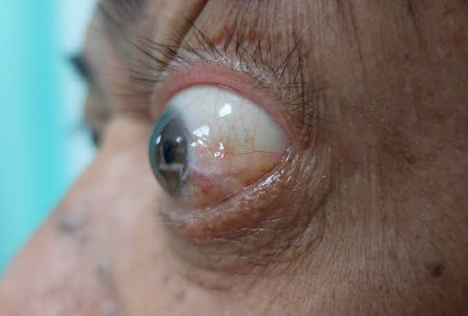
Proptosis, often interchangeably referred to as exophthalmos, is a critical ophthalmological sign that denotes the anterior displacement of the eyeball from the orbital cavity. While it can present as a benign finding, proptosis frequently signals an underlying pathology within the orbit, the bony structure encasing the eye. Its presence warrants thorough investigation due to the potential for vision-threatening complications and severe cosmetic disfigurement.
Defining Proptosis
Proptosis is clinically defined as the abnormal protrusion of one or both eyeballs beyond the orbital rim. While normal variations in eye prominence exist among individuals, a diagnosis of proptosis is typically made when the measurement, usually by an exophthalmometer, exceeds standard norms (e.g., >20-21mm in Caucasians, with a difference of >2mm between the two eyes indicating unilateral proptosis). It is crucial to distinguish proptosis from buphthalmos (enlargement of the entire globe, as seen in congenital glaucoma) or high myopia, where the eye appears prominent but is not truly displaced from the orbit. The direction of displacement, whether axial (straight forward) or non-axial (e.g., downward, lateral), often provides important clues to the underlying cause.
Types of Proptosis
Proptosis can be classified based on several characteristics, each offering insights into its potential etiology and severity:
- Unilateral vs. Bilateral:
- Unilateral Proptosis: Involves only one eye. Common causes include orbital tumors, inflammation (e.g., orbital pseudotumor, cellulitis), or vascular lesions.
- Bilateral Proptosis: Affects both eyes simultaneously. The most prevalent cause is Thyroid-Associated Ophthalmopathy (TAO), but it can also result from systemic conditions like lymphoma or carotid-cavernous fistulas.
- Axial vs. Non-Axial (Displacement Direction):
- Axial Proptosis: The globe protrudes straight forward, along the visual axis. This often suggests a lesion located within the muscle cone (e.g., optic nerve glioma, cavernous hemangioma).
- Non-Axial Proptosis: The globe is displaced in an eccentric direction (e.g., downward and outward, upward and inward). This indicates a mass or pathology outside the muscle cone, displacing the globe in the opposite direction of the lesion. For instance, a lesion in the superotemporal orbit would cause inferonasal proptosis.
- Acute vs. Chronic:
- Acute Proptosis: Develops rapidly over days to weeks. Often associated with inflammatory, infectious, or hemorrhagic conditions (e.g., orbital cellulitis, orbital hemorrhage, acute pseudotumor).
- Chronic Proptosis: Develops slowly over months to years. Typically seen with benign tumors (e.g., dermoid cyst, meningioma), slowly progressive inflammatory conditions, or long-standing TAO.
- Pulsatile vs. Non-Pulsatile:
- Pulsatile Proptosis: The globe visibly pulses synchronous with the patient’s heartbeat. This is highly indicative of a vascular anomaly, such as a carotid-cavernous fistula or an orbital varix, where arterial blood flow is directly transmitted to the orbital contents. A bruit (a whooshing sound) may also be audible over the orbit.
- Non-Pulsatile Proptosis: No visible pulsation of the globe. This is the more common presentation for most causes of proptosis, including tumors, inflammation, and infections.
- Reducible vs. Non-Reducible (or Irreducible):
- Reducible Proptosis: The proptotic eye can be pushed back into the orbit with gentle pressure. This suggests a lesion that is soft or compressible, such as an orbital varix (especially on Valsalva maneuver) or a lipoma.
- Non-Reducible Proptosis: The eye cannot be pushed back into the orbit. This is characteristic of solid tumors, severe inflammation, or conditions where orbital contents are fixed.
- Intermittent Proptosis: Occurs or worsens under specific conditions, such as straining (Valsalva maneuver) or changes in head position (e.g., crying in children). This is almost pathognomonic for an orbital varix.
Causes of Proptosis
The causes of proptosis are diverse, ranging from common benign conditions to rare life-threatening malignancies. Identifying the underlying etiology is paramount for effective management.
- Thyroid-Associated Ophthalmopathy (TAO)/Graves’ Ophthalmopathy: The most common cause of bilateral proptosis in adults. It is an autoimmune condition where antibodies target orbital fibroblasts, leading to inflammation, edema, and fibrosis of orbital fat and extraocular muscles.
- Orbital Tumors:
- Benign Tumors:
- Vascular: Cavernous hemangioma (most common primary orbital tumor in adults), capillary hemangioma (common in children), orbital varix.
- Neural: Optic nerve glioma (common in children), optic nerve sheath meningioma.
- Cystic: Dermoid cyst, epidermoid cyst, mucocele (from paranasal sinuses).
- Other: Pleomorphic adenoma of the lacrimal gland, lipoma, neurofibroma.
- Malignant Tumors:
- Primary Orbital Malignancies: Rhabdomyosarcoma (most common in children), orbital lymphoma, lacrimal gland carcinoma.
- Metastatic Tumors: Breast, lung, prostate, kidney, and gastrointestinal cancers can metastasize to the orbit.
- Extension from Adjacent Structures: Squamous cell carcinoma of the skin/sinuses, retinoblastoma disseminating into the orbit.
- Benign Tumors:
- Inflammatory Conditions:
- Orbital Cellulitis: A severe infection of the orbital tissues, usually bacterial, causing acute, painful proptosis, redness, swelling, and fever. Can be preseptal (anterior to orbital septum) or postseptal (behind septum, true orbital cellulitis).
- Idiopathic Orbital Inflammation (Orbital Pseudotumor): Non-infectious inflammatory process affecting orbital tissues. Can mimic tumor or infection.
- Vasculitis: Granulomatosis with polyangiitis (Wegener’s), sarcoidosis, IgG4-related orbital disease.
- Vascular Anomalies:
- Carotid-Cavernous Fistula (CCF): Abnormal communication between the carotid artery and the cavernous sinus, leading to arterial blood shunting into the orbital venous system, causing proptosis, chemosis, redness, and often a pulsatile bruit.
- Orbital Varices: Abnormally dilated orbital veins that can cause intermittent proptosis.
- Trauma:
- Orbital Hemorrhage: Bleeding into the orbit, causing acute proptosis and ecchymosis.
- Orbital Fracture: Especially ‘blow-out’ fractures, can lead to muscle entrapment or emphysema, though these more commonly cause enophthalmos.
- Infections:
- Fungal Infections: Mucormycosis, especially in immunocompromised individuals.
- Parasitic Infections: Hydatid cyst.
- Cystic Lesions: Other than dermoid/epidermoid, mucoceles originating from adjacent sinuses can expand into the orbit.
Clinical Features of Proptosis
The clinical presentation of proptosis extends beyond mere eye protrusion, encompassing a range of symptoms and signs that reflect the underlying etiology and its impact on ocular health and function.
- Symptoms (Patient Complaints):
- Cosmetic Concern: Often the primary complaint, especially in chronic, non-vision-threatening cases.
- Diplopia (Double Vision): Due to restricted eye movements from muscle inflammation, compression, or entrapment (e.g., in TAO, orbital tumors).
- Blurred Vision or Vision Loss: Indicates compression of the optic nerve (optic neuropathy), severe corneal exposure, or retinal involvement.
- Ocular Pain or Discomfort: Common in acute inflammatory or infectious conditions (e.g., orbital cellulitis, pseudotumor) or rapidly expanding masses.
- Redness, Tearing, Irritation: Due to conjunctival inflammation, chemosis (swelling of conjunctiva), or exposure keratopathy.
- Sensation of Pulsation or Whooshing Sound (Bruit): Highly suggestive of a vascular malformation like a carotid-cavernous fistula.
- General Symptoms: Fever (in infection), weight loss (in malignancy), symptoms of thyroid dysfunction.
- Signs (Objective Findings on Examination):
- Exophthalmometry: Measurement of the degree of proptosis using a Hertel exophthalmometer. Essential for quantifying protrusion and monitoring changes.
- Eyelid Retraction (Dalrymple’s Sign): Upper eyelid elevation, giving a “staring” appearance, characteristic of TAO.
- Lagophthalmos: Incomplete closure of the eyelids, leading to exposure of the globe, especially during sleep.
- Chemosis: Swelling of the conjunctiva, often severe in inflammatory or vascular causes.
- Ocular Motility Restriction (Strabismus): Limited movement of the eye in certain directions, leading to diplopia. Often seen in TAO (inferior rectus restriction is common), orbital tumors, or inflammatory conditions.
- Optic Disc Edema or Atrophy (Optic Neuropathy): Swelling of the optic nerve head (early sign) or pallor (late sign) due to compression, indicating potential vision loss.
- Corneal Exposure: Due to lagophthalmos, leading to dryness, punctate epithelial erosions, persistent epithelial defects, or even ulceration.
- Palpable Mass, Pulsations, or Thrill: Direct palpation of the orbit may reveal a mass, its consistency, or a palpable thrill (vibration) in vascular lesions.
- Conjunctival Injection and Vasodilatation: Engorged blood vessels on the conjunctiva, especially prominent in CCF.
- Proptosis Direction: Assessment of whether the proptosis is axial or non-axial.
Effects of Proptosis
Unmanaged proptosis can lead to significant and potentially irreversible complications, affecting both vision and quality of life.
- Ocular Surface Disease and Exposure Keratopathy: The most common complication. Due to lagophthalmos and incomplete blinking, the cornea is exposed to air, leading to dryness, irritation, and epithelial damage. This can progress to corneal ulceration, infection, and permanent scarring, severely impairing vision.
- Optic Neuropathy: Compression of the optic nerve by orbital tumors, enlarged muscles (in TAO), or orbital edema can lead to ischemia and direct damage to nerve fibers. This manifests as progressive vision loss, dyschromatopsia (impaired color vision), and visual field defects, eventually leading to permanent blindness if not treated.
- Diplopia and Strabismus: Restriction of eye movements from muscle damage, compression, or inflammation causes misalignment of the eyes, resulting in double vision. This significantly impacts daily activities and quality of life.
- Cosmetic Disfigurement: The prominent appearance of the eyes, often coupled with eyelid retraction and redness, can lead to severe psychological distress, affecting self-esteem and social interactions.
- Pain and Discomfort: While not always present, pain can be a major effect, particularly in acute inflammatory or infectious conditions, or with rapidly expanding masses.
- Increased Intraocular Pressure: Orbital congestion can impede venous outflow, leading to elevated intraocular pressure, which can exacerbate optic nerve damage, particularly in patients with pre-existing glaucoma.
Investigations of Proptosis
A methodical approach to investigation is crucial to identify the underlying cause of proptosis, assess its severity, and guide appropriate management.
- Comprehensive Ophthalmic Examination:
- Visual Acuity and Color Vision: To assess visual function and detect early optic nerve involvement.
- Pupil Assessment: Relative Afferent Pupillary Defect (RAPD) suggests optic nerve dysfunction.
- Ocular Motility: To detect restriction of eye movements causing diplopia.
- Slit-Lamp Examination: To evaluate the ocular surface for signs of exposure keratopathy, and the anterior segment for inflammation.
- Fundoscopy: To examine the optic nerve head for signs of edema or atrophy, and the retina for venous congestion or folds.
- Exophthalmometry: Standardized measurement of globe protrusion using a Hertel exophthalmometer. Repeated measurements are crucial for monitoring.
- Perimetry (Visual Fields): To detect visual field defects indicative of optic neuropathy.
- Intraocular Pressure: To rule out secondary glaucoma.
- Imaging Studies: These are indispensable for visualizing orbital structures and identifying the pathology.
- Computed Tomography (CT) Scan: Excellent for bony detail, calcification, acute hemorrhage, and defining the extent of orbital tumors or inflammatory processes. Useful for planning surgical approaches.
- Magnetic Resonance Imaging (MRI) Scan: Superior for soft tissue differentiation, optic nerve pathology, orbital fat, and inflammatory conditions. Provides high-resolution images of extraocular muscles, tumors, and vascular malformations. Often preferred for suspected optic nerve compression or inflammatory conditions.
- Orbital Ultrasound (B-scan): A non-invasive and cost-effective initial imaging modality. Useful for differentiating cystic from solid lesions, identifying vascular flow in varices, and assessing extraocular muscle thickness in TAO.
- CT Angiography (CTA) / MR Angiography (MRA): Specifically indicated for suspected vascular lesions like carotid-cavernous fistulas or orbital varices, to delineate the vascular anatomy.
- Laboratory Tests:
- Thyroid Function Tests (TFTs): TSH, free T3, free T4, and thyroid autoantibodies (TSH receptor antibodies [TRAb], anti-thyroglobulin, anti-thyroid peroxidase) are essential for diagnosing and monitoring Thyroid-Associated Ophthalmopathy.
- Inflammatory Markers: Erythrocyte Sedimentation Rate (ESR) and C-reactive Protein (CRP) to assess systemic inflammation.
- Autoimmune Panel: Antinuclear antibodies (ANA), anti-neutrophil cytoplasmic antibodies (ANCA), and other specific markers may be required if systemic autoimmune disease (e.g., sarcoidosis, vasculitis) is suspected.
- Complete Blood Count (CBC) with Differential: To check for signs of infection or hematological malignancies.
- Cultures: If infection is suspected (e.g., orbital cellulitis).
- Orbital Biopsy: When imaging and laboratory tests are inconclusive, or malignancy is suspected, a biopsy of the orbital lesion is often necessary for definitive histological diagnosis. This is critical for guiding specific treatment, especially for tumors or atypical inflammatory conditions.
Management Protocol for Proptosis
The management of proptosis is highly individualized and depends entirely on the underlying cause, the severity of symptoms, and the risk of vision loss. A multidisciplinary approach often involving ophthalmologists, endocrinologists, neurosurgeons, ENT surgeons, oncologists, and radiologists is frequently required.
General Principles:
- Treat the Underlying Cause: This is the primary goal.
- Protect the Eye: Prevent complications like exposure keratopathy.
- Preserve Vision: Address optic nerve compression promptly.
- Improve Cosmesis: For functional and psychological well-being.
Specific Management Based on Cause:
- Thyroid-Associated Ophthalmopathy (TAO):
- Medical Management:
- Local Symptomatic Relief: Lubricating eye drops/ointments, eyelid taping at night, moisture chambers for exposure keratopathy.
- Corticosteroids: Oral or intravenous steroids (e.g., methylprednisolone pulses) for active inflammation and optic neuropathy.
- Immunosuppressants: Second-line agents like mycophenolate mofetil, cyclosporine, or rituximab for severe or resistant cases.
- Targeted Therapies: Teprotumumab (an IGF-1R inhibitor) for moderate-to-severe active TAO, significantly reducing proptosis and improving symptoms.
- Selenium Supplementation: May be beneficial in mild cases.
- Surgical Management (typically performed in the inactive phase of the disease):
- Orbital Decompression: Removal of orbital bone and/or fat to enlarge the orbital volume, allowing the eye to recede. Indicated for optic neuropathy, severe exposure keratopathy, or significant cosmetic concerns.
- Strabismus Surgery: To correct diplopia once eye movements are stable.
- Eyelid Surgery: To correct eyelid retraction or lagophthalmos.
- Radiotherapy: Low-dose orbital radiation can be used for active inflammation, particularly affecting the extraocular muscles or optic nerve.
- Medical Management:
- Orbital Tumors:
- Surgical Excision: The primary treatment for most benign orbital tumors (e.g., cavernous hemangioma, dermoid cyst, optic nerve meningioma, lacrimal gland pleomorphic adenoma) that are symptomatic, growing, or causing vision compromise.
- Biopsy followed by Chemotherapy/Radiotherapy: For malignant tumors (e.g., lymphoma, rhabdomyosarcoma, metastatic lesions). Systemic chemotherapy or targeted radiation therapy are often the mainstays.
- Observation: For small, asymptomatic, non-progressive benign lesions.
- Inflammatory Conditions:
- Orbital Cellulitis: Requires immediate intravenous broad-spectrum antibiotics. Surgical drainage of an orbital abscess may be necessary.
- Idiopathic Orbital Inflammation (Orbital Pseudotumor): Primarily treated with high-dose oral corticosteroids. Immunosuppressants or radiotherapy may be used for steroid-resistant or recurrent cases.
- Systemic Inflammatory Conditions: Management of underlying systemic disease (e.g., sarcoidosis, vasculitis) with systemic immunosuppression.
- Vascular Anomalies:
- Carotid-Cavernous Fistula: Managed by endovascular embolization, blocking the abnormal communication.
- Orbital Varices: Often managed conservatively. Surgical excision or sclerotherapy may be indicated for significant symptoms or complications.
- Trauma:
- Orbital Hemorrhage: May resolve spontaneously or require surgical decompression if vision is threatened due to high orbital pressure.
- Fractures: Managed based on the type and severity of the fracture, and associated complications.
- Supportive Care:
- Ocular Lubrication: Frequent use of artificial tears and lubricating ointments, especially at night, for exposure keratopathy.
- Eyelid Taping/Moisture Chambers: To protect the ocular surface, particularly during sleep.
- Elevation of Head: To reduce orbital venous congestion.
Conclusion
Proptosis is a complex clinical sign that demands careful and systematic evaluation. Its correct diagnosis relies on a thorough clinical examination, advanced imaging studies, and targeted laboratory tests. Given the array of potential underlying causes, ranging from benign to life-threatening, a precise diagnosis is paramount for effective treatment planning. Management protocols are highly specific to the etiology and aim not only to resolve the protrusion but also to preserve visual function, alleviate discomfort, and restore aesthetic appearance. A collaborative, multidisciplinary approach is often the key to successfully managing patients with proptosis, ensuring optimal outcomes and improved quality of life.


