The advent of X-rays marked a pivotal moment in science and medicine, revolutionizing diagnostic capabilities and opening new frontiers in physics. From their serendipitous discovery to the intricate principles governing their production and the remarkable evolution of the technology, X-rays continue to be an indispensable tool.
The Serendipitous Discovery of X-rays
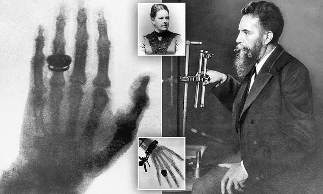
The discovery of X-rays on November 8, 1895, by German physicist Wilhelm Conrad Röntgen stands as one of the most significant accidental scientific breakthroughs. Röntgen, then head of the Physics Department at the University of Würzburg, was engrossed in experiments involving cathode rays. These rays, known to be streams of electrons, were produced in a highly evacuated glass tube (a Crookes tube) that had a high voltage applied across its electrodes.
On that fateful evening, Röntgen had completely enclosed the Crookes tube in thick black cardboard to block out any visible light from the discharge within. He noticed a faint, flickering greenish luminescence on a barium platinocyanide screen that lay on a bench several feet away. What startled him was that this luminescence persisted even when the tube was heavily shielded and the room was completely dark – a phenomenon impossible for the known cathode rays, which could only travel a few centimeters in air.
Intrigued, Röntgen began systematic investigations, placing various objects between the tube and the fluorescent screen. He observed that some materials, like paper and wood, were transparent to these new rays, while others, like lead, were opaque. Most remarkably, when he held his hand between the tube and the screen, he saw the shadowy outlines of his bones, a chilling yet exhilarating revelation. This was the world’s first “radiograph,” albeit a live one.
Röntgen worked in isolation for several weeks, thoroughly documenting the properties of these mysterious rays before making his discovery public. Due to their unknown nature, he provisionally named them “X-rays,” with “X” denoting the unknown variable, a nomenclature that has persisted globally. The immediate impact of his discovery was profound, leading to a Nobel Prize in Physics in 1901 and swiftly transforming medical diagnosis and treatment.
The Fundamental Principle of X-ray Production
X-rays are a form of electromagnetic radiation, residing on the high-energy, short-wavelength end of the spectrum, beyond ultraviolet light. Their production relies on a fundamental physical principle: the rapid deceleration of high-speed electrons. This process occurs within a specialized vacuum tube, known as an X-ray tube.
At its core, an X-ray tube consists of:
- A Cathode: Typically a tungsten filament, which, when heated to incandescence (thermionic emission), releases a cloud of electrons.
- An Anode: A metallic target, usually made of tungsten or an alloy of tungsten and rhenium, which is positively charged and serves as the target for the accelerated electrons.
- A High Voltage (Kilovoltage Peak – kVp): A large potential difference is applied between the cathode and anode, creating an intense electric field that accelerates the electrons from the cathode towards the anode at tremendous speeds (often half the speed of light).
- A Vacuum: The entire cathode-anode assembly is enclosed within a vacuum envelope (typically glass or metal) to prevent the electrons from colliding with gas molecules, which would impede their acceleration and cause tube damage.
When the high-speed electrons from the cathode strike the focal spot on the anode, their kinetic energy is converted into two primary forms: heat (over 99%) and X-rays (less than 1%). The X-rays are produced via two distinct mechanisms:
a) Bremsstrahlung (Braking Radiation)
This is the predominant mechanism for X-ray production in medical imaging (accounting for approximately 80-90% of the X-ray beam). When an incident high-speed electron from the cathode approaches the nucleus of an anode atom, the strong positive electric field of the nucleus attracts the negatively charged electron. This interaction causes the electron to decelerate (“brake”) and change direction. As it loses kinetic energy, this energy is emitted as an X-ray photon. The amount of deceleration and, consequently, the energy of the emitted photon, vary widely depending on the electron’s proximity to the nucleus. This results in a continuous spectrum of X-ray energies, characteristic of Bremsstrahlung radiation.
b) Characteristic Radiation
While less dominant in overall beam quantity, characteristic radiation provides specific, discrete X-ray energies. This occurs when an incident high-speed electron directly interacts with and ejects an inner-shell electron (e.g., K-shell or L-shell) from an anode atom. This leaves a vacancy in the inner shell, creating an unstable atomic state. To restore stability, an outer-shell electron from a higher energy level immediately drops into the vacant inner shell. As the outer-shell electron moves to a lower energy state, it emits an X-ray photon with energy precisely equal to the difference in binding energies between the two electron shells involved. These emitted photons have energies characteristic of the specific anode material (e.g., tungsten’s K-shell characteristic X-rays are around 59-69 keV), hence the name “characteristic radiation.” This process requires the incident electron to have kinetic energy greater than the binding energy of the electron it aims to eject.
Early X-ray Tubes and Their Development
The journey from Röntgen’s crude experimental setup to today’s sophisticated X-ray tubes reflects a century of engineering ingenuity driven by the quest for greater control, predictability, and efficiency in X-ray production.
a) Röntgen’s Crookes Tube (Cold Cathode Gas-Filled Tube)
Röntgen’s initial apparatus was essentially a modified Crookes tube. These were gas-filled tubes (containing residual gas molecules, not a high vacuum) with a “cold cathode” – meaning the cathode was not heated to emit electrons. Instead, electrons were produced by the ionization of residual gas molecules within the tube under high voltage. The “target” was often a small platinum or aluminum disc. These tubes suffered from several limitations:
- Unpredictable Output: The amount of gas within the tube would change over time due to absorption by the glass walls or electrodes, leading to erratic X-ray output and fluctuations in beam quality.
- Limited Lifespan: The variable gas pressure and arc discharges would degrade the tube quickly.
- Difficulty in Control: It was challenging to independently control the X-ray quantity and quality.
Early innovators focused on improving vacuum pumps to achieve higher vacuums and experimented with different target materials to enhance X-ray yield. Efforts were also made to incorporate concave cathodes to focus the electron beam onto a smaller spot on the anode, leading to sharper images.
b) The Coolidge Tube (Hot Cathode Tube)
The most significant leap in X-ray tube technology came in 1913 with the invention of the “hot cathode” X-ray tube by William D. Coolidge at General Electric. This invention revolutionized X-ray production and laid the foundation for all modern X-ray tubes. Coolidge’s key innovations included:
- Heated Filament (Thermionic Emission): Instead of relying on gas ionization, the Coolidge tube used a heated tungsten filament as the cathode. When heated, the filament would “boil off” electrons (thermionic emission) in a controlled manner. This allowed for a much more stable and predictable electron stream.
- High Vacuum: The Coolidge tube operated under a much higher vacuum than its predecessors, virtually eliminating residual gas and preventing the erratic behavior seen in gas-filled tubes.
- Independent Control: The high vacuum and controlled thermionic emission allowed for independent control over the number of electrons (and thus X-ray quantity, by adjusting filament current/mA) and the energy of the electrons (and thus X-ray quality, by adjusting kVp). This was a game-changer for diagnostic imaging.
- Stationary Anode: Early Coolidge tubes typically featured a stationary anode, usually a large piece of tungsten embedded in a copper block for heat dissipation.
c) Rotating Anode Tube
While the Coolidge tube was a vast improvement, stationary anodes struggled to dissipate the immense heat generated during X-ray production, especially for higher power applications and longer exposures. This led to the development of the rotating anode tube in the 1920s. In a rotating anode tube, the anode disc (typically tungsten or a molybdenum-backed tungsten-rhenium alloy) rotates rapidly during exposure. This rotation spreads the heat generated by the electron beam over a much larger surface area, dramatically increasing the tube’s heat capacity and allowing for higher tube currents (mA) and shorter exposure times. This innovation was crucial for improving image quality, reducing patient dose, and enabling a wider range of diagnostic procedures.
Factors Affecting Quality and Quantity of X-ray Production
Controlling the characteristics of the X-ray beam is paramount for producing diagnostic images of optimal quality while minimizing patient dose. Two primary properties of an X-ray beam are its quantity (intensity or exposure rate) and its quality (penetrability or energy).
a) Factors Affecting X-ray Quantity (Intensity/Exposure)
X-ray quantity refers to the number of X-ray photons in the beam. It directly influences the exposure of the image receptor and, consequently, the brightness or density of the image.
- Milliamperage (mA) and Exposure Time (s) (mAs): The tube current (mA) is a direct measure of the number of electrons flowing from the cathode to the anode per second. A higher mA setting means more electrons are boiled off the filament and strike the target, resulting in the production of more X-ray photons. Similarly, extending the exposure time (s) allows for more electrons to strike the target. Therefore, the product of mA and time (mAs) is directly proportional to the total number of X-ray photons produced. Doubling the mAs doubles the X-ray quantity.
- Kilovoltage Peak (kVp): Increasing the kVp significantly increases X-ray quantity. While kVp primarily controls beam quality, a higher kVp means electrons strike the anode with greater kinetic energy, leading to more efficient X-ray production (both Bremsstrahlung and characteristic). The relationship is approximately kVp², meaning a small increase in kVp can lead to a substantial increase in X-ray quantity.
- Target Material (Anode Atomic Number, Z): Materials with higher atomic numbers (Z), such as tungsten (Z=74), are more efficient at producing X-rays, especially Bremsstrahlung radiation, due to their stronger nuclear electric fields. A higher Z target will produce a greater quantity of X-rays compared to a lower Z target under the same conditions.
- Filtration: The primary purpose of filtration (typically aluminum) is to remove low-energy, non-diagnostic X-ray photons from the beam. While it improves beam quality (hardening), it does so by reducing the overall X-ray quantity. Added filtration will decrease the number of photons reaching the image receptor.
- Distance: The intensity of the X-ray beam decreases with increasing distance from the source. This is governed by the Inverse Square Law, which states that the intensity is inversely proportional to the square of the distance from the source. Doubling the distance reduces the intensity to one-fourth. While not a factor in X-ray production, it critically affects the quantity of X-rays reaching the patient and image receptor.
b) Factors Affecting X-ray Quality (Penetrability/Energy)
X-ray quality refers to the penetrating power or the average energy of the X-ray beam. A higher quality beam is more penetrating and less likely to be absorbed by the patient, reaching the image receptor.
- Kilovoltage Peak (kVp): Kilovoltage peak is the primary controlling factor for X-ray beam quality. A higher kVp increases the kinetic energy with which electrons strike the anode. This results in the production of higher maximum energy X-ray photons, especially for Bremsstrahlung radiation, and a shift in the overall X-ray spectrum towards higher average energies. Consequently, the beam becomes more penetrating or “harder.”
- Filtration: Filtration significantly affects beam quality. By preferentially absorbing lower-energy (soft) X-ray photons, filters effectively remove these less useful components of the beam. This process “hardens” the beam, increasing its average energy and penetrability. While it reduces quantity, it improves the diagnostic utility of the remaining photons by reducing patient dose from non-imaging radiation.
- Voltage Waveform: The type of high-voltage generator used (e.g., single-phase, three-phase, high-frequency) influences the efficiency and quality of X-ray production. More constant voltage waveforms (like those from high-frequency generators) result in a higher effective kVp and a more penetrating, higher-quality beam compared to pulsating waveforms, as electrons are consistently accelerated with higher energy.
In conclusion, the discovery of X-rays by Wilhelm Conrad Röntgen irrevocably altered the landscape of science and medicine. The fundamental principles of X-ray production, rooted in the controlled collision of high-speed electrons with a target material, have been meticulously refined through the development of sophisticated X-ray tubes, most notably the Coolidge tube and its rotating anode successor. Understanding and precisely controlling the factors influencing X-ray quantity and quality are paramount for diagnostic imaging, ensuring both optimal image detail and patient safety in this indispensable medical technology.


