Bedside radiography is a vital diagnostic tool that enables healthcare professionals to obtain high-quality images of patients who are unable to be transported to a radiology department due to their medical condition or immobility.
Defining Bedside Radiography
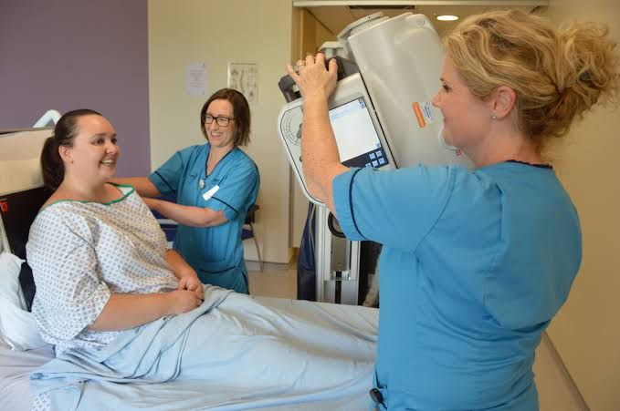
Bedside radiography refers to the process of capturing medical images, such as X-rays, at the patient’s bedside. This technique is employed when a patient is too ill, immobile, or unstable to be transported to a radiology department for imaging. Bedside radiography ensures that patients receive the necessary diagnostic imaging without compromising their health or safety.
Guide to Bedside Radiography
- Preparation: Before commencing bedside radiography, ensure that all necessary equipment is available and in good working condition. This includes an X-ray machine, protective lead aprons, and a mobile image receptor. It is essential to communicate with the patient and their healthcare team to understand the patient’s medical condition and any specific requirements.
- Patient positioning: Position the patient on the bed in a way that allows for optimal image capture. The patient should be comfortable and well-supported to prevent movement during the procedure. For example, a patient with a suspected fracture should be placed in a position that immobilizes the affected area while ensuring that the surrounding anatomy is visible in the image.
- Image acquisition: Once the patient is positioned correctly, the X-ray machine should be set up according to the specific requirements of the examination. This includes selecting the appropriate exposure factors, such as kilovoltage (kV) and milliampere-seconds (mAs). The radiographer should then ensure that they and the patient are protected from radiation exposure by wearing lead aprons and positioning the image receptor correctly.
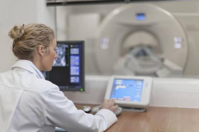
- Image assessment: After the X-ray has been captured, the image should be assessed for quality and diagnostic accuracy. This may involve reviewing the image on a portable monitor or sending it to a radiologist for further analysis.
Defining Traction
Traction is a therapeutic technique used in orthopedic and rehabilitation medicine to immobilize, align, or mobilize injured or diseased parts of the body. It involves applying a steady force to a specific body part, either manually or mechanically, to achieve the desired outcome.
Different Types of Tractions
- Skin traction: In skin traction, adhesive straps or bandages are applied directly to the skin to exert a pulling force on the affected body part. This type of traction is typically used for short-term immobilization or alignment of fractures and dislocations.
- Skeletal traction: Skeletal traction involves the insertion of pins, wires, or screws into the bones to create a secure anchor for the traction apparatus. This type of traction is used for more severe injuries, such as unstable fractures or dislocations, and can be applied for extended periods.
- Continuous traction: Continuous traction is a method of applying a constant force to the affected body part, typically using a weight or spring-loaded device. This type of traction is often used in hospital settings to maintain alignment and immobilization during the healing process.
- Intermittent traction: Intermittent traction involves applying a force to the affected body part for short periods, followed by a period of rest. This type of traction is commonly used in rehabilitation settings to promote muscle strengthening and joint mobility.
Factors to Consider during Ward Radiography
- Patient safety: Ensuring the patient’s safety is of paramount importance during ward radiography. This includes assessing the patient’s medical condition, ensuring they are adequately supported, and minimizing radiation exposure.
- Image quality: The quality of the captured images is crucial for accurate diagnosis and treatment planning. Factors that can affect image quality include patient positioning, exposure factors, and the condition of the equipment.
- Infection control: Bedside radiography can pose a risk of infection transmission, particularly if the patient has open wounds or invasive devices. It is essential to follow strict infection control protocols, such as wearing gloves and using sterile equipment.
- Staff safety: Radiographers and other healthcare professionals should be aware of the potential risks associated with bedside radiography, including radiation exposure and the risk of injury when handling heavy equipment. Appropriate personal protective equipment (PPE) and safe work practices should be employed to minimize these risks.
Conclusion
Bedside radiography is an essential diagnostic tool that enables healthcare professionals to obtain high-quality images of patients who are unable to be transported to a radiology department. Understanding the principles of bedside radiography, traction, and the factors to consider during ward radiography is crucial for providing safe and effective care to patients with a wide range of medical conditions. By following a step-by-step approach and adhering to best practices, healthcare professionals can ensure that patients receive the necessary diagnostic imaging without compromising their health or safety.


