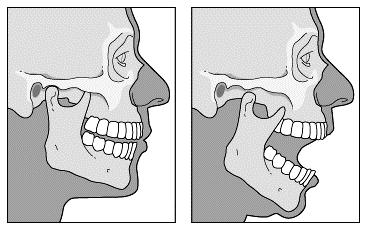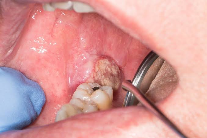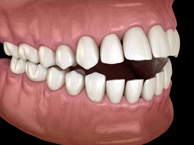
The temporomandibular joint (TMJ) is a complex and crucial articulation connecting the mandible (lower jaw) to the temporal bone of the skull. Essential for mastication, speech, and facial expression, its proper function is vital for daily life. Disruptions to this joint, such as dislocation or conditions causing trismus, can significantly impair a patient’s quality of life, necessitating prompt and appropriate management.
Signs and Symptoms of Temporomandibular Joint Dislocation
Temporomandibular joint dislocation occurs when the mandibular condyle translates anteriorly beyond the articular eminence and into the infratemporal fossa, becoming locked in this position and preventing the jaw from closing. This condition can be acute or chronic/recurrent.
1. Cardinal Signs:
- Inability to Close the Mouth: This is the most striking and immediate sign. The patient’s mouth remains wide open, often with an exaggerated anterior open bite, meaning the front teeth cannot meet.
- Jaw Deviation: The mandible may deviate to one side if the dislocation is unilateral, or appear centrally displaced if bilateral. In bilateral dislocations, the jaw typically protrudes, and both condyles are palpable anterior to the articular eminences.
- Facial Elongation: The face may appear noticeably elongated due to the dropped jaw.
- Palpable Condyle Displacement: Upon palpation in the preauricular area, the normal depression over the joint space may be absent or replaced by a noticeable bulge anterior to the ear, indicating the dislocated condyle(s).
2. Accompanying Symptoms:
- Intense Pain: Patients typically experience significant sharp pain in the preauricular region, often radiating to the temple, ear, or neck. The pain is exacerbated by any attempt to move the jaw.
- Difficulty Speaking (Dysarthria): The wide-open mouth position makes articulation challenging, leading to muffled or incomprehensible speech.
- Difficulty Swallowing (Dysphagia): Saliva may pool in the mouth, leading to drooling, as swallowing becomes difficult or impossible without closing the mouth.
- Malocclusion: The patient will report that their teeth do not fit together properly.
- Muscle Spasm: The muscles of mastication (masseter, temporalis, medial and lateral pterygoids) often go into spasm, further locking the dislocated jaw and contributing to pain.
- Anxiety and Distress: The inability to close the mouth, combined with pain and difficulty communicating, can cause considerable distress for the patient.
3. Clinical Presentation Variations:
- Acute Dislocation: Typically occurs following an exaggerated mouth opening (e.g., yawning, laughing, dental procedures, intubation, vomiting). The onset of symptoms is sudden and dramatic.
- Chronic/Recurrent Dislocation: Some individuals are prone to repeated dislocations due to ligamentous laxity, shallow articular eminences, or prior trauma. The symptoms are similar, but the patient may be familiar with the sensation and even attempt self-reduction.
Management of Temporomandibular Joint Dislocation
Management of TMJ dislocation primarily involves prompt reduction of the joint, followed by measures to prevent recurrence.
1. Immediate Assessment and Preparation:
- Confirm Diagnosis: Visually confirm the signs of dislocation.
- Rule Out Fracture: While dislocation is often obvious clinically, it is crucial to rule out an associated condylar or mandibular fracture, especially after trauma. Imaging (e.g., panoramic radiograph, CT scan) may be necessary if a fracture is suspected.
- Patient Positioning: Seat the patient in an upright position or semi-reclined in a sturdy chair with adequate head support, allowing the operator to stand comfortably in front.
- Analgesia and Sedation: Pain and muscle spasm can hinder reduction. Administer intravenous (IV) analgesics (e.g., opioid) and/or benzodiazepines (e.g., midazolam, diazepam) to relax the muscles and alleviate pain. Local anesthesia (e.g., injecting lidocaine into the joint capsule or surrounding muscles) can also be used.
- Operator Protection: Wear gloves.
2. Manual Reduction Techniques:
The goal of reduction is to move the condyle inferiorly and posteriorly, allowing it to slide back over the articular eminence and into the glenoid fossa.
- The Hippocratic Method (Classical Method): This is the most common manual reduction technique.
- Operator Position: Stand in front of the seated patient, with your elbows resting on the patient’s shoulders or forearms to achieve leverage and stability.
- Hand Placement: Grasp the patient’s mandible firmly with both hands. Place your thumbs on the occlusal surfaces of the mandibular molars (or on the retromolar pad if no teeth are present) and wrap your fingers around the inferior border of the mandible. For safety, wrap your thumbs in gauze to prevent accidental biting.
- Downward and Backward Pressure: Apply firm, sustained downward pressure on the posterior molars with your thumbs. This disengages the condyle from the anterior aspect of the articular eminence.
- Posterior and Upward Rotation: Once the condyle is inferior to the eminence, gently apply posterior pressure with your thumbs while simultaneously lifting the chin with your fingers. This allows the condyle to slide back into the glenoid fossa. A distinct “clunk” may be felt and heard as the joint reduces.
- Post-Reduction: Once reduced, the patient will be able to close their mouth.
- Other Techniques:
- Syringe Method: A 3-5 mL syringe barrel is placed between the posterior molars on one side, and the patient is encouraged to bite down on it repeatedly. This distracts the joint and often allows spontaneous reduction. Primarily for unilateral dislocations.
- Gag Reflex Method: For select patients, stimulating the gag reflex can induce involuntary muscle contractions that may reduce the dislocation.
- Bimanual Digital Reduction (Finger Wrap Method): Similar to Hippocratic method but can be done with fingers on the retromolar pad (useful for edentulous patients).
- External Reduction: In some cases, external manipulation of the condyle combined with intraoral pressure can be effective.
3. Post-Reduction Care:
- Jaw Immobilization/Support: Immediately after reduction, apply a Barton bandage or elastic chin strap to provide gentle support and limit mouth opening for 24-48 hours. This allows muscle spasm to resolve and tissues to heal.
- Soft Diet: Advise the patient to consume a soft diet for 1-2 weeks to minimize jaw movement.
- Avoid Wide Opening: Instruct the patient to avoid wide yawning, excessive laughter, or large bites for at least 3-4 weeks. They should consciously support their jaw when yawning (e.g., by placing a fist under the chin).
- Pain Management: Prescribe NSAIDs for residual pain and inflammation.
- Follow-up: Schedule a follow-up appointment to assess healing and joint stability.
4. Management of Recurrent Dislocation:
For patients with chronic or recurrent dislocations, additional strategies may be needed:
- Non-Surgical:
- Botox Injections: Botulinum toxin (Botox) can be injected into the lateral pterygoid muscles to weaken them, reducing the muscle’s ability to pull the condyle anteriorly. This is a temporary solution.
- Physical Therapy: Exercises to strengthen jaw muscles and improve stability.
- Occlusal Splints: May help in some cases by repositing the mandible.
- Surgical: If conservative measures fail, surgical interventions may be considered:
- Eminence Reduction (Eminectomy/Eminoplasty): Resection or recontouring of the articular eminence to create a less obstructive path for the condyle.
- Condylar Neck Fracture/Osteotomy: Creating a deliberate fracture or osteotomy in the condylar neck to limit its anterior movement or alter its angulation.
- Capsular Plication/Ligament Repair: Tightening the joint capsule or repairing stretched ligaments.
- Mandel’s Procedure (Zygomatic Arch Infratemporal Osteotomy): A more invasive procedure to restrict condylar movement.
Differentiating Trismus Due to Dental Causes from Other Causes
Trismus, or “lockjaw,” refers to the inability to open the mouth fully. It is a symptom, not a diagnosis, and its causes range from localized dental issues to severe systemic conditions. Differentiating the cause is critical for appropriate management.
1. Trismus Due to Dental/Odontogenic Causes:
These are most commonly associated with infection or trauma localized to the oral cavity or jaws.
- Pericoronitis: Inflammation and infection of the soft tissue surrounding a partially erupted tooth, most commonly the mandibular third molar. Swelling and pain in the area can restrict jaw movement.
- Dental Abscess/Cellulitis: Infections originating from teeth (e.g., periodontal abscess, periapical abscess) can spread to adjacent fascial spaces (e.g., masticator space, submasseteric space), causing inflammation and spasm of the muscles of mastication.
- Post-Extraction Complications:
- Infection: Localized infection after tooth extraction.
- Dry Socket (Alveolar Osteitis): Although less common as a direct cause of severe trismus, associated pain and inflammation can limit opening.
- Hematoma: Trauma during extraction can cause a hematoma in surrounding muscles, leading to swelling and pain-induced trismus.
- Local Anesthetic Complications: Direct trauma to muscles or nerves during injection, or subsequent infection.
- TMJ Disorders (Internal Derangement/Myofascial Pain): While distinct from dislocation (where the mouth is open), internal derangement (e.g., disc displacement without reduction) or severe myofascial pain of the masticatory muscles can cause painful limitation of mouth opening.
- Mandibular Fractures: Fractures of the condyle, angle, or ramus can result in pain, muscle spasm, and mechanical interference, leading to trismus.
- Odontogenic Cysts/Tumors: Though less common, large cysts or tumors in the jaw or surrounding soft tissues can mechanically impede jaw opening.
2. Trismus Due to Non-Dental/Systemic/Other Causes:
These causes are often more severe and require broader medical investigation.
- Tetanus: A severe bacterial infection caused by Clostridium tetani, producing toxins that affect the nervous system, leading to widespread muscle spasms, including lockjaw as an early and prominent symptom. This is a medical emergency.
- Central Nervous System (CNS) Disorders:
- Stroke: Can affect motor control, leading to muscle rigidity.
- Epilepsy: Certain seizure types can present with sustained muscle contractions.
- Dystonia: Neurological movement disorder causing involuntary muscle contractions.
- Drug-Induced Dystonia: Certain medications, particularly antipsychotics (e.g., phenothiazines), can cause acute dystonic reactions, including trismus.
- Systemic Conditions:
- Malignant Hyperthermia: A rare, life-threatening reaction to certain anesthetic drugs, characterized by severe muscle rigidity, often including masseter spasm.
- Infections (Non-Odontogenic):
- Parotitis (Mumps or Bacterial): Inflammation of the parotid gland can cause swelling and pain that restricts jaw movement.
- Tonsillitis/Peritonsillar Abscess (Quinsy): Severe throat infections, especially a peritonsillar abscess, can cause significant pain and spasm of the medial pterygoid muscle due to its proximity, leading to trismus.
- Meningitis: Severe neck stiffness can indirectly limit jaw mobility.
- Trauma (Non-Mandibular): Head and neck trauma not involving the mandible directly but causing muscle injury, swelling, or nerve damage can lead to trismus.
- Radiation Therapy: Patients who have undergone radiation therapy to the head and neck region for cancer treatment can develop radiation-induced fibrosis of the masticatory muscles, resulting in chronic and progressive trismus.
- Psychogenic: In rare cases, trismus can be a manifestation of psychological distress or conversion disorder.
3. Diagnostic Approach:
- Thorough History: Onset, duration, associated pain, fever, swelling, recent dental procedures, trauma, systemic illnesses, medications, vaccination history.
- Clinical Examination:
- Extraoral: Facial symmetry, swelling, redness, warmth, tenderness over muscles (masseter, temporalis, pterygoids), lymphadenopathy, range of motion.
- Intraoral: Dental caries, signs of infection (pus, erythema), pericoronitis, mucosal lesions, foreign bodies, tonsillar pathology, oral hygiene. Assess maximum interincisal opening.
- Imaging: Panoramic radiograph (OPG), CT scan (especially for suspected abscess, fracture, or complex pathology), MRI (for soft tissue lesions, TMJ internal derangement).
- Laboratory Tests: CBC (for infection markers), CRP, ESR. For suspected tetanus, specific toxin tests are available.
- Neurological Examination: If CNS causes are suspected.
Identifying Indications for Referral
Prompt and appropriate referral is crucial to ensure optimal patient outcomes, especially when cases are complex, refractory to initial treatment, or involve serious underlying conditions.
1. Indications for Referral for TMJ Dislocation:
- Failed Manual Reduction: If initial attempts at manual reduction are unsuccessful, especially after adequate analgesia and sedation, referral to an oral and maxillofacial surgeon (OMFS) or emergency department specialist is warranted. This could indicate muscle spasm refractory to conservative measures, an anatomical anomaly, or an unrecognised fracture.
- Associated Fractures: Any suspicion or confirmed presence of a condylar or mandibular fracture concomitant with dislocation requires immediate referral to OMFS.
- Recurrent Dislocations: Patients experiencing multiple episodes of TMJ dislocation despite conservative management and patient education should be referred to OMFS for evaluation of underlying predisposing factors (e.g., loose ligaments, flattened articular eminence) and consideration of surgical intervention.
- Chronic Pain or Dysfunction Post-Reduction: If a patient continues to experience significant pain, clicking, locking, or functional limitation after a reduction, referral to OMFS or a TMJ specialist is indicated for further diagnosis and management (e.g., physical therapy, splint therapy, or advanced imaging).
- Pediatric Cases: TMJ dislocations in children may be more complex due to growth considerations and should generally be managed by an OMFS or pediatric specialist.
2. Indications for Referral for Trismus:
- Unidentified Cause: If the cause of trismus cannot be readily determined after initial history and clinical examination, or if it does not fit a common dental etiology, referral to an OMFS, infectious disease specialist, or neurologist is necessary.
- Worsening Symptoms Despite Initial Management: If trismus or associated symptoms (pain, swelling, systemic signs) worsen despite appropriate initial dental management (e.g., antibiotics for infection), it suggests inadequate treatment or a more serious underlying issue.
- Signs of Spreading Infection or Airway Compromise: Any signs of rapidly spreading infection (e.g., cellulitis extending into the neck or mediastinum), fever, increasing difficulty breathing (dyspnea), or difficulty swallowing (odynophagia) are absolute indications for immediate referral to an emergency department or OMFS for urgent airway assessment and management. This could indicate deep space infection.
- Systemic Involvement: If systemic symptoms are prominent (e.g., high fever, malaise, extreme fatigue, neurological signs), or if tetanus is suspected, immediate medical referral to an emergency physician or infectious disease specialist is critical.
- Suspected Non-Dental Cause: If the history or examination points towards a non-odontogenic cause (e.g., neurological disorder, drug-induced trismus, parotitis, peritonsillar abscess, or malignancy), referral to the appropriate medical specialist (neurologist, ENT, internal medicine) is essential.
- Trauma with Suspected Fracture: Any trismus following trauma, especially if a mandibular or facial fracture is suspected, requires urgent referral to OMFS or trauma surgery.
- Need for Advanced Imaging or Surgical Intervention: If the diagnosis requires advanced imaging (e.g., CT, MRI) not available in the primary care setting, or if surgical drainage of an abscess or other surgical intervention is anticipated, referral to an OMFS is indicated.
- Chronic Trismus: For trismus persisting for an extended period, particularly following radiation therapy, referral to an OMFS, physical therapist specializing in head and neck rehabilitation, or a pain management specialist is important for long-term management and improvement of quality of life.
Conclusion
Effective management of TMJ dislocation and accurate diagnosis of trismus require a systematic approach, combining meticulous clinical assessment with an understanding of diverse etiologies. While primary care providers can often manage straightforward cases, recognizing the complexities and knowing when to refer to specialists is paramount for ensuring patient safety and optimal functional recovery. This educational guide serves as a foundational resource for healthcare professionals in navigating these challenging oral-maxillofacial conditions.


