Fetal circulation is a remarkable physiological adaptation designed to support life in an intrauterine environment where the lungs are non-functional for gas exchange and nutrient acquisition depends entirely on the placenta. This intricate system ensures that oxygenated blood bypasses the inactive lungs and is efficiently delivered to vital organs, particularly the brain and heart.
The Unique Landscape of Fetal Circulation
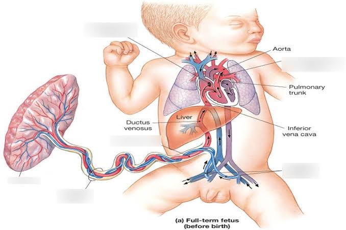
Unlike adult circulation, fetal circulation is characterized by the presence of several shunts—anatomical bypasses that allow blood to divert away from areas not yet fully functional (like the lungs) or to directly deliver oxygenated blood to critical organs. The placenta serves as the crucial interface for gas and nutrient exchange, effectively acting as the fetal “lungs” and “digestive system.” Oxygenated blood originates from the placenta, travels via the umbilical vein, and eventually integrates into the systemic circulation. Deoxygenated blood returns to the placenta via the umbilical arteries.
Sites of Mixing of Oxygenated and Deoxygenated Blood in a Fetus
In a fetus, the concept of “oxygenated” and “deoxygenated” blood is distinct from the adult. Perfectly oxygenated blood only exists in the umbilical vein as it leaves the placenta. As this blood travels through the fetal body, it continuously mixes with less oxygenated or deoxygenated systemic venous return, resulting in varying degrees of oxygen saturation throughout the circulation. The key sites where significant mixing occurs are:
- Ductus Venosus: The umbilical vein, carrying highly oxygenated blood (approx. 80-85% saturation) from the placenta, primarily enters the portal vein. However, a significant portion bypasses the hepatic capillaries via the ductus venosus, directly connecting the umbilical vein to the Inferior Vena Cava (IVC). Here, the highly oxygenated umbilical venous blood mixes with moderately deoxygenated blood returning from the lower body of the fetus (from the IVC itself). This mixing ensures that the most oxygenated blood stream available to the fetus is directed towards the heart.
- Right Atrium (RA): The IVC, now carrying a mixture of oxygenated umbilical blood (via ductus venosus) and deoxygenated systemic blood, enters the right atrium. In the RA, this mixed blood further combines with deoxygenated blood returning from the fetal head and upper limbs via the Superior Vena Cava (SVC) and from the heart itself via the coronary sinus. Due to the anatomical arrangement and the presence of the Eustachian valve (valve of the IVC), the streaming of blood in the fetal RA is crucial: the more oxygenated IVC blood is preferentially directed across the foramen ovale into the left atrium, while the less oxygenated SVC blood tends to flow into the right ventricle. Despite this streaming, significant mixing of blood of different oxygen saturations occurs within the RA.
- Left Atrium (LA): Blood enters the left atrium primarily from two sources: the more oxygenated blood shunted from the right atrium via the foramen ovale, and a small amount of relatively deoxygenated blood returning from the pulmonary veins (as fetal lungs receive very little blood flow). Mixing occurs between these two streams.
- Ductus Arteriosus: The majority of the blood pumped by the right ventricle enters the pulmonary artery. However, because pulmonary vascular resistance is very high in utero, most of this blood (around 90%) is diverted away from the lungs. This diversion occurs through the ductus arteriosus, a shunt connecting the pulmonary artery directly to the aorta, typically distal to the origin of the great vessels supplying the head and upper limbs. Here, relatively deoxygenated blood from the pulmonary artery mixes with the more oxygenated blood already present in the aorta (which originated from the left ventricle). This mixing ensures that the lower body receives blood that, while mixed, is still sufficient for its needs, and it allows the blood that ultimately returns to the placenta via the umbilical arteries to be largely deoxygenated.
Needs of These Sites in a Fetus
The presence of these shunts and mixing sites is paramount for fetal survival and development in a non-pulmonary environment:
- Bypass of Non-Functional Lungs: The primary function of the fetal shunts (foramen ovale and ductus arteriosus) is to divert blood away from the fetal lungs. In utero, the lungs are fluid-filled and collapsed, offering very high vascular resistance. Only a small percentage (approx. 8-10%) of the right ventricular output flows through the pulmonary circulation to nourish the developing lung tissue.
- The foramen ovale allows oxygenated blood from the right atrium (primarily from the IVC) to bypass the right ventricle and pulmonary circulation, flowing directly into the left atrium and then to the left ventricle. This ensures that the most oxygenated blood is preferentially delivered to the left side of the heart, which then pumps it to the brain and heart itself, critical organs requiring maximal oxygenation.
- The ductus arteriosus shunts the majority of the blood ejected from the right ventricle (into the pulmonary artery) directly into the aorta. This prevents blood from flowing into the high-resistance pulmonary circuit and instead directs it into the systemic circulation, specifically to the lower body and the umbilical arteries for placental gas exchange.
- Efficient Oxygen Delivery to Vital Organs: The ductus venosus ensures that the most oxygenated blood from the placenta largely bypasses the hepatic sinusoids, which would otherwise reduce its oxygen content. By directly channeling it into the IVC and then preferentially across the foramen ovale to the left heart, it ensures that the brain, heart, and upper body receive the highest possible oxygen saturation, crucial for their rapid development.
- Management of Vascular Resistances: Fetal circulation operates with very high pulmonary vascular resistance and relatively low systemic vascular resistance (due to the presence of the low-resistance placental circulation). The shunts are essential for maintaining these pressure gradients and ensuring appropriate blood flow distribution. The ductus arteriosus, for instance, allows the right ventricle to eject blood into a low-resistance circuit (aorta via the ductus) rather than the high-resistance pulmonary circuit.
Changes Occurring in Human Circulation After Birth
Birth triggers a rapid and dramatic transformation in the circulatory system, adapting it from a placental-dependent model to an independent, pulmonary-dependent one. These changes are largely driven by the first breath and the clamping of the umbilical cord:
- First Breath and Lung Expansion:
- Decreased Pulmonary Vascular Resistance: As the infant takes its first breath, the lungs expand, liquid is replaced by air, and pulmonary arterioles dilate in response to increased oxygen tension. This causes a dramatic drop in pulmonary vascular resistance.
- Increased Pulmonary Blood Flow: With the significant drop in pulmonary resistance, blood now rushes into the pulmonary arteries and capillaries, leading to a substantial increase in blood flow through the lungs.
- Increased Left Atrial Pressure: The increased blood flow through the lungs leads to a significant increase in pulmonary venous return to the left atrium, causing the pressure in the left atrium to rise sharply.
- Clamping of the Umbilical Cord:
- Cessation of Placental Blood Flow: Clamping the umbilical cord eliminates the low-resistance placental circulation.
- Increased Systemic Vascular Resistance: The loss of the placental circulation instantly increases systemic vascular resistance (SVR), leading to a rise in systemic blood pressure.
- Decreased Right Atrial Pressure: The cessation of blood flow from the umbilical vein (via ductus venosus) to the IVC leads to a decrease in venous return to the right atrium, causing a slight drop in right atrial pressure.
- Closure of Fetal Shunts: The pressure changes resulting from lung expansion and cord clamping are the primary triggers for the closure of the fetal shunts:
- Foramen Ovale: The rising left atrial pressure (due to increased pulmonary venous return) and the relatively falling right atrial pressure (due to reduced umbilical venous return) cause the flap-like septum primum to be pressed against the septum secundum, functionally closing the foramen ovale. This functional closure occurs within minutes to hours after birth. Anatomical closure, involving fusion of the septa to form the fossa ovalis, typically takes several weeks to months, sometimes remaining “probe patent” without clinical significance.
- Ductus Arteriosus: The increased systemic oxygen tension (from lung breathing) and the abrupt withdrawal of placental prostaglandins (which kept the ductus patent in utero) cause the muscular wall of the ductus arteriosus to constrict. This functional closure usually occurs within 10-15 hours of birth. Over the next few weeks, anatomical closure occurs through fibrosis and obliteration, forming the ligamentum arteriosum.
- Ductus Venosus: With the cessation of umbilical venous flow after cord clamping, the ductus venosus constricts. Functional closure occurs within hours to days. Over the next 1-3 weeks, it obliterates to form the ligamentum venosum.
- Umbilical Vessels: The umbilical arteries constrict and functionally close minutes after birth, obliterating over weeks to become the medial umbilical ligaments. The umbilical vein obliterates to form the ligamentum teres hepatis.
Embryological Basis of Congenital Anomalies of the Cardiovascular System (CVS)
Understanding fetal circulation and its postnatal changes is fundamental to comprehending the embryological origins of various congenital heart defects (CHDs). Many CHDs arise from the failure of a fetal structure to involute or close properly, or from abnormal development of structures essential for the formation and separation of the heart chambers and great vessels.
- Persistent Foramen Ovale (PFO): This is the failure of the foramen ovale to anatomically close, remaining probe patent. While common and often asymptomatic, in some cases, it can allow right-to-left shunting (e.g., during activities that increase right atrial pressure, like coughing or straining), potentially leading to paradoxical emboli (blood clots from the venous system entering the arterial circulation and causing stroke) or transient hypoxemia. It represents a failure of complete fusion of the septum primum and secundum after functional closure.
- Patent Ductus Arteriosus (PDA): This anomaly results from the failure of the ductus arteriosus to functionally and anatomically close after birth. It is more common in premature infants due to their immature response to oxygen and higher prostaglandin levels. A PDA leads to left-to-right shunting of blood from the high-pressure aorta to the low-pressure pulmonary artery, increasing pulmonary blood flow. Over time, this can lead to pulmonary hypertension, heart failure, and eventually Eisenmenger syndrome (reversal of shunt due to irreversible pulmonary damage). Its embryological basis is the persistence of the fetal shunt.
- Atrial Septal Defects (ASDs): These are holes in the interatrial septum. While a PFO is a type of ASD, “true” ASDs involve more significant defects in the septal wall itself, not just a patent foramen ovale flap.
- Secundum ASD: The most common type, often results from excessive resorption of the septum primum or an underdeveloped septum secundum, leading to a permanent opening at the site of the foramen ovale. It differs from PFO in that it’s a true defect in the septal tissue rather than just failure of a flap valve to fuse.
- Primum ASD: Occurs when the septum primum fails to fuse with the endocardial cushions, resulting in a defect near the atrioventricular valves.
- Sinus Venosus ASD: Located near the entry of the SVC or IVC, often associated with anomalous pulmonary venous return. All ASDs lead to left-to-right shunting, increasing right heart volume and pulmonary blood flow.
- Ventricular Septal Defects (VSDs): These are holes in the interventricular septum, allowing communication between the two ventricles. VSDs are the most common congenital heart defects. They result from incomplete development or fusion of the various components that form the interventricular septum (muscular septum, membranous septum, aorticopulmonary septum). VSDs lead to left-to-right shunting, increasing pulmonary blood flow and leading to pulmonary hypertension and heart failure if large.
- Transposition of the Great Arteries (TGA): This severe CHD occurs due to the failure of the aorticopulmonary septum to spiral during development, instead growing straight down. This results in the aorta originating from the right ventricle and the pulmonary artery from the left ventricle, creating two parallel, independent circulatory systems. Survival postnatally depends entirely on the presence of mixing sites, typically a PDA or a PFO (or a large VSD), to allow some oxygenated blood to reach the systemic circulation. This highlights how crucial the shunts (normally temporary) become for survival in specific congenital anomalies.
- Tetralogy of Fallot (TOF): This complex cyanotic heart defect is characterized by four components:
- Pulmonary stenosis (narrowing of the pulmonary outflow tract)
- Right ventricular hypertrophy (enlargement of the RV wall due to increased workload)
- Overriding aorta (aorta sitting directly over the VSD, receiving blood from both ventricles)
- Ventricular Septal Defect (VSD) TOF arises from an unequal division of the conus arteriosus by the aorticopulmonary septum. The degree of pulmonary stenosis determines the severity of the right-to-left shunting (via the VSD) and hence the degree of cyanosis. In utero, the ductus arteriosus helps maintain systemic blood flow, but after birth, the limited pulmonary flow and right-to-left shunting cause hypoxemia.
- Coarctation of the Aorta (CoA): This is a localized narrowing of the aorta, most commonly just distal to the origin of the left subclavian artery, often at the insertion of the ductus arteriosus (juxtaductal). In the fetus, the ductus arteriosus can bypass this narrowed segment, allowing blood to reach the lower body. After birth, as the ductus closes, the coarctation becomes a significant obstruction to systemic blood flow, leading to high blood pressure in the upper extremities and low or absent pulses in the lower extremities. Its embryological basis is still debated but may involve abnormal migration of ductal tissue into the aorta.
Conclusion
Fetal circulation represents a remarkable feat of physiological engineering, meticulously designed to sustain life in a unique intrauterine environment. The intricate network of shunts and mixing sites ensures efficient oxygen delivery to vital organs while bypassing the non-functional lungs. The rapid and synchronized closure of these fetal shunts after birth is a testament to the body’s adaptive capacity, seamlessly transitioning the circulatory system to an adult pattern. A deep understanding of these fetal adaptations and their postnatal changes is paramount for medical professionals, offering crucial insights into the embryological origins and clinical manifestations of numerous congenital cardiovascular anomalies, thereby guiding diagnosis, treatment, and long-term management strategies.


