Hemopoiesis, often referred to as hematopoiesis, is a sophisticated biological process responsible for the continuous production, development, and differentiation of all types of blood cells from a common progenitor cell. This intricate system is vital for maintaining physiological homeostasis, ensuring oxygen transport, immune defense, and hemostasis. The sites where this remarkable process occurs exhibit a dynamic shift throughout an individual’s lifespan, adapting to the developmental and physiological demands of the body. Furthermore, the precise orchestration of erythrogenesis, the specific lineage producing red blood cells, exemplifies the complexity and controlled nature of hemopoiesis, meticulously guided by various growth and differentiation inducers.
Sites of Hemopoiesis in the Body During Different Stages of Life
The primary sites of hemopoiesis undergo a fascinating transformation from embryonic development through adulthood, ensuring a consistent supply of blood cells tailored to the body’s evolving needs.
- Embryonic and Fetal Life:
- Yolk Sac (Mesoblastic Stage – 0-3 months gestation): Hemopoiesis initially commences in the mesoderm of the yolk sac. Here, primitive erythroid cells (megaloblasts) are first formed, primarily responsible for oxygen transport in the early embryo. This stage is characterized by the formation of blood islands.
- Liver (Hepatic Stage – 2-7 months gestation): As the embryo develops, the liver becomes the predominant site of hemopoiesis, peaking significantly during the second trimester. It produces a wider range of blood cell types, including erythrocytes, granulocytes, and megakaryocytes. The spleen also contributes to hemopoiesis during this period, though to a lesser extent than the liver.
- Bone Marrow (Myeloid Stage – From 5 months gestation onwards and continuing postnatally): Around the fifth month of gestation, the bone marrow gradually takes over as the primary site of hemopoiesis. By the time of birth, the bone marrow in virtually all bones is actively producing blood cells, fully assuming its role as the central hemopoietic organ.
- Infancy and Childhood:
- During infancy and childhood, the red bone marrow found in the medullary cavities of virtually all bones (long bones, flat bones, irregular bones) is actively hemopoietic. This widespread activity is necessary to support rapid growth and development.
- Adulthood:
- As an individual matures, the need for widespread hemopoiesis diminishes, and the active red marrow gradually recedes or undergoes fatty involution (yellow marrow) in the shafts of long bones.
- In healthy adults, hemopoiesis is primarily confined to the axial skeleton:
- Vertebrae
- Sternum
- Ribs
- Pelvic bones (iliac crest)
- Skull bones
- Additionally, the epiphyses (proximal ends) of large long bones, such as the femur and humerus, retain some red marrow activity.
- In situations of severe demand (e.g., chronic anemia, marrow failure), extramedullary hemopoiesis can reactivate in organs like the liver and spleen, reverting to their fetal hemopoietic functions.
Flow Sheet Diagram of Various Stages of Erythrogenesis with Relevant Features and Sizes
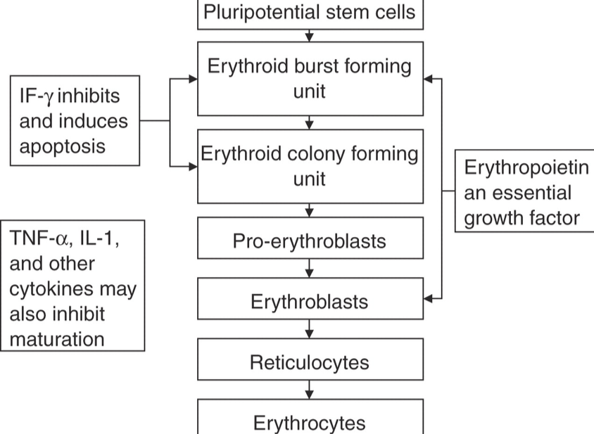
Erythrogenesis, or erythropoiesis, is the process by which multipotent hematopoietic stem cells (HSCs) differentiate and mature into functional red blood cells (erythrocytes). This remarkable journey involves a series of morphological and biochemical transformations, culminating in a highly specialized cell optimized for oxygen transport. The entire maturation process from progenitor cell to mature erythrocyte takes approximately 5-7 days.
Flow Sheet and Explanations:
- Pluripotential Hematopoietic Stem Cell (HSC) / Common Myeloid Progenitor (CMP):
- Features: These are the foundational cells, characterized by their self-renewal capacity and multipotency (HSC) or multipotent but committed to myeloid lineages (CMP). They are morphologically indistinguishable from small lymphocytes by light microscopy.
- Size: ~10-15 µm.
- Differentiation: Under specific cytokine influence (e.g., IL-3, GM-CSF), CMPs differentiate into progenitor cells specifically committed to the erythroid lineage.
- Erythroid Burst-Forming Unit (BFU-E):
- Features: The earliest committed erythroid progenitor cell. It is highly sensitive to erythropoietin (EPO) and produces large, diffuse erythroid colonies in vitro. They retain some proliferative capacity.
- Size: Not morphologically identifiable, but progenitor cells are generally larger than mature erythrocytes.
- Differentiation: Gives rise to CFU-E.
- Erythroid Colony-Forming Unit (CFU-E):
- Features: A more mature erythroid progenitor cell than BFU-E, highly sensitive to EPO but less proliferative. Produces smaller, tightly packed colonies in vitro. Represents the first stage committed solely to erythroid differentiation.
- Size: Not morphologically identifiable.
- Differentiation: Gives rise to the first morphologically recognizable erythroid precursor.
- Proerythroblast (Pronormoblast):
- Features: The first morphologically identifiable erythroid precursor cell in the bone marrow.
- Nucleus: Large, round to oval, centrally located with fine, delicate chromatin and 1-2 prominent nucleoli. Occupies about 80% of the cell.
- Cytoplasm: Deeply basophilic (blue) due to abundant RNA and ribosomes (for protein synthesis, including hemoglobin globins), no granules.
- Size: Large, ~12-20 µm.
- Mitosis: Highly mitotic.
- Features: The first morphologically identifiable erythroid precursor cell in the bone marrow.
- Basophilic Erythroblast (Early Normoblast):
- Features: Smaller than proerythroblast.
- Nucleus: Becomes smaller, chromatin condenses and appears coarser, nucleoli usually disappear.
- Cytoplasm: Intensely basophilic, reflecting increased ribosome synthesis and early, minimal hemoglobin synthesis, which is not yet visually apparent.
- Size: ~10-16 µm.
- Mitosis: Actively mitotic.
- Features: Smaller than proerythroblast.
- Polychromatophilic Erythroblast (Intermediate Normoblast):
- Features: This stage is characterized by the beginning of significant hemoglobin synthesis.
- Nucleus: Smaller, eccentric, with heavily condensed, “checkerboard” or “wagon-wheel” chromatin pattern.
- Cytoplasm: Shows a mixed staining pattern—polychromatic—due to the simultaneous presence of basophilic RNA (reducing) and acidophilic hemoglobin (increasing). This gives it a grayish-blue or grayish-pink appearance.
- Size: ~8-12 µm.
- Mitosis: Last stage capable of mitosis.
- Features: This stage is characterized by the beginning of significant hemoglobin synthesis.
- Orthochromatophilic Erythroblast (Late Normoblast):
- Features:
- Nucleus: Very small, dense, pyknotic (dark, degenerate) nucleus that is often eccentric as it prepares for extrusion. Chromatin is fully condensed, appearing as a solid black mass.
- Cytoplasm: Predominantly acidophilic (pink/orange) due to massive accumulation of hemoglobin, with only a slight residual basophilia.
- Size: ~7-10 µm (similar to mature RBC size).
- Mitosis: No longer capable of mitosis. The nucleus is extruded from the cell at the end of this stage.
- Features:
- Reticulocyte:
- Features: An anucleated cell, slightly larger than a mature erythrocyte.
- Cytoplasm: Still contains residual ribosomal RNA and mitochondria, which appear as a fine, dark blue reticular (mesh-like) network when stained with supravital dyes (e.g., new methylene blue). This RNA content allows for continued, albeit diminished, hemoglobin synthesis. About 1-2% of circulating RBCs are reticulocytes.
- Size: ~8-10 µm.
- Circulation: Released from the bone marrow into the peripheral blood. Matures within 1-2 days in the circulation.
- Features: An anucleated cell, slightly larger than a mature erythrocyte.
- Mature Erythrocyte (Red Blood Cell – RBC):
- Features: Biconcave disc, anucleated. Lacks mitochondria and ribosomes.
- Cytoplasm: Full of hemoglobin, imparting a characteristic salmon-pink color. Highly flexible to navigate narrow capillaries.
- Size: ~6-8 µm.
- Function: Primary function is oxygen and carbon dioxide transport. Lifespan of approximately 120 days.
- Features: Biconcave disc, anucleated. Lacks mitochondria and ribosomes.
Different Growth and Differentiation Inducers Involved in Erythropoiesis
Erythropoiesis is a tightly regulated process, ensuring an adequate supply of red blood cells to meet the body’s oxygen demands. This regulation involves a complex interplay of various growth factors, cytokines, and hormones, which act as crucial inducers and modulators.
- Erythropoietin (EPO):
- Primary Inducer: EPO is the most critical and primary regulator of erythropoiesis.
- Source: Primarily produced by the peritubular capillary endothelial cells in the kidneys in response to tissue hypoxia (low oxygen levels). A small amount is also produced by the liver.
- Function: EPO stimulates the proliferation and differentiation of erythroid progenitor cells (BFU-E and CFU-E) in the bone marrow. It prevents apoptosis (programmed cell death) of erythroid precursors, promoting their survival and maturation. It also accelerates hemoglobin synthesis.
- Interleukin-3 (IL-3):
- Source: Produced by T-lymphocytes and stromal cells in the bone marrow.
- Function: A multi-lineage growth factor, IL-3 supports the early proliferation and differentiation of various hematopoietic stem cells, including BFU-E, acting synergistically with EPO to promote robust erythroid expansion.
- Stem Cell Factor (SCF) / Kit Ligand:
- Source: Produced by bone marrow stromal cells.
- Function: SCF is a potent synergistic factor for the early stages of hemopoiesis. It enhances the proliferation of various progenitor cells, including BFU-E, by binding to the c-Kit receptor. It increases the responsiveness of progenitor cells to other colony-stimulating factors.
- Granulocyte-Macrophage Colony-Stimulating Factor (GM-CSF):
- Source: Produced by T-cells, macrophages, and stromal cells.
- Function: While primarily known for stimulating granulocyte and macrophage precursors, GM-CSF also exhibits some synergistic effects on early erythroid progenitors (BFU-E), contributing indirectly to erythropoiesis.
- Insulin-like Growth Factor-1 (IGF-1):
- Source: Primarily produced in the liver and various other tissues.
- Function: IGF-1 acts synergistically with EPO, enhancing the proliferation and differentiation of erythroid progenitors. It supports the maturation of erythroid cells.
- Thyroid Hormones (Thyroxine, T3):
- Source: Thyroid gland.
- Function: Thyroid hormones generally stimulate metabolic activity, including erythropoiesis. They enhance the sensitivity of erythroid progenitor cells to EPO. Hypothyroidism can lead to anemia due to reduced erythropoiesis.
- Androgens (e.g., Testosterone):
- Source: Testes (males), adrenal glands, ovaries (females).
- Function: Androgens have a stimulatory effect on erythropoiesis, contributing to higher red blood cell counts in males. They are believed to increase EPO production by the kidneys and enhance the responsiveness of erythroid precursors to EPO.
- Glucocorticoids (e.g., Cortisol):
- Source: Adrenal cortex.
- Function: Glucocorticoids can stimulate erythropoiesis to some extent, particularly in pharmacological doses, by enhancing the production of other growth factors or directly promoting erythroid cell survival.
- Vitamins and Minerals (Essential Nutrients, not direct inducers but critical for process):
- Iron: Crucial for heme synthesis, a core component of hemoglobin.
- Vitamin B12 (Cobalamin) and Folate (Folic Acid): Essential for DNA synthesis and cell division in rapidly proliferating erythroid precursors. Deficiencies lead to megaloblastic anemia.
- Vitamin B6 (Pyridoxine): Involved in the synthesis of the heme portion of hemoglobin.
- Vitamin C (Ascorbic Acid): Enhances iron absorption and utilization.
In conclusion, hemopoiesis is a remarkable biological symphony, with its sites shifting gracefully across the human lifespan to meet the body’s changing demands. The journey of erythrogenesis, from a pluripotent stem cell to a mature red blood cell, is a testament to the precision of cellular differentiation, meticulously governed by a complex network of growth factors and indispensable nutrients. Understanding these processes is fundamental to comprehending blood disorders and developing therapeutic interventions.


