Radiography, the art and science of capturing images of the internal structures of the body using X-rays, relies heavily on meticulous technique to achieve diagnostic quality. Among the various exposure techniques available, the High Kilovoltage Peak (kVp) technique stands as a crucial methodology employed to optimize image quality while minimizing patient dose in specific clinical scenarios.
Definition of High kV Technique
In diagnostic radiography, kilovoltage peak (kVp) refers to the maximum voltage applied across the X-ray tube during an exposure. This voltage dictates the energy and penetrating power of the X-ray photons produced. A higher kVp generates X-rays with greater energy, enabling them to penetrate denser or thicker tissues more effectively.
The High kV Technique is a radiographic imaging approach characterized by the deliberate use of significantly elevated kilovoltage settings, typically ranging from 90 kVp to 150 kVp or even higher, depending on the specific examination and equipment. This contrasts with conventional techniques that often operate in the 60-80 kVp range. The primary rationale behind employing high kVp is to increase the penetrating power of the X-ray beam, thereby reducing the need for high milliampere-seconds (mAs) settings.
From a physics perspective, increasing kVp results in a broader spectrum of X-ray energies, with a greater proportion of high-energy photons. These higher-energy photons are more likely to pass through the patient’s body without interaction (transmission), or undergo Compton scattering, and are less likely to be absorbed via the photoelectric effect, which is responsible for much of the subject contrast in lower kVp images. While this reduces differential absorption (and thus inherent subject contrast), it ensures that a sufficient number of photons reach the image receptor, especially when imaging dense or large anatomical structures, or when contrast media are used. The subsequent reduction in mAs directly translates to a lower patient radiation dose and shorter exposure times, mitigating motion blur.
Radiographic Examinations for High kV Technique
The High kV technique is particularly beneficial for examinations where increased penetration is essential, where a wide exposure latitude is desired, or when visualizing structures obscured by dense tissues or contrast media. Key radiographic examinations well-suited for the High kV technique include:
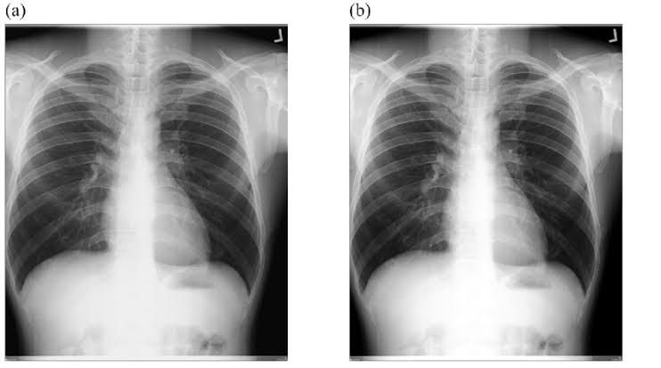
- Chest Radiography (especially PA and Lateral views): High kV (typically 120-150 kVp) is commonly used to penetrate the mediastinum (heart, great vessels, trachea) and visualize the lung parenchyma simultaneously. It allows for a wider dynamic range, ensuring that both the lung fields (low attenuation) and the heart/mediastinum (high attenuation) are adequately penetrated and visible on a single image. This technique helps to “burn through” the heart shadow to demonstrate the thoracic spine and retrocardiac lung region.
- Barium Studies (Upper Gastrointestinal Series, Small Bowel Follow-Through, Barium Enema): When imaging organs filled with barium contrast, high kVp (typically 90-120 kVp) is preferred. The high atomic number of barium (Z=56) provides significant inherent contrast. Using high kVp ensures sufficient penetration of the barium column while still allowing visualization of the mucosal patterns and surrounding soft tissues. It prevents “over-penetration” of the barium, which would obscure detail, by allowing the X-rays to pass through the barium with appropriate density differentiation to visualize the walls of the structures.
- Intravenous Urography (IVU) / Intravenous Pyelography (IVP): Similarly to barium studies, High kV (90-100 kVp) is used for visualizing the urinary tract after the injection of iodinated contrast media. This kVp range provides optimal penetration of the contrast-filled kidneys, ureters, and bladder, allowing for clear visualization against surrounding soft tissues.
- Spine Radiography (particularly Lateral Cervical, Thoracic, and Lumbar Spine): Due to the varying densities present in the spine (dense vertebral bodies, less dense intervertebral discs, and surrounding soft tissues), high kV (e.g., 90-100 kVp) can improve penetration of the vertebral bodies, especially in the thoracic and lumbar regions, which are often superimposed by shoulders and abdomen respectively. This helps to visualize the vertebral body, disc spaces, and spinous processes more uniformly.
- Abdomen Radiography (General Survey): While often performed at lower kVp, some abdominal views, especially for larger patients or when looking for subtle calcifications, may benefit from higher kVp (80-90 kVp) to ensure adequate penetration and reduced mAs.
- Tomography (Conventional): Historically, conventional tomography, though largely replaced by CT, often utilized high kVp techniques to penetrate overlying and underlying structures, blurring them out to visualize a specific plane of interest.
Positioning and Exposure Technique for High kV Technique
Achieving diagnostic quality with the High kV technique requires meticulous attention to both patient positioning and exposure parameters.
1. Patient Positioning
Proper patient positioning remains paramount regardless of the kVp chosen. The principles are universal:
- Accuracy: Ensure the anatomical region of interest is correctly aligned with the central ray and centered on the image receptor.
- Immobilization: Minimize patient motion through clear instructions, breathing techniques (e.g., suspended respiration for chest/abdomen), or immobilization devices if necessary. Shorter exposure times with high kV do inherently reduce motion blur, but cooperation is still vital.
- Collimation: Precise collimation to the area of interest is crucial. With high kVp, scatter radiation increases significantly, and tight collimation helps to mitigate this, thereby improving image contrast and reducing patient dose.
- Source-to-Image Distance (SID): Maintain standard SIDs (e.g., 72 inches for chest, 40 inches for abdomen/spine) to ensure consistent magnification and spatial resolution. High kV does not inherently alter SID requirements.
2. Exposure Technique Specifics
The core of the High kV technique lies in its unique balance of kVp and mAs.
- Kilovoltage Peak (kVp) Selection:
- Primary Factor: kVp is the controlling factor for beam quality (energy and penetration) and influences image contrast. In high kV techniques, the aim is to use the highest practical kVp while maintaining acceptable image contrast.
- Typical Ranges:
- Chest: 120-150 kVp
- Barium Studies: 90-120 kVp
- IVU/IVP: 90-100 kVp
- Spine: 90-100 kVp
- Impact on Contrast: Higher kVp inherently produces a lower subject contrast due to increased Compton scattering and reduced photoelectric absorption. However, modern digital imaging systems with their wide dynamic range and advanced post-processing algorithms can often compensate for this, allowing the use of higher kVp while maintaining diagnostic contrast.
- Milliampere-seconds (mAs) Adjustment:
- Inverse Relationship: The fundamental principle of the High kV technique is to significantly reduce mAs as kVp is increased to maintain optimal image density (brightness).
- The 15% Rule: A commonly applied rule of thumb is the “15% Rule.” Increasing kVp by 15% while halving the mAs will produce an image with similar overall density but with lower contrast and a lower patient dose. Conversely, decreasing kVp by 15% and doubling mAs will produce a similar density but higher contrast. This rule highlights the substantial mAs reduction possible with higher kVp.
- Dose Reduction: Lower mAs directly translates to less radiation dose delivered to the patient.
- Motion Blur: Shorter exposure times (due to lower mAs) minimize the risk of motion blur, which is particularly advantageous for chest radiography (cardiac motion) and pediatric imaging.
- Grids:
- Necessity: High kV techniques generate a significantly larger proportion of scatter radiation due to increased Compton interactions. Therefore, high-ratio grids (e.g., 10:1 or 12:1) are essential for nearly all high kV examinations to absorb scatter and maintain image contrast.
- Grid Focus: Ensure the correct grid focus distance is used to prevent grid cut-off.
- Automatic Exposure Control (AEC):
- Cell Selection: When using AEC, careful selection of the appropriate ionization chambers is critical based on the anatomical region and the technique being employed. For chest X-rays using high kV, often all three cells are activated to average the signal. For barium studies, the central cell or specific cells covering the contrast-filled area would be chosen.
- Backup Timer: Always set an appropriate backup timer to prevent overexposure in case of AEC malfunction.
- Image Receptors:
- Digital Radiography (DR/CR): With the widespread use of digital imaging systems, the high kV technique is even more versatile. Digital receptors have a wide exposure latitude, meaning they can capture a broader range of X-ray intensities. Post-processing algorithms can then adjust contrast and brightness to optimize the final image display, compensating for the inherently lower subject contrast produced by higher kVp.
- Quantum Mottle: While lower mAs reduces dose, it can also lead to increased quantum mottle (graininess) if mAs is reduced excessively. A balance must be struck to ensure sufficient signal-to-noise ratio.
Advantages and Disadvantages of High kV Technique
The High kV technique offers distinct benefits and drawbacks that must be weighed in clinical practice.
1. Advantages
- Reduced Patient Radiation Dose: This is arguably the most significant advantage. By decreasing the mAs, the total radiation exposure to the patient is substantially lowered. This aligns with the ALARA (As Low As Reasonably Achievable) principle of radiation protection.
- Increased Penetration: High-energy photons can more effectively penetrate dense or large body parts, internal prostheses, or contrast-filled organs. This ensures that all anatomical structures within the field of view are adequately visualized, reducing the likelihood of repeat exposures due to under-penetration.
- Wider Exposure Latitude: The technique is more forgiving of minor exposure errors. Because the primary factor affecting density (mAs) is lower, small variations in patient thickness or positioning are less likely to result in a diagnostically unacceptable image. This broadens the “acceptable” range of exposures.
- Reduced Motion Blur: The ability to significantly reduce mAs leads to shorter exposure times. This minimizes the impact of involuntary patient motion (e.g., heart beat, respiratory motion) or voluntary motion, resulting in sharper images.
- Increased Consistency in Image Density: Due to the wider exposure latitude, image density (brightness) tends to be more consistent across a range of patient sizes and pathologies.
- Extended X-ray Tube Life: Lower mAs settings mean less heat is generated at the anode of the X-ray tube per exposure, contributing to a longer operational life for the X-ray tube.
- Improved Visualization of Structures Behind Dense Anatomy: For example, in chest radiography, high kVp helps to visualize the spine and retrocardiac lung behind the heart shadow.
2. Disadvantages
- Increased Scatter Radiation: Higher kVp results in a greater proportion of Compton interactions, leading to more scatter radiation being produced within the patient. This scatter reaches the image receptor and degrades image contrast.
- Reduced Subject Contrast: Due to the increased scatter and reduced photoelectric effect (which yields high contrast), the inherent subject contrast of the anatomical part is lower. While digital systems can compensate through post-processing, subtle differences in tissue densities might be less apparent compared to lower kVp techniques if post-processing is not optimized.
- Increased Potential for Fog: The higher amount of scatter radiation can lead to increased image fog (a generalized darkening of the image), further reducing contrast if effective scatter reduction methods (grids, collimation) are not rigorously applied.
- Requires High-Ratio Grids: To combat the increased scatter, high-ratio grids are essential. These grids are more costly and can sometimes introduce grid lines if not properly aligned.
- Less Diagnostic Detail (in some cases): While overall penetration is better, the lower subject contrast might make it harder to differentiate very subtle pathology that relies on minute differences in tissue attenuation, unless digital post-processing is highly effective.
- Risk of Patient Dose Creep (if not careful): If radiographers do not appropriately reduce mAs when increasing kVp, there is a risk of inadvertently increasing patient dose without gaining significant image quality benefits. Strict adherence to technique charts and the 15% rule is vital.
Conclusion
The High kV technique represents a sophisticated and indispensable tool in the radiographer’s arsenal. By judiciously increasing the penetrating power of the X-ray beam, it offers significant advantages in reducing patient radiation dose, minimizing motion blur, and providing a wider exposure latitude for precise imaging of challenging anatomical areas or contrast-filled structures. While it introduces challenges related to increased scatter and reduced inherent subject contrast, these can be effectively managed through the use of high-ratio grids, meticulous collimation, and the advanced capabilities of modern digital imaging systems. Mastery of the High kV technique is fundamental for producing high-quality diagnostic images efficiently and safely, upholding the highest standards of patient care in diagnostic radiography.


