The intricate balance of the human immune system is fundamental to health, enabling defense against pathogens while coexisting peacefully with the body’s own tissues and beneficial microbes. At the core of this balance lies the concept of immunological tolerance. When this tolerance breaks down or is insufficient, significant medical challenges arise, ranging from autoimmune diseases to the rejection of life-saving transplanted organs or tissues. Consequently, understanding and actively inducing tolerance, particularly in clinical settings, represents a major goal in modern medicine.
Defining Immunological Tolerance
Immunological tolerance is the state of unresponsiveness of the immune system to substances or tissues that have the capacity to elicit an immune response. Crucially, it is an active process mediated by specific cellular and molecular mechanisms, not merely a passive lack of response. This deliberate non-reactivity is essential for several physiological reasons:
- Self-Tolerance: The most fundamental form of tolerance is the ability of the immune system to distinguish “self” antigens (components of the body’s own cells and tissues) from “non-self” antigens (originating from pathogens, allergens, or foreign tissues). Failure of self-tolerance leads to autoimmunity, where the immune system attacks the body’s own structures.
- Tolerance to Commensals: The body harbors vast communities of symbiotic microorganisms, particularly in the gut and on the skin. The immune system must tolerate these beneficial or harmless microbes while remaining vigilant against pathogens.
- Transplant Tolerance: In the context of transplantation, tolerance refers to the acceptance of donor organs, tissues, or cells by the recipient’s immune system without the need for continuous, high-dose immunosuppressive drugs. Achieving transplant tolerance is the ultimate goal of transplantation immunology, as it would eliminate the significant side effects and complications associated with long-term immunosuppression, such as increased susceptibility to infection, malignancy, cardiovascular disease, and organ toxicity.
Immunological tolerance is established and maintained through several mechanisms, broadly categorized into two main types:
- Central Tolerance: Occurs during the development of T and B lymphocytes in the primary lymphoid organs (thymus for T cells, bone marrow for B cells). Lymphocytes that react strongly to self-antigens encountered during maturation are typically eliminated (clonal deletion) or rendered inactive. The thymus plays a critical role in T cell central tolerance through a process involving expression of self-antigens and negative selection.
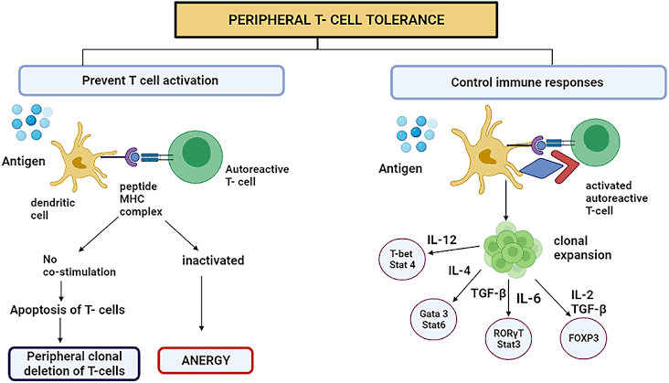
- Peripheral Tolerance: Operates in the secondary lymphoid organs (lymph nodes, spleen) and peripheral tissues after lymphocytes have left the primary lymphoid organs. This is crucial for tolerating antigens not encountered in the primary lymphoid organs (e.g., tissue-specific antigens, antigens from commensal microbes, or transplanted organs). Mechanisms of peripheral tolerance include:
- Anergy: A state of functional inactivation induced when lymphocytes receive the primary activating signal (recognition of antigen) but lack necessary co-stimulatory signals.
- Regulatory T Cells (Tregs): A specialized subset of T cells that actively suppress the activation and proliferation of other immune cells. Tregs are vital for maintaining tolerance to self and beneficial non-self antigens.
- Clonal Deletion: Activation-induced cell death of lymphocytes stimulated without appropriate co-stimulation or in the presence of certain inhibitory signals.
- Immune Privilege: Certain sites in the body (e.g., eye, brain, testes) have evolved mechanisms to limit immune responses, protecting delicate tissues from inflammatory damage.
In the context of clinical transplantation, the goal is often to induce a state of specific peripheral tolerance to the donor alloantigens (antigens from a genetically different individual), ideally without compromising the recipient’s ability to respond to pathogens.
Rationale for Using Co-stimulation Blockade to Induce Clinical Tolerance
The activation of T lymphocytes, central mediators of transplant rejection, is a tightly regulated process requiring multiple signals. The primary signal (°1) is the recognition of the donor’s alloantigen (presented on major histocompatibility complex, MHC molecules) by the T cell receptor (TCR). However, for full activation, proliferation, and differentiation into effector cells capable of mediating rejection, a T cell also requires a co-stimulatory signal (°2). This second signal is typically delivered through the interaction of molecules on the T cell surface (e.g., CD28) with their ligands on antigen-presenting cells (APCs) (e.g., B7 molecules like CD80 and CD86).
The rationale for using co-stimulation blockade as a strategy to induce clinical tolerance is based on a fundamental principle of T cell biology: encountering antigen (Signal 1) in the absence of adequate co-stimulation (Signal 2) does not lead to productive activation but rather induces a state of anergy (functional unresponsiveness) or promotes differentiation into non-effector or even regulatory T cells. This mimics a natural mechanism by which the immune system prevents reactivity to self-antigens presented by non-professional APCs that lack co-stimulatory molecules.
By administering agents that block key co-stimulatory pathways, clinicians aim to:
- Prevent Full T Cell Activation: Agents such as fusion proteins (e.g., Belatacept, which is a modified CTLA4-Ig molecule) or antibodies targeting co-stimulatory molecules (like CD40L, though agents targeting this pathway have faced clinical challenges) bind to or block these molecules. This prevents the required Signal 2 from being delivered to the T cell even if it recognizes the donor antigen (Signal 1).
- Induce Anergy: Without the co-stimulatory signal, T cells that encounter donor antigens are rendered anergic. These cells cannot mount an effective immune response and may even actively suppress responses by other T cells.
- Promote Regulatory T Cell Development: Blocking co-stimulation can, in certain contexts (especially when antigen is presented in a specific manner), favor the development or expansion of regulatory T cells (Tregs). These Tregs can then suppress the activity of other T cells specifically reactive to the donor antigens, contributing to tolerance.
- Target Specificity: Unlike broad immunosuppressants that suppress all immune responses, co-stimulation blockade preferentially affects T cells encountering the specific donor antigens being presented, particularly during the early phases post-transplant. This offers the potential for achieving donor-specific tolerance, where the recipient remains capable of responding to infections and other foreign antigens.
- Reduce Systemic Immunosuppression: Successful induction of tolerance via co-stimulation blockade could potentially lead to a reduction or even elimination of calcineurin inhibitors and other broad immunosuppressants, thereby mitigating their significant short-term and long-term toxicities and improving patient outcomes.
In essence, co-stimulation blockade exploits the physiological requirement for Signal 2 in T cell activation. By strategically interrupting this pathway, the goal is to reprogram the T cell response to the transplanted organ from one of rejection towards one of tolerance or non-reactivity, thereby facilitating graft acceptance with reduced systemic immunosuppression.
Rationale for Using Lymphocyte Depletion Followed by Reconstitution with Donor Bone Marrow or Stem Cells to Induce Clinical Tolerance
This strategy, often referred to in the context of hematopoietic stem cell transplantation (HSCT) or bone marrow transplantation (BMT), represents a more profound approach to tolerance induction. It aims to replace the recipient’s entire immune system, which is primed to reject the donor tissue, with a new immune system derived from the donor, which will recognize the donor tissues as “self.” The rationale is based on establishing a state of chimerism, where cells from two genetically distinct individuals coexist within a single organism.
The process typically involves two main phases:
- Lymphocyte (and Hematopoietic Cell) Depletion (Conditioning): The recipient’s immune system is largely eliminated through chemotherapy, radiation therapy, or potent lymphocyte-depleting antibodies (e.g., anti-thymocyte globulin, Alemtuzumab). The rationale for this conditioning phase is multi-faceted:
- Ablation of Host Immunity: Eliminating or significantly reducing the host lymphocytes, particularly T cells, removes the primary cellular components responsible for mediating rejection of the subsequent donor cell infusion or the transplanted organ (if combined with organ transplant).
- Creation of “Space” in the Bone Marrow: Conditioning prepares the recipient’s bone marrow niche to receive and support the engraftment and proliferation of donor hematopoietic stem cells.
- Immunomodulation: Some conditioning regimens may also induce a state of temporary tolerance or modulate the host environment to be more receptive to the donor cells.
- Reconstitution with Donor Hematopoietic Stem Cells (HSCs) or Bone Marrow: Following depletion, the recipient is infused with HSCs or bone marrow derived from the organ donor (in the case of combined organ and bone marrow transplant) or a separate, compatible donor. The rationale here is to:
- Establish Donor Hematopoiesis: The infused HSCs engraft in the recipient’s bone marrow and differentiate into all lineages of blood cells, including lymphocytes, monocytes, dendritic cells, etc. Over time, the recipient’s immune system is replaced by one derived from the donor.
- Induce Central Tolerance in the Recipient Thymus: As donor T cell progenitors mature in the recipient’s thymus (which contains recipient thymic stromal cells expressing recipient self-antigens), these developing donor T cells undergo central tolerance education. They are exposed to recipient self-antigens during negative selection. T cells that react strongly to recipient antigens are deleted or inactivated. Thus, the new, donor-derived T cell repertoire becomes tolerant to recipient tissues.
- Establish Peripheral Tolerance (Mixed Chimerism): If a state of stable mixed chimerism is achieved (where both recipient and donor hematopoietic cells coexist), this provides a continuous source of donor antigens presented by donor-derived APCs, as well as recipient antigens presented by recipient-derived cells. This complex environment further promotes peripheral tolerance mechanisms (anergy, regulation by Tregs) towards both donor and recipient tissues, while preserving the ability to react to novel foreign antigens. Achieving stable mixed chimerism without full donor chimerism is often preferred in solid organ transplantation to minimize the risk of graft-versus-host disease (GVHD), where donor immune cells attack recipient tissues.
The rationale is that by replacing the host immune system with a donor-derived system that has been tolerized to recipient tissues, a state of true, durable, and potentially drug-free immunological tolerance can be achieved for any simultaneously or subsequently transplanted donor organ or tissue. This strategy is particularly powerful because it addresses tolerance at the fundamental level of immune system development and recognition. While associated with significant risks (e.g., infection during depletion, GVHD, failure of engraftment), successful implementation holds the promise of lifelong tolerance, freeing patients from the burden of chronic immunosuppression.
Conclusion
Achieving clinical immunological tolerance remains a pivotal goal in transplantation and the management of autoimmune diseases. It represents a paradigm shift from suppressing unwanted immune responses nonspecifically to actively reprogramming the immune system to accept specific tissues while retaining immunocompetence against pathogens. Strategies like co-stimulation blockade target specific activation pathways to induce anergy or regulatory responses, offering the potential for donor-specific tolerance with reduced systemic toxicity. In contrast, lymphocyte depletion followed by donor stem cell reconstitution aims for a more fundamental reset of the immune system, establishing central and peripheral tolerance through chimerism. Both approaches, though different in their mechanisms and risks, reflect the ongoing scientific effort to harness the complex processes of immunological tolerance to improve patient outcomes and minimize the long-term complications associated with conventional immunosuppression. Continued research is essential to refine these strategies, enhance their efficacy, reduce side effects, and make clinical tolerance induction a safer and more widely applicable reality.


