Pterygium is a common ocular surface condition encountered in clinical practice, particularly in regions with high sun exposure. While generally benign, it can cause discomfort, irritation, and potentially impact vision, necessitating appropriate diagnosis and management.
Defining Pterygium
The term “pterygium” (pronounced tuh-RIJ-ee-uhm) originates from the Greek word “pterygos,” meaning “wing,” aptly describing its appearance.
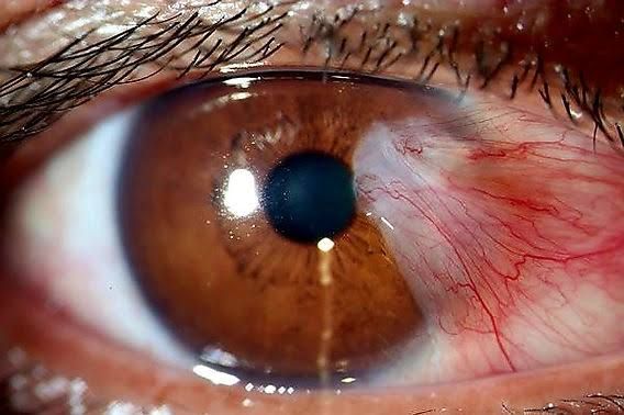
Professionally defined, a pterygium is a triangular or wing-shaped fibrovascular growth of conjunctival tissue that extends onto the cornea. It typically originates from the limbus (the border between the cornea and the sclera) and slowly progresses across the corneal surface.
It is essential to differentiate a true pterygium from a pseudopterygium. A pseudopterygium is a fold of conjunctiva that adheres to the cornea secondary to inflammation or trauma (e.g., burns, ulcers) and can occur anywhere on the limbus. A key clinical differentiator is that a probe can be passed under the neck of a pseudopterygium, whereas this is not possible with a true pterygium.
Pterygia are considered benign growths, although they are degenerative and hyperplastic changes of the conjunctiva and underlying connective tissue (stroma) in response to chronic environmental insults.
Understanding Incidence and Etiology
Understanding who is affected and what causes pterygium is fundamental to prevention and risk assessment.
- Incidence: Pterygium is significantly more prevalent in populations exposed to higher levels of ultraviolet (UV) radiation. It is commonly found in tropical and subtropical climates near the equator. While it can occur at any age, it is most frequently diagnosed in adults, particularly those over 30 years old. Some studies suggest a slightly higher incidence in males, potentially linked to increased outdoor occupational exposure. The incidence is lower in temperate regions and among individuals who habitually use protective eyewear.
- Etiology: The primary and most well-established etiological factor is chronic exposure to ultraviolet (UV) radiation, specifically UV-B light. This is supported by the geographical distribution of the condition and the observation that outdoor workers are at higher risk. UV radiation is thought to cause damage to DNA and proteins in conjunctival cells, leading to abnormal proliferation and migration onto the cornea. Other contributing factors include:
- Chronic exposure to wind, dust, sand, and other environmental irritants, which can cause surface dryness and inflammation, potentially exacerbating the effects of UV damage.
- Genetic predisposition: Family history may play a role in some cases, suggesting a genetic component to susceptibility.
- Other irritants: Smoke and pollutants may also contribute.
In essence, pterygium develops as a protective reaction to chronic environmental stress, but it results in pathological tissue changes.
Recognizing Clinical Features (Signs and Symptoms)
Pterygium presents with a characteristic appearance and can cause a range of symptoms depending on its size and progression.
- Signs (What you observe during examination):
- Appearance: A fleshy, triangular or wing-shaped growth.
- Location: Most commonly originates from the nasal limbus and extends horizontally onto the cornea. Temporal pterygia are less common. Bilateral involvement is frequent, although often asymmetrical.
- Progression: Typically slow-growing, but the rate can vary.
- Vascularity: The growth is often vascularised, appearing pink or red, especially when inflamed.
- Structure: It has a distinct head (the leading edge on the cornea), a neck (at the limbus), and a body (the conjunctival portion).
- Stocker’s Line: An iron deposition line may be visible on the cornea just ahead of the pterygium’s head. This indicates corneal tissue changes and is often associated with progression.
- Symptoms (What the patient reports):
- Cosmetic Concern: Often the initial reason for seeking consultation, especially for smaller lesions.
- Irritation/Foreign Body Sensation: Feeling of something in the eye, especially with dryness or inflammation.
- Redness and Inflammation: The pterygium and surrounding conjunctiva can become injected and irritated, particularly after exposure to sun, wind, or dust.
- Dryness or Tearing: Disruption of the normal tear film distribution over the irregular surface can lead to paradoxically dry eye symptoms or excessive tearing.
- Blurred Vision: Occurs if the pterygium body grows into the visual axis, directly obscuring vision. It can also induce astigmatism by pulling and changing the curvature of the cornea, affecting vision even before reaching the center.
- Diplopia (Double Vision): Extremely rare, occurring only in massive pterygia that restrict eye movement by tethering the globe.
Conducting a Professional History Taking
A systematic history taking is crucial for understanding the patient’s experience, identifying risk factors, and guiding the examination and management plan.
- Presenting Complaint: Ask the patient why they have sought consultation. Is it cosmetic? Irritation? Vision change?
- Onset and Duration: When did they first notice the growth? Has it changed in size or appearance? How quickly?
- Symptom Analysis:
- Characterize symptoms: Describe the nature of irritation, redness, dryness, tearing, and pain (pain is uncommon unless significantly inflamed or infected, which is rare).
- Vision Changes: Is vision blurred? In one eye or both? Does it fluctuate? Have they noticed a change in their glasses prescription?
- Aggravating/Relieving Factors: What makes symptoms worse (e.g., sun, wind, dust, dry environments, prolonged screen use)? What makes them better (e.g., artificial tears, staying indoors)?
- Risk Factor Assessment:
- Environmental Exposure: Detailed history of outdoor activities, occupation (e.g., farming, construction, fishing, sailing), duration of exposure to sun, wind, dust, sand.
- Protective Measures: Do they use sunglasses, hats, or other eye protection regularly when outdoors?
- Geographical History: Have they lived in sunny or tropical climates for extended periods?
- Past Ocular History: Any history of previous ocular surgery, trauma, infections, or other conditions (e.g., dry eye, blepharitis).
- Medical History: Systemic diseases. While not directly linked to pterygium, it’s part of a complete history.
- Medications: Current topical and systemic medications, especially any eye drops used for the pterygium or other conditions.
- Social History: Smoking (irritant?), lifestyle, occupation.
Document all findings clearly and concisely.
Performing a Thorough Clinical Examination
A comprehensive eye examination, focusing particularly on the anterior segment, is essential for diagnosing pterygium, assessing its severity, and planning management.
- Visual Acuity: Measure best-corrected visual acuity (BCVA) for distance and near in each eye. This is critical to assess if the pterygium is impacting vision.
- External Examination: Note general appearance, head position, any obvious redness or asymmetry.
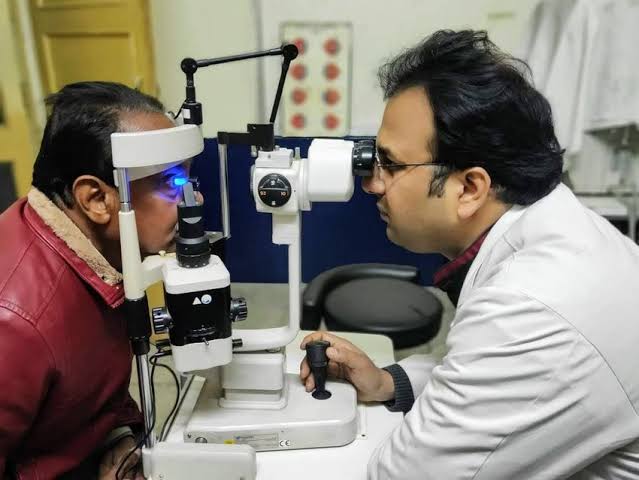
- Slit Lamp Biomicroscopy: This is the cornerstone of the examination.
- Location and Extent: Identify the location (nasal/temporal). Measure the extent of the pterygium’s growth onto the cornea (e.g., in millimeters from the limbus or estimate based on corneal diameter). Note if it is encroaching on or within the pupillary area (visual axis).
- Morphology: Describe the shape (triangular, wing-shaped), thickness, vascularity (mild vs. marked injection), and whether it appears fleshy or relatively avascular.
- Apex and Leading Edge: Examine the head of the pterygium on the cornea. Is it flat or raised? Is there a visible opaque line (Stocker’s line) ahead of it? Look for signs of active growth.
- Corneal Effects: Assess for induced astigmatism (though this is often better quantified with refraction/keratometry). Note any corneal scarring or opacity related to the pterygium.
- Associated Features: Examine the surrounding conjunctiva for signs of inflammation, dryness, or other lesions (e.g., pinguecula). Evaluate the tear film (break-up time, tear meniscus) as dry eye often coexists.
- Differentiation from Pseudopterygium: Attempt to gently probe under the neck of the lesion with a fine instrument (like a blunt spatula or flattened forceps blade) at the limbus. If it’s a true pterygium, the probe cannot pass under the neck.
- Refraction and Keratometry/Topography: Perform a refraction to assess for astigmatism, particularly irregular astigmatism induced by the pterygium pulling on the cornea. Corneal topography or keratometry provides objective measurement of corneal curvature changes.
- Intraocular Pressure (IOP): Measure IOP as part of a standard eye exam, and because steroid drops, if used for inflammation, can affect IOP.
- Fundus Examination: Complete the eye exam by examining the posterior segment.
- Documentation: Record all findings meticulously, including measurements, sketches, or clinical photographs for future comparison to monitor progression.
Outlining Management Strategies
Management of pterygium depends on its size, symptoms, rate of growth, impact on vision, and patient preference.
- Prevention: The most important step, particularly in susceptible individuals, is prevention. Advise patients on consistent use of UV-blocking sunglasses and wide-brimmed hats when outdoors, especially during peak sun hours.
- Observation: Small, asymptomatic pterygia that are not growing and do not affect vision can often be managed with observation. Regular follow-up is recommended to monitor for progression.
- Medical Management (Symptomatic Relief):
- Lubricating Eye Drops/Gels/Ointments: First-line treatment for irritation, dryness, and foreign body sensation. Frequent use helps maintain a smooth ocular surface.
- Topical Vasoconstrictors: Can reduce redness for cosmetic purposes, but are not curative and can cause rebound redness with prolonged use. Generally not recommended for routine management.
- Topical Corticosteroids: Low-potency steroid drops (e.g., loteprednol, fluorometholone) can be used short-term to reduce inflammation and redness associated with acute exacerbations. Caution: Must be used judiciously due to potential side effects like elevated IOP, cataract formation, and increased risk of infection. Monitor IOP if using steroids.
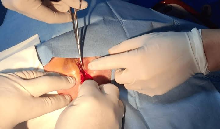
- Surgical Management: Surgical excision is indicated in specific circumstances:
- Visual Impairment: If the pterygium encroaches on or is near the visual axis, or induces significant symptomatic astigmatism not correctable with spectacle changes.
- Significant or Frequent Symptoms: Chronic redness, irritation, or foreign body sensation unresponsive to medical therapy.
- Significant Cosmetic Concern: If the appearance significantly impacts the patient’s quality of life.
- Documented Growth: Rapid or significant growth threatening vision.
- Restriction of Eye Movement: If the pterygium is large enough to limit motility (rare).
- Techniques: Simple “bare sclera” excision has a high recurrence rate (25-50%). Preferred modern techniques aim to prevent recurrence:
- Conjunctival Autografting: Excision of the pterygium followed by transplanting a piece of healthy conjunctiva from elsewhere on the patient’s eye (e.g., superior bulbar conjunctiva) to cover the bare sclera. This is the most common and effective method, significantly reducing recurrence rates (<5-10%).
- Amniotic Membrane Grafting: Similar to autografting, but uses preserved human amniotic membrane. Useful when healthy conjunctiva is limited or for large defects.
- Adjunctive Therapies: Agents like Mitomycin C (an antiproliferative) or anti-VEGF injections may be used during or after surgery to further reduce recurrence risk, especially in recurrent or aggressive cases, but carry potential complications.
- Post-operative Care: Typically involves topical antibiotics and corticosteroids, along with close follow-up to monitor healing and for signs of recurrence. Recurrence, when it happens, is often more aggressive and difficult to manage than the primary lesion.
Identifying Indications for Referral
While primary care practitioners or optometrists may encounter and even initially manage symptomatic relief for simple pterygia, referral to an ophthalmologist is often necessary or advisable in several situations:
- Confirmed or Suspected Diagnosis: Any suspected pterygium warrants evaluation by an eye care specialist (ophthalmologist or optometrist depending on scope of practice) for accurate diagnosis and assessment of severity.
- Visual Impairment: If the patient reports decreased visual acuity or if the pterygium examination suggests encroachment on the visual axis or significant induced astigmatism.
- Significant Growth: Documented or suspected rapid progression in size.
- Severe or Persistent Symptoms: Symptoms (redness, irritation, pain) are not controlled by simple lubrication or short-term low-potency steroids.
- Cosmetic Concern Requiring Surgery: Patients seeking surgical removal for cosmetic reasons require surgical consultation.
- Suspicion of Malignancy: Although rare, pterygia can sometimes mimic or coexist with conjunctival intraepithelial neoplasia (CIN) or even squamous cell carcinoma. Any atypical appearance (e.g., unusual thickness, papillomatous surface, prominent blood vessels outside the usual pattern) necessitates referral for biopsy.
- Difficulty Differentiating from Pseudopterygium: If the diagnosis is uncertain, referral to an ophthalmologist is appropriate.
- Recurrent Pterygium: Management of recurrent pterygia is often more complex and requires specialized surgical skills and consideration of adjunctive therapies.
- Consideration for Surgery: Once surgical management is considered necessary based on the criteria above, referral to an ophthalmologist specializing in ocular surface surgery is indicated.
Conclusion:
Pterygium is a common degenerative condition linked primarily to UV exposure. A professional approach involves a thorough history to identify risk factors and symptoms, a detailed clinical examination using techniques like slit lamp biomicroscopy to assess the lesion’s characteristics and impact, and a structured management plan. While observation and medical therapy suffice for smaller, asymptomatic lesions, surgical intervention is indicated for visual impairment, significant symptoms, or rapid growth. Prevention through UV protection is paramount. Recognizing the indications for referral to an ophthalmologist ensures patients receive appropriate specialized care and surgical management when necessary, ultimately aiming to preserve vision and alleviate discomfort.


