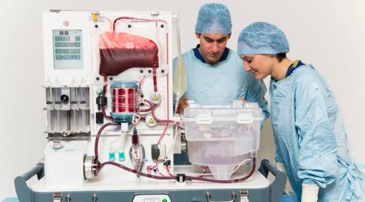
Organ transplantation is a cornerstone of modern medicine, offering life-saving treatment for patients with end-stage organ failure. The success of transplantation critically relies on the ability to maintain organ viability from the time it is procured from a donor until it is implanted into a recipient. This process, known as organ preservation, is complex and multifaceted.
Basic Principles of Organ Preservation
The fundamental goal of organ preservation is to minimize cellular injury that occurs from the moment blood supply is interrupted (ischemia) until blood flow is restored in the recipient (reperfusion). Organs begin to deteriorate rapidly without oxygen and nutrients. Preservation techniques aim to significantly slow down the metabolic processes within the organ’s cells, thereby reducing the demand for oxygen and energy, and limiting the accumulation of harmful metabolic byproducts.
Key principles underpinning successful organ preservation include:
- Hypothermia: Reducing the temperature of the organ is the primary and most effective method to lower cellular metabolic rate. Typically, organs are cooled to hypothermic temperatures, ranging from 4°C to 8°C. At these temperatures, cellular enzymatic activity slows dramatically, reducing oxygen consumption to about 10% of normal levels. This extends the time the organ can tolerate being without blood flow.
- Chemical Environment: Providing a specific chemical milieu is crucial. Simply cooling the organ is insufficient. The surrounding or perfusing solution must prevent cellular swelling (edema), maintain electrolyte balance, buffer against acidosis, provide substrates for minimal metabolic activity, and potentially include agents that scavenge harmful free radicals or stabilize cell membranes. This is where organ preservation fluids play a vital role.
- Minimizing Ischemic Time: Despite optimal preservation, ischemic time (the period without blood flow) is a critical factor. Shorter ischemic times are consistently associated with better outcomes. Preservation techniques aim to mitigate the damage occurring during this period.
- Gentle Handling: Physical trauma during procurement, preservation, and transport must be avoided, as it can cause additional injury to the delicate organ tissue.
Basic Principles of Organ Preservation Fluids
Organ preservation fluids are specially formulated solutions designed to protect cells from the damaging effects of cold ischemia. They are introduced into the organ’s vascular system (flushed through the arteries and veins) immediately after procurement and often surround the organ during storage. The specific composition of these fluids is critical for their function.
Key components and their roles typically include:
- Impermeant Molecules (e.g., Lactobionate, Gluconate, Raffinose): These are large molecules that cannot easily cross cell membranes. Their primary role is to create an osmotic gradient across the cell membrane, preventing water from entering the cells and causing them to swell (cellular edema), which is a common consequence of cold ischemia and impaired ion pumps.
- Buffers (e.g., Histidine, Phosphate, Bicarbonate): Metabolic processes, even slowed at cold temperatures, produce acids. Buffers help maintain a stable pH within the tissue, preventing cellular damage caused by acidosis. Histidine is particularly effective at low temperatures and can also act as a free radical scavenger.
- Electrolytes (Specific Concentrations of Sodium, Potassium, Magnesium): The concentration of electrolytes in preservation fluids is carefully controlled to mimic the intracellular environment (high Potassium, low Sodium, high Magnesium) rather than the extracellular environment (high Sodium, low Potassium). This helps to support the function of ion pumps that are impaired by cold and ischemia, further preventing swelling and maintaining cellular integrity.
- Osmotic Agents (e.g., Mannitol, Sucrose): Similar to impermeant molecules, these help maintain osmotic balance and reduce cell swelling. Mannitol can also act as a free radical scavenger.
- Substrates and Additives (e.g., Adenosine, Glutathione, Allopurinol, Steroids):
- Energy Substrates (e.g., Adenosine): Provide precursors for ATP regeneration upon reperfusion.
- Antioxidants/Free Radical Scavengers (e.g., Glutathione, Allopurinol): Help neutralize harmful reactive oxygen species (free radicals) generated during ischemia and, critically, during reperfusion.
- Membrane Stabilizers (e.g., Steroids, Lidocaine): May help protect cell membranes from damage.
- Colloids (e.g., Hydroxyethyl Starch, Albumin – less common in initial flush solutions, more in perfusion fluids): Can help maintain oncotic pressure in some perfusion solutions.
Examples of widely used preservation fluids include University of Wisconsin (UW) solution, Custodiol® (HTK – Histidine-Tryptophan-Ketoglutarate) solution, and IGL-1 solution. Each has a slightly different composition optimized for different organs or preservation methods.
Limits of Organ Preservation
Despite significant advancements, organ preservation has inherent limitations that restrict how long an organ can remain viable outside the body and influence its function post-transplant.
The primary limitations include:
- Accumulation of Ischemic Damage: While hypothermia slows metabolism, it does not stop it entirely. Cellular processes continue at a reduced rate, leading to the gradual depletion of energy stores (ATP) and accumulation of toxic metabolic byproducts (e.g., lactic acid). Ion gradients across cell membranes collapse, leading to uncontrolled calcium influx, which activates destructive enzymes. This damage progresses over time.
- Cold Injury: Hypothermia itself can cause cellular stress and injury. Changes in cell membrane fluidity, cytoskeletal structure, and enzymatic function occur at low temperatures, independent of ischemia.
- Ischemia-Reperfusion Injury (IRI): This is arguably the most significant limit and source of damage. IRI occurs when oxygenated blood is restored to an organ that has been ischemic. While necessary for function, reperfusion triggers a complex cascade of events:
- Oxidative Stress: The sudden influx of oxygen leads to a burst of free radical production.
- Inflammation: Activation of the immune system causes inflammatory cells to infiltrate the organ, releasing cytokines and proteases that damage tissue.
- Complement Activation: The complement system, part of the innate immune response, is activated, contributing to vascular and cellular injury.
- Endothelial Dysfunction: The lining of blood vessels is particularly susceptible, leading to impaired blood flow regulation, increased permeability, and potential thrombosis. IRI damages cells that may have survived the hypothermic ischemic period, significantly impacting early graft function.
- Preservation Time Limits (Cold Ischemia Time – CIT): Due to the cumulative damage from ischemia, cold injury, and the impending reperfusion injury, there are practical limits to how long an organ can be preserved. These limits vary significantly depending on the organ’s sensitivity to ischemia:
- Heart and Lungs: Highly sensitive, typically limited to 4-6 hours.
- Liver: Moderately sensitive, typically 8-12 hours, sometimes up to 15-20 hours with optimal preservation.
- Kidney: More tolerant, often preserved for 24-36 hours, sometimes longer with machine perfusion.
- Pancreas: Similar to kidney, often 12-20 hours. Beyond these times, the risk of severe IRI and primary non-function (the organ failing to work immediately) increases dramatically.
- Donor Factors: The health and circumstances of the donor (age, cause of death, presence of comorbidities, duration of hot ischemia before cooling) significantly influence the organ’s initial quality and its tolerance to preservation, complicating the limits and risks.
Attendant Risk of Organ Dysfunction Over Time
The injuries sustained during the preservation period, particularly IRI, have lasting consequences that contribute to organ dysfunction not just immediately after transplant but also over the long term.
Key risks of dysfunction over time related to preservation include:
- Delayed Graft Function (DGF): This is a common complication in kidney transplantation, defined as the need for dialysis within the first week post-transplant. DGF is a direct consequence of severe preservation injury and IRI. While many kidneys with DGF eventually recover function, it is associated with longer hospital stays, increased costs, a higher incidence of acute rejection episodes, and a greater risk of chronic graft loss. Preservation injury to the kidney tubules is a major contributor to DGF.
- Primary Non-Function (PNF): In rare but devastating cases, the organ may fail to function at all after transplantation. This is usually due to catastrophic preservation injury or severe hyperacute/acute rejection, often exacerbated by pre-existing damage or prolonged/suboptimal preservation.
- Increased Risk of Acute Rejection: While rejection is primarily an immunological process, preservation injury can make the organ more susceptible to the recipient’s immune system. Damaged cells express stress signals that can activate immune responses.
- Chronic Graft Dysfunction and Loss: Preservation injury, particularly IRI, sets in motion processes that can lead to chronic damage, including inflammation, fibrosis (scarring), and vascular changes within the organ. This chronic damage progressively impairs organ function over months and years, ultimately contributing to chronic allograft nephropathy (in kidneys), chronic rejection, and is a leading cause of long-term graft failure. The initial insult from cold ischemia and reperfusion can initiate a cycle of ongoing injury and repair that is ultimately detrimental to long-term organ survival.
- Increased Susceptibility to Other Insults: An organ that has sustained significant preservation injury may be less resilient to subsequent challenges such as infection, calcineurin inhibitor toxicity (common immunosuppressants), or recurrent disease.
Basic Principles of Pulsatile Kidney Perfusion
Pulsatile kidney perfusion is a method of machine preservation that involves continuously pumping a cold preservation solution through the kidney’s vasculature in a pulsatile (non-continuous) manner, mimicking physiological blood flow to some extent. It is a more active preservation method compared to static cold storage (SCS).
Basic principles of pulsatile kidney perfusion:
- Continuous Flow: Unlike SCS where the fluid is simply flushed in and the organ stored, machine perfusion actively circulates the fluid through the organ’s blood vessels.
- Hypothermic Temperature: The perfusate is kept cold, typically between 4°C and 8°C, to maintain the benefits of reduced metabolic rate.
- Specialized Perfusate: While similar in principle to SCS fluids, perfusates for machine perfusion are often lower viscosity and specifically formulated for continuous flow through the microvasculature. Examples include Belzer MPS (Machine Perfusion Solution) and Kidney Perfusion Solution-1 (KPS-1). These maintain osmotic balance, provide buffers, and include essential electrolytes and potentially other protective agents.
- Pulsatile Flow: The fluid is pumped in pulses rather than a steady stream. This pulsatile nature is thought to have several advantages:
- May help to distribute the perfusate more evenly throughout the microcirculation.
- Could potentially reduce interstitial edema compared to continuous flow or static storage.
- May exert shear stress on endothelial cells, potentially maintaining their health and function better than in static storage.
- Assessment of Viability: A key benefit of pulsatile perfusion is the ability to monitor parameters during the preservation process. The machine typically measures perfusion pressure and flow rate. From these, vascular resistance (Pressure/Flow) can be calculated. Higher resistance can indicate vascular damage, edema, or clots within the organ, serving as an indicator of potential viability and predicting the likelihood of DGF. This allows for a more objective assessment of organ quality pre-transplant compared to subjective evaluation in SCS.
- Extended Preservation Time: Pulsatile perfusion is particularly beneficial for kidneys from expanded criteria donors (ECDs) or those expected to have longer cold ischemia times. The continuous flow providing nutrients, removing waste, and potentially reducing edema can mitigate some of the progressive damage seen in prolonged SCS, allowing for safer preservation for longer durations.
In essence, pulsatile kidney perfusion provides a more dynamic and potentially more protective environment than static cold storage, particularly for marginal kidneys or when longer preservation times are anticipated. While more complex and costly than SCS, its ability to reduce DGF and provide viability assessment makes it a valuable tool in modern kidney transplantation.


