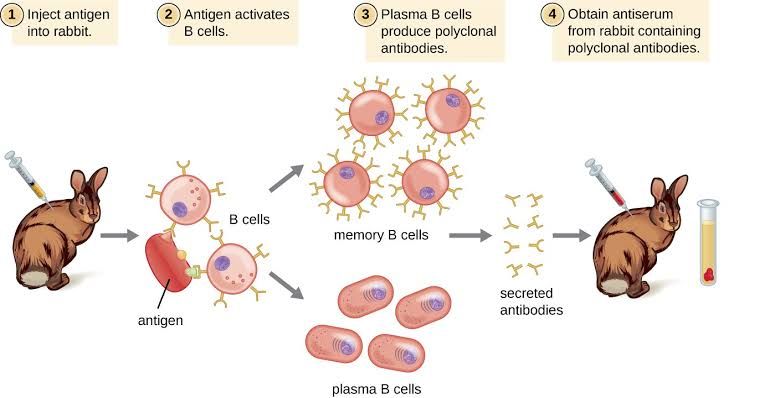
Polyclonal antibodies, particularly xenogenic preparations like rabbit anti-thymocyte globulin (rATG) and equine anti-thymocyte globulin (eATG), represent a foundational class of immunosuppressive agents. Widely used in medical practice, notably in solid organ and hematopoietic stem cell transplantation to prevent rejection and graft-versus-host disease (GVHD), and in conditions like severe aplastic anemia, their efficacy stems from their potent ability to deplete lymphocytes. Unlike monoclonal antibodies which target a single epitope, polyclonal antibodies are a heterogeneous mix targeting multiple antigens on the surface of target cells, resulting in complex and often powerful immunomodulatory effects.
Understanding Xenogenic Polyclonal Anti-Human Lymphocyte Sera
“Xenogenic” indicates that the antibodies are produced in a species different from the recipient (e.g., horse or rabbit antibodies used in humans). “Anti-human lymphocyte sera” signifies that the antibodies are raised against human lymphocytes, making them capable of targeting these specific cells. The resulting product is a complex mixture of antibodies directed against numerous surface markers found on T lymphocytes, but often also on B lymphocytes, NK cells, plasma cells, and even non-lymphoid cells like platelets and neutrophils.
Basic Steps in the Preparation of Xenogenic Polyclonal Anti-Human Lymphocyte Sera
The production of therapeutic-grade polyclonal anti-human lymphocyte sera is a complex, multi-step process requiring stringent quality control and adherence to Good Manufacturing Practices (GMP). The fundamental steps involve immunizing a non-human animal with human lymphocytes or related cell lines and purifying the resulting antibodies.
- Step 1: Preparation of the Immunogen
- The process begins with obtaining appropriate human lymphocytes or lymphoid cell lines. These cells serve as the antigen used to stimulate an immune response in the host animal. The source and preparation of these cells are critical, as they determine the range of antigens the resulting antibodies will target. Often, thymocytes, peripheral blood lymphocytes, or established T-cell lines are used. The cells are carefully prepared to maintain viability and antigenic integrity.
- Step 2: Immunization of the Host Animal
- Large animals, typically horses (for eATG) or rabbits (for rATG), are selected as the host species due to their ability to produce large volumes of serum and mount a robust immune response.
- The prepared human lymphocytes (the immunogen) are administered to the host animals via multiple injections over a specific period. The immunization protocol typically involves an initial dose followed by booster injections at regular intervals.
- Adjuvants, such as Freund’s adjuvant (though increasingly replaced by less inflammatory alternatives in modern manufacturing), are often used to enhance the immune response and ensure high antibody titers are generated. The goal is to stimulate the animal’s B cells to produce a broad spectrum of antibodies against the various antigens present on the human lymphocytes.
- Step 3: Monitoring the Immune Response
- Throughout the immunization period, blood samples are regularly collected from the host animals.
- These samples are tested to measure the level (titer) and potency of the anti-human lymphocyte antibodies being produced. Assays such as flow cytometry (measuring antibody binding to human lymphocytes) or lymphocyte cytotoxicity assays are used to assess the strength and specificity of the immune response. This monitoring helps determine the optimal time for serum collection.
- Step 4: Collection of Antiserum
- Once adequate antibody titers are achieved, a large volume of blood is collected from the hyperimmunized animals.
- The blood is allowed to clot, and the serum, which contains the antibodies, is separated from the cellular components. This raw antiserum contains the polyclonal antibodies directed against the human lymphocytes, as well as other antibodies and proteins naturally present in the animal’s serum.
- Step 5: Purification and Processing
- This is a critical and complex stage aimed at isolating the therapeutic antibodies and removing unwanted components.
- Immunoglobulin Fractionation: The globulin fraction, containing the antibodies (primarily IgG), is isolated from the bulk serum proteins using techniques such as salt precipitation (e.g., ammonium sulfate precipitation), chromatography, or a combination thereof.
- Removal of Unwanted Antibodies: Steps may be taken to remove antibodies directed against non-lymphocyte human cells (e.g., erythrocytes, platelets) by adsorbing the serum with these cell types. This helps reduce the risk of side effects like anemia and thrombocytopenia.
- Viral Inactivation/Removal: Rigorous steps are implemented to inactivate or remove potential animal viruses or other pathogens that could contaminate the product. This typically involves methods like heat treatment, solvent/detergent treatment, or nanofiltration.
- Sterile Filtration: The preparation is passed through sterilizing filters to remove bacteria and other microorganisms.
- Step 6: Formulation, Quality Control, and Packaging
- The purified and processed antibody preparation is formulated into a stable and administrable form, typically a sterile liquid solution or lyophilized powder for intravenous infusion.
- Extensive quality control testing is performed on the final product batch. This includes assessing:
- Potency: Ensuring the preparation can effectively bind to and deplete human lymphocytes (often measured by effects on lymphocyte subsets in in vitro assays).
- Safety: Testing for sterility, absence of pyrogens, viral contaminants, and abnormal toxicity.
- Purity: Assessing the composition and absence of significant impurities.
- Identity: Confirming the product is indeed xenogenic anti-human lymphocyte globulin.
- Finally, the product is filled into vials or bottles and packaged for distribution.
This meticulous process ensures that the final therapeutic product is potent, pure, and safe for human administration.
Mechanisms of Lymphocyte Depletion
Polyclonal antibodies induce lymphocyte depletion through multiple, synergistic mechanisms. Their ability to bind to a wide array of surface antigens means they can trigger diverse cytotoxic pathways and modulate cellular function.
- Complement-Dependent Cytotoxicity (CDC): Polyclonal antibodies, particularly those of the IgG and IgM subclasses present in the preparation, can bind to target lymphocytes and activate the classical complement pathway. Antibody binding leads to the recruitment of complement proteins (C1q, C2, C4), initiating a cascade that culminates in the formation of the Membrane Attack Complex (MAC, C5b-C9) on the cell surface. MAC insertion disrupts the cell membrane, causing osmotic lysis and cell death. Rabbit complement is generally more efficient at fixing to bound rabbit IgG than horse complement to bound horse IgG, which is thought to contribute to potential differences in the immediate lymphocyte depletion patterns between rATG and eATG.
- Antibody-Dependent Cell-Mediated Cytotoxicity (ADCC): Antibodies bound to the surface of lymphocytes expose their Fc regions. These Fc regions can be recognized and bound by Fc receptors (FcγR) expressed on effector cells such as Natural Killer (NK) cells, macrophages, and neutrophils. Cross-linking of Fc receptors on the effector cell triggers the release of cytotoxic molecules (like perforin and granzymes) or pro-inflammatory cytokines by the effector cell, leading to the apoptosis or lysis of the antibody-coated target lymphocyte.
- Opsonization and Phagocytosis: Antibody-coated lymphocytes, particularly when also tagged by complement fragments (like C3b), are efficiently recognized and engulfed by phagocytic cells, primarily macrophages, via Fc receptors and complement receptors. This process, known as opsonization, tags the lymphocytes for destruction and clearance by the reticuloendothelial system (e.g., in the spleen and liver).
- Induction of Apoptosis: Binding of polyclonal antibodies to certain surface receptors on lymphocytes can directly trigger intracellular signaling pathways that lead to programmed cell death or apoptosis. For example, binding to molecules involved in signaling or adhesion may initiate these pro-apoptotic signals.
- Modulation or Blocking of Cell Surface Receptors: While primarily known for depletion, polyclonal antibodies also bind to receptors involved in cell signaling, adhesion, and activation (e.g., CD2, CD3, CD28, LFA-1). Binding can modulate the function of these receptors, potentially blocking activation signals, inhibiting cell-cell interactions, or causing receptor internalization (antigenic modulation). These effects can contribute to immunosuppression even independently of direct cell lysis.
The combined action of these mechanisms results in a profound and relatively prolonged depletion of circulating lymphocytes, particularly T cells, which are the primary mediators of transplant rejection and GVHD. The duration of depletion varies depending on the dose, the specific product (rATG vs. eATG), and patient factors, often lasting for weeks to months.
Known Binding Sites of Polyclonal Antibodies
As polyclonal preparations, ATG products contain a heterogeneous mix of antibodies recognizing numerous different epitopes on various cell surface molecules. While the exact composition varies between manufacturers and even batches, antibodies against the following markers commonly found on lymphocytes and other blood cells are typically present:
- T Cell Markers: CD2, CD3 (part of the T cell receptor complex), CD4 (T helper cells), CD5, CD7, CD8 (cytotoxic T cells/suppressor cells).
- Activation/Adhesion Markers: CD11a/CD18 (LFA-1), CD25 (IL-2 receptor alpha chain), CD28 (co-stimulatory receptor), CD29, CD44, CD49 (integrins).
- Major Histocompatibility Complex (MHC) Markers: HLA Class I and Class II (involved in antigen presentation).
- Pan-Leukocyte/Cluster of Differentiation (CD) Markers: CD45 (Leukocyte Common Antigen) and its isoforms (CD45RA, CD45RO).
- Other Markers: CD52 (present on lymphocytes, monocytes, granulocytes), and potentially others depending on the immunogen and purification process.
It is important to note that while primarily targeting lymphocytes, these preparations also contain antibodies that bind to markers present on other blood cells, such as platelets (leading to thrombocytopenia), neutrophils (leading to neutropenia), and endothelial cells, contributing to both therapeutic effects (e.g., modulation of adhesion) and side effects (e.g., infusion reactions, cytopenias).
Dosing Strategies for the Use of Polyclonal Antibodies
Dosing strategies for polyclonal antibodies, specifically rATG and eATG, are highly variable and depend on several factors, including the clinical indication, the specific product being used, patient characteristics, and institutional protocols derived from clinical trial data and experience. There is no single universal dose or regimen.
- Factors Influencing Dosing:
- Indication: The required level and duration of immunosuppression differ significantly between solid organ transplantation (induction vs. rejection), hematopoietic stem cell transplantation (lymphoablation/GVHD prophylaxis), and aplastic anemia. Aplastic anemia typically requires a higher cumulative dose than transplant induction.
- Product Type (rATG vs. eATG): Rabbit and equine ATG products differ in their potency, pharmacokinetics, and potential for complement fixation. Doses, particularly for induction therapy in transplantation, are generally lower for rATG (e.g., 1-3 mg/kg/day) compared to eATG (e.g., 10-15 mg/kg/day).
- Patient Factors: Patient weight (dosing is usually mg/kg), age, renal function (ATG is primarily cleared renally), and pre-existing levels of lymphocytes or sensitization can influence optimal dosing and tolerance.
- Concomitant Immunosuppression: ATG is rarely used alone; the combination therapy being used will influence the required ATG dose and duration.
- Monitoring Parameters: Clinical response, absolute lymphocyte count (ALC), and sometimes specific T cell subset counts (e.g., CD3+ cells) are monitored to guide dosing and assess the degree of depletion. Tolerance and side effects also heavily influence dosing decisions.
- Typical Dosing Regimens (Illustrative Examples):
- Solid Organ Transplantation (Induction):
- rATG: Common regimens range from 1 to 3 mg/kg administered intravenously daily for 3 to 7 days, often initiated around the time of transplantation. The total cumulative dose is typically lower than for aplastic anemia.
- eATG: Historically used at higher daily doses, such as 10-15 mg/kg/day intravenously for 3 to 5 days.
- Treatment of Acute Rejection (Solid Organ Transplantation): Dosing is often similar to or slightly higher than induction regimens, aiming for rapid lymphocyte depletion to reverse the rejection process.
- Aplastic Anemia:
- eATG: Historically, a standard regimen involved a total dose of 160 mg/kg administered over 4 days (40 mg/kg/day).
- rATG: Regimens for aplastic anemia often involve cumulative doses higher than transplant induction, such as 2.5-3.75 mg/kg/day for 5-10 days, adapted based on lymphocyte counts and clinical response.
- Hematopoietic Stem Cell Transplantation: Used in conditioning regimens for some patients to enhance engraftment and reduce GVHD risk, with doses and durations varying based on intensity of conditioning and GVHD risk stratification.
- Solid Organ Transplantation (Induction):
- Administration Considerations:
- ATG is administered via slow intravenous infusion, typically over several hours (e.g., 4-12 hours).
- Pre-medication (e.g., corticosteroids, antihistamines, antipyretics) is standard practice to mitigate infusion-related reactions, which are common due to cytokine release triggered by rapid lymphocyte lysis (cytokine storm).
- Monitoring for infusion reactions, vital signs, and allergic responses is crucial, especially during the first dose.
- Monitoring and Adjustments:
- Regular monitoring of complete blood counts, particularly lymphocyte counts (ALC and sometimes CD3+ counts by flow cytometry), is essential to assess the extent of depletion and guide subsequent doses.
- Dose reductions or delays may be necessary in cases of profound cytopenias (especially thrombocytopenia or severe neutropenia), significant infection, or severe non-hematologic toxicity.
- Monitoring for potential infectious complications (bacterial, viral, fungal), particularly reactivation of latent viruses (e.g., CMV, EBV), is critical due to the profound immunosuppression.
In summary, dosing strategies for polyclonal antibodies are clinically nuanced and tailored to the specific situation, balancing the need for effective immunosuppression and lymphocyte depletion with the risk of significant side effects. Close clinical and laboratory monitoring is integral to safe and effective use.
Conclusion
Polyclonal antibodies like rATG and eATG remain valuable therapeutic agents in modern medicine, particularly in transplantation and hematologic diseases. Their efficacy is rooted in their complex preparation process, yielding a mixture of antibodies capable of triggering multiple potent lymphocyte depletion mechanisms through binding to a wide array of cell surface antigens. Effective and safe use of these agents requires a thorough understanding of these mechanisms, recognition of their broad binding specificities, and careful application of tailored dosing strategies guided by clinical indication, patient response, and ongoing monitoring for both efficacy and toxicity. Despite the advent of more targeted monoclonal antibodies, the unique properties and established clinical utility of polyclonal antibodies ensure their continued role in immunosuppressive therapy.


