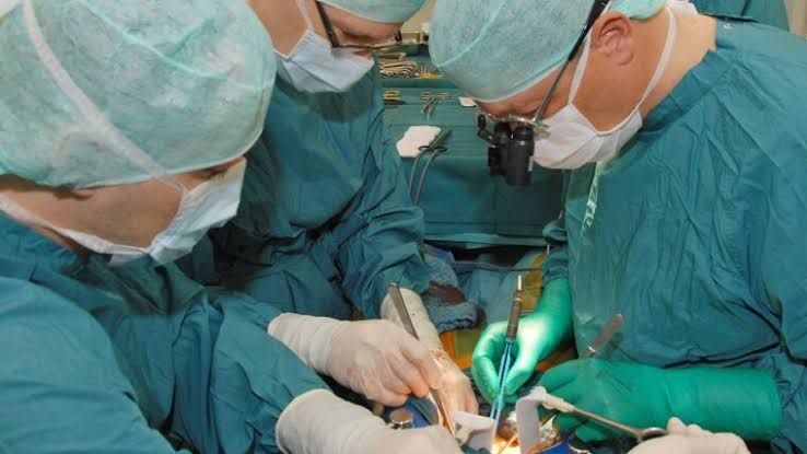
Living organ donation is a remarkable medical advancement that offers a vital alternative for individuals awaiting life-saving transplants. Unlike deceased donor transplantation, living donation involves the surgical removal of an organ or a portion of an organ from a healthy, living individual for immediate transplantation into a recipient. This approach significantly reduces waiting times, allows for planned procedures, and can often lead to better long-term outcomes for the recipient. However, the process is complex, requiring rigorous donor evaluation, a highly skilled surgical team, and precise surgical techniques to ensure the safety of the donor and the viability of the retrieved organ.
General Principles of Living Donor Organ Recovery
Regardless of the specific organ being recovered, several fundamental principles underpin all living donor surgical procedures:
- Comprehensive Donor Evaluation: Prior to surgery, potential living donors undergo an extensive medical, psychological, and social evaluation. This ensures they are in excellent health, fully understand the risks and benefits, and are donating voluntarily. The primary objective is always the safety and well-being of the donor.
- Multidisciplinary Surgical Team: Living donor recovery requires a specialized team including transplant surgeons, anesthesiologists, surgical nurses, and support staff experienced in transplant procedures. Coordination and communication within the team are paramount.
- Pre-operative Preparation: This includes detailed anatomical imaging (CT scans, MRIs, angiography) to map the donor’s specific vascular and biliary anatomy, which is crucial for surgical planning. Standard pre-operative measures such as fasting, bowel preparation (depending on the procedure), and administration of appropriate medications are also performed.
- Aseptic Technique: Strict adherence to sterile technique is critical throughout the procedure to prevent infection, which could jeopardize both donor recovery and graft viability.
- Gentle Tissue Handling: The organ or segment being recovered must be handled with extreme care to minimize trauma and preserve its function for transplantation. This includes carefully dissecting blood vessels and ducts to ensure adequate length and integrity for the recipient’s anastomosis.
- Minimizing Warm Ischemia Time: Warm ischemia time refers to the time the organ is without blood supply at body temperature. While less critical in living donation compared to deceased donation due to immediate cold perfusion after removal, minimizing this period is still important to maintain graft quality.
- Pain Management and Post-operative Care: Effective pain control and meticulous post-operative monitoring are essential for the donor’s rapid and safe recovery.
Living Donor Kidney Recovery (Nephrectomy)
Living donor kidney donation is the most common type of living organ donation. The procedure involves removing one healthy kidney from the donor, typically the left kidney due to its longer renal vein, which facilitates the recipient’s surgery. The donor retains their remaining kidney, which is sufficient to maintain normal renal function. Kidney recovery can be performed using either an open surgical approach or a minimally invasive laparoscopic technique.
(a) Open Donor Nephrectomy
The open approach, while less common now than laparoscopic, was the standard for many years and is still used in specific cases. It involves a larger incision but offers direct tactile feedback and visualization.
Steps for Open Donor Nephrectomy:
- Incision: A flank incision (often subcostal or slightly oblique) is made, typically 15-30 cm in length, depending on the donor’s anatomy and the surgeon’s preference.
- Muscle Division: The muscles of the flank are carefully divided or retracted to access the retroperitoneal space where the kidney is located.
- Retroperitoneal Dissection: The peritoneum is reflected away to expose the kidney, adrenal gland, ureter, and the great vessels (aorta and inferior vena cava).
- Identification and Mobilization: The kidney is identified and carefully mobilized from the surrounding fat and connective tissue (Gerota’s fascia). The adrenal gland, situated superiorly, is identified and preserved.
- Ureter Dissection: The ureter is identified as it courses inferiorly from the renal pelvis. It is carefully dissected free from surrounding tissues down to the level of the iliac vessels, ensuring its blood supply from the retroperitoneum and renal vessels is preserved as much as possible until it is ligated and divided.
- Vascular Dissection: The renal artery and renal vein supplying the kidney are meticulously identified and dissected free from the surrounding tissue. The goal is to obtain sufficient vessel length for the transplant surgeon during the recipient operation. Any accessory vessels are also identified.
- Ligation and Division: Once the kidney is fully mobilized and the vessels and ureter are free, the renal artery, renal vein, and ureter are sequentially ligated and divided. This is a critical step performed rapidly to minimize warm ischemia time. Vascular staplers or ligatures may be used for the artery and vein, while the ureter is typically ligated and divided sharply.
- Kidney Removal: After the vessels and ureter are severed, the kidney is carefully lifted out of the incision.
- Hemostasis and Closure: The surgical field is thoroughly inspected for bleeding, which is controlled using sutures, clips, or cautery. Drains may be placed. The muscle layers, fascia, and skin are then closed in layers.
(b) Laparoscopic Donor Nephrectomy
Laparoscopic donor nephrectomy is now the preferred method in most transplant centers due to its minimally invasive nature, leading to less post-operative pain, shorter hospital stays, and faster recovery for the donor compared to the open approach. It can be performed entirely laparoscopically or with hand assistance.
Steps for Laparoscopic Donor Nephrectomy:
- Port Placement: Small incisions (typically 0.5-1.5 cm) are made in the abdomen or flank to insert trocars, which are tubes allowing the passage of endoscopic instruments and a camera. The number and position of ports vary depending on the surgeon’s technique and whether robotic assistance is used.
- Pneumoperitoneum: The abdomen is insufflated with carbon dioxide gas to create a working space and visualize organs.
- Identification and Mobilization: Using the camera (laparoscope) for visualization, the kidney and surrounding structures are identified within the retroperitoneal space. The peritoneum is typically incised laparoscopically to access this space. The kidney is carefully mobilized from surrounding tissues using laparoscopic dissecting instruments.
- Ureter and Vessel Dissection: The ureter, renal artery, and renal vein (and any accessory vessels) are identified and meticulously dissected using specialized laparoscopic instruments. This requires precise coordination and dexterity. Special care is taken to preserve optimal vessel length.
- Preparation for Division: Once the structures are sufficiently dissected, they are prepared for division using laparoscopic staplers or clips. Staples are commonly used for the renal artery and vein.
- Retrieval Site Incision: A slightly larger incision (typically 5-10 cm) is made elsewhere on the abdomen (often in the midline below the umbilicus or in the iliac fossa) to allow the kidney to be retrieved.
- Placement in Retrieval Bag: The kidney is carefully placed into a specialized retrieval bag within the abdomen to protect it and facilitate its removal through the smaller incision.
- Vascular and Ureteric Division: The renal artery, renal vein, and ureter are then divided using laparoscopic staplers or clips. This is often done just before or as the kidney is being guided into the retrieval site.
- Kidney Retrieval: The retrieval bag containing the kidney is pulled through the retrieval site incision.
- Hemostasis and Closure: The laparoscopic working space is inspected for bleeding. The small port incisions are closed, and the larger retrieval site incision is closed in layers.
Hand-Assisted Laparoscopic Donor Nephrectomy: This technique combines elements of both methods. A slightly larger port site is used, allowing the surgeon to insert one hand into the abdomen to provide tactile feedback and assist with tissue retraction and dissection while using laparoscopic instruments with the other hand and camera assistance.
Both open and laparoscopic techniques require significant surgical skill and experience. The choice of technique depends on surgeon expertise, donor anatomy, and potential medical considerations.
Live Donor Liver Donation Procedure
Living donor liver donation involves removing a segment of the donor’s liver for transplantation into the recipient. The liver has a unique ability to regenerate, allowing both the donor’s remaining liver and the transplanted segment in the recipient to grow back to nearly full size within a few months. Liver donation is surgically more complex than kidney donation due to the liver’s intricate vascular supply and biliary drainage system. The specific segment removed depends on the recipient’s size and needs, with the right lobe being common for adult-to-adult transplants and the left lobe or a smaller segment (e.g., left lateral segment) used for pediatric recipients or smaller adults.
Steps for Live Donor Hepatectomy (Example: Right Lobe Donation):
- Pre-operative Planning: Extensive imaging (CT, MRI, angiography) is performed to precisely map the donor’s liver anatomy, including variations in hepatic arteries, portal veins, and bile ducts. This detailed roadmap is essential for surgical planning and execution.
- Incision: A large incision is typically made across the upper abdomen, often in a “Mercedes-Benz” shape (inverted Y) or a large subcostal incision, to provide adequate exposure.
- Exposure and Mobilization: The abdomen is opened, and the liver is exposed. The ligaments suspending the liver are divided to allow for mobilization of the right lobe.
- Dissection at the Hilum (Porta Hepatis): The structures entering the liver at the hilum (portal vein, hepatic artery, common bile duct) are meticulously dissected. The branches supplying the chosen segment (e.g., right branch of the portal vein, right hepatic artery, right hepatic duct) are identified. Care is taken to preserve the structures supplying the remaining liver segment (e.g., left branches).
- Dissection of Hepatic Veins: The hepatic veins, which drain blood from the liver into the inferior vena cava (IVC), are identified. The major hepatic veins draining the chosen segment (e.g., right hepatic vein) are dissected where they enter the IVC. Preservation of the middle hepatic vein and left hepatic vein, which typically drain the remaining liver, is critical.
- Parenchymal Transection: This is the process of dividing the liver tissue along the planned transection line. Various techniques may be used, including ultrasonic aspirators (CUSA), surgical staplers, or energy devices, often in combination. The transection must be precise to follow the internal anatomical divisions and minimize blood loss and bile leakage. Small vessels and bile ducts encountered during transection are carefully ligated or clipped.
- Division of Inflow and Outflow: Once the parenchymal transection is complete, the large inflow vessels (right portal vein branch, right hepatic artery) and outflow vessels (right hepatic vein) supplying the segment are ligated and divided. This is a critical step, often performed using vascular staplers or ligatures.
- Segment Removal: The isolated liver segment is carefully removed from the abdomen.
- Hemostasis and Closure of Remaining Liver: The raw surface of the remaining liver is thoroughly inspected for bleeding and bile leaks. Hemostasis is secured with sutures, clips, or cautery. Fibrin sealant or other hemostatic agents may be applied.
- Drain Placement and Closure: Drains are typically placed near the transected liver surface to monitor for bleeding or bile leakage. The abdominal wall is closed in layers.
Live donor liver donation is one of the most demanding surgical procedures. It requires exceptional anatomical knowledge, meticulous dissection, and precise execution to ensure the safety of the donor while obtaining a viable graft for the recipient.
Conclusion
Living donor organ recovery procedures for both kidneys and liver segments are complex, highly specialized surgeries performed by expert transplant teams. Whether utilizing open or minimally invasive techniques, the primary focus remains on donor safety, minimizing surgical risk, and recovering a healthy organ or segment for transplantation. Understanding the detailed steps involved in these procedures highlights the skill, precision, and dedication required from the surgical team and underscores the profound gift made by the living donor. Continued advancements in surgical techniques aim to further improve donor outcomes while maintaining excellent results for transplant recipients.


