Kidney transplantation is a complex surgical procedure offering life-changing benefits to patients with end-stage renal disease. While the vascular anastomoses (connecting the renal artery and vein) are critical for blood flow to the transplanted kidney, the successful implantation of the donor ureter into the recipient’s urinary tract is equally vital for establishing effective urine drainage and preventing significant post-transplant complications. A well-executed ureteral anastomosis ensures uninterrupted urine flow, reduces the risk of urinary leak, stricture formation, and vesicoureteral reflux (backward flow of urine), all of which can compromise graft function and patient outcomes.
A cornerstone of successful ureteral implantation is the meticulous preservation of the ureter’s blood supply. The donor ureter receives its blood supply during the organ recovery process, primarily from small vessels originating from the renal artery and sometimes the renal vein, forming an adventitial plexus around the ureter. Unlike the native ureter, which receives segmental blood supply from various points along its length, the transplanted ureter relies almost entirely on this proximal supply derived from the renal hilum. Excessive dissection or stripping of the adventitia during recovery or implantation jeopardizes this delicate vascular network, leading to ischemia, necrosis, and subsequent complications like leaks or strictures. Therefore, handling the ureter with care, preserving surrounding tissue during dissection, and ensuring a tension-free anastomosis are paramount to maintaining viability.
The primary objective of ureter implantation is to create a durable, watertight connection that allows unimpeded urine flow from the transplanted kidney while minimizing the risk of vesicoureteral reflux. The most common technique is ureteroneocystostomy, implanting the donor ureter directly into the recipient bladder. However, alternative techniques are necessary in specific clinical scenarios. This guide outlines the standard procedure and addresses alternative approaches and related considerations.
The Standard Technique: Ureteroneocystostomy
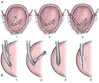
Ureteroneocystostomy, the direct anastomosis of the donor ureter to the recipient bladder, is the preferred method in most kidney transplant recipients with a suitable bladder. The goal is to create a low-pressure, non-refluxing junction. Two main techniques have been widely used: the Leadbetter-Politano method (a tunnel technique involving passing the ureter through the bladder wall and then submucosally before entering the lumen) and the extravesical (Lich-Gregoir) technique (creating a detrusor tunnel on the external bladder surface). The tunnel technique is often favored for its anti-reflux properties, although complication rates can be comparable between the two in experienced hands.
Step-by-Step Guide to Ureteroneocystostomy (Tunnel Technique Principles):
- Bladder Preparation:
- Before implantation, the recipient bladder is prepared. It is typically distended with sterile saline through a bladder catheter. This distension helps identify the posterior or posterolateral aspect of the bladder wall, which is the usual site for implantation, and makes the bladder wall easier to work with.
- The bladder is exposed within the iliac fossa incision.
- Creation of the Anti-Reflux Tunnel: This is a critical step aimed at mimicking the physiological mechanism of the native ureterovesical junction, preventing the backward flow of urine from the high-pressure bladder into the low-pressure ureter and kidney.
- An incision is made through the bladder musculature (detrusor muscle) on the chosen implantation site, typically 4-5 cm long. This incision goes down to, but not through, the bladder mucosa.
- Using fine instruments (e.g., Metzenbaum scissors or blunt dissectors), a submucosal tunnel is carefully created by dissecting between the detrusor muscle and the underlying mucosa. This dissection should extend for approximately 2-3 cm. The length and integrity of this tunnel are key to its anti-reflux function.
- A separate small incision (approximately 1 cm) is made through the mucosa at the distal end of the submucosal tunnel, allowing access to the bladder lumen.
- Ureteric Preparation and Passage:
- The donor ureter is brought down to the bladder implantation site. Its viability is assessed (color, bleeding from cut edge). The very end of the ureter, if potentially ischemic, may be trimmed.
- The distal tip of the ureter is often spatulated (cut obliquely) to widen the opening and create a larger surface area for the anastomosis, reducing the risk of stricture at the new orifice.
- A clamp (e.g., Babcock clamp) or suture placed through the spatulated end is passed from inside the bladder lumen out through the mucosal incision, then through the created submucosal tunnel, and finally out through the detrusor incision.
- The spatulated end of the ureter is then grasped and gently pulled through the submucosal tunnel and into the bladder lumen. Care must be taken to avoid twisting or stretching the ureter, which could compromise its blood supply. The ureter should lie within the tunnel without tension.
- Ureterovesical Anastomosis:
- Once the ureter is positioned within the bladder lumen via the tunnel, the spatulated end is sutured to the bladder mucosa surrounding the distal mucosal opening of the tunnel. This anastomosis typically uses fine absorbable sutures (e.g., 5-0 or 6-0 monofilament). The sutures are placed to oppose the full thickness of the spatulated ureteric wall to the bladder mucosa, creating a watertight seal. Typically, four to six sutures are placed around the circumference of the anastomosis.
- Tunnel Closure (Optional but common):
- After completing the mucosal anastomosis, the edges of the detrusor incision are often loosely approximated over the ureter as it lies in the tunnel. This adds further support and reinforces the anti-reflux mechanism. Care is taken not to constrict the ureter. Some surgeons also fix the ureter to the bladder wall at the point of entry through the detrusor to prevent retraction.
- Bladder Closure:
- The bladder is closed in one or two layers using absorbable sutures. A bladder catheter is left in place to drain urine and keep the bladder decompressed during the initial healing phase (usually 5-7 days).
The functional principle of the anti-reflux tunnel is based on the fact that when the bladder fills and intravesical pressure rises, the supratrigonal bladder muscle contracts. This contraction compresses the segment of the ureter as it passes obliquely through the submucosal tunnel, effectively closing it and preventing the backflow of urine into the ure relatively low-pressure ureter and kidney collecting system.
Indications for Stent Placement
Placement of a temporary internal ureteral stent (usually a double-J stent) across the ureterovesical anastomosis is a common practice in kidney transplantation, although the routine use varies between centers. The primary indications and perceived benefits include:
- Support for the Anastomosis: Provides a scaffold for the healing suture line, reducing mechanical stress.
- Prevention of Urinary Leak: By allowing urine to flow through the stent, it bypasses the fresh suture line, reducing pressure and tension that could lead to a leak, especially in the early post-operative period when edema is present.
- Maintenance of Ureteral Patency: Helps keep the ureteral lumen open during the initial healing phase when swelling and edema around the anastomosis are common.
- Aid in Identifying Ureteral Orifice: Makes the new ureteral opening easier to locate during cystoscopy when the stent is removed.
- Monitoring Urine Output: Ensures drainage from the transplanted kidney.
While routine stenting is practiced by many, it is not without potential complications, including stent-related bladder irritation (frequency, urgency), hematuria, infection (stent can act as a nidus), and stent migration. Therefore, the decision to stent is often based on surgeon preference, institutional protocol, and specific risk factors for complications (e.g., difficult anastomosis, potentially compromised ureter, re-transplantation). Stents are typically removed cystoscopically several weeks after transplantation (e.g., 4-12 weeks).
Addressing the Difficult Bladder
In certain situations, the recipient’s bladder may not be suitable for a standard ureteroneocystostomy. This is referred to as a “difficult bladder” and presents a significant challenge requiring alternative strategies for ureteric drainage. Conditions leading to a difficult bladder include:
- Severe Neurogenic Bladder: Dysfunction leading to high pressures or poor emptying.
- Contracted Bladder: Small bladder capacity secondary to chronic inflammation, interstitial cystitis, or prior surgery/radiation.
- Prior Pelvic Irradiation: Causes fibrosis and poor healing potential of the bladder wall.
- Prior Complex Bladder Surgery: Including augmentation cystoplasty, which alters bladder anatomy and function.
- Prior Cystectomy: The entire bladder has been removed, necessitating drainage into an alternative conduit.
When a difficult bladder is encountered, implantation into the bladder may be impossible or carry an unacceptably high risk of complications. In these cases, alternative techniques for urine drainage from the transplanted kidney must be employed. The choice of alternative depends on the underlying bladder issue and remaining anatomy.
Alternative Ureter Implantation Techniques
When ureteroneocystostomy is not feasible, two primary alternative techniques are considered for establishing urinary drainage: Ureteroureterostomy and Implantation into a Urinary Conduit.
1. Ureteroureterostomy:
This technique involves anastomosing the donor ureter directly to the recipient’s native ureter. This is a viable option when the native ureter is healthy, unobstructed, and of sufficient length to reach the donor ureter without tension. It is often preferred when the bladder is unsuitable but a healthy native ureter is present, or sometimes in re-transplantation where previous bladder implantation was complicated.
Technique Steps:
- Preparation of Recipient Ureter: The recipient’s native ureter is identified and mobilized in the retroperitoneum above the iliac vessels. Its viability and patency are assessed. A segment is prepared for anastomosis.
- Preparation of Donor Ureter: The donor ureter is brought into proximity with the prepared native ureter, ensuring adequate length and preserved blood supply.
- Anastomosis:
- The recipient ureter is typically spatulated. The donor ureter is also spatulated to create a larger opening.
- An end-to-side anastomosis (donor ureter end spatulated, anastomosed to a longitudinal opening in the side of the recipient ureter) or an end-to-end anastomosis (both ureters spatulated and joined) is performed. End-to-side is often favored to avoid stricture risk at a narrow end-to-end junction.
- The anastomosis is created using fine absorbable sutures (e.g., 5-0 or 6-0) with careful attention to creating a watertight and tension-free connection. Sutures are placed to approximate the full thickness of the ureteric walls.
- Stenting: A double-J stent is almost always placed across a ureteroureterostomy to support healing and ensure drainage.
- Drainage: A surgical drain is typically placed near the anastomosis site to monitor for potential urine leakage.
Advantages: Avoids working with a difficult bladder, potentially simpler procedure than conduit formation or manipulation. Disadvantages: Relies on the health and accessibility of the native ureter, risk of stricture or leak at the anastomosis, risk to the native ureter’s function.
2. Implantation into a Urinary Conduit:
This technique is necessary when the recipient has undergone cystectomy or has a severely diseased or non-functional bladder requiring diversion into a urinary conduit (e.g., ileal conduit, colon conduit). In this scenario, the transplanted ureter is directly anastomosed to the created conduit segment.
Technique Steps:
- Conduit Identification: The previously created urinary conduit (often an isolated segment of ileum or colon brought to the abdominal surface as a stoma) is identified within the abdomen.
- Preparation of Conduit Segment: A suitable site on the conduit wall is selected for the anastomosis. An opening is created in the conduit wall.
- Preparation of Donor Ureter: The donor ureter is brought to the prepared conduit site, ensuring adequate length and blood supply. The ureter is spatulated.
- Anastomosis: The spatulated end of the donor ureter is anastomosed directly to the opening in the conduit wall. This is typically a direct, full-thickness anastomosis using fine absorbable sutures. The anastomosis must be watertight and tension-free.
- Stenting: A double-J stent is usually placed across the anastomosis into the conduit and often extended through the stoma, or a feeding tube is placed temporarily through the conduit for drainage.
- Drainage: A surgical drain is placed near the anastomosis.
Advantages: Provides a necessary drainage pathway when the bladder is absent or unusable.
Disadvantages: Higher complexity, risk of complications related to the conduit itself (stricture at the anastomosis, conduit-related issues like pyelonephritis, stoma complications), potential for urine to be in contact with bowel mucosa.
Conclusion
Successful ureter implantation is a critical step in kidney transplantation. While ureteroneocystostomy with an anti-reflux tunnel is the standard technique, understanding the anatomy, particularly the precarious blood supply of the donor ureter, is paramount to preventing ischemic complications. The decision regarding routine stent placement is often center-dependent but guided by factors influencing the risk of early complications. In cases of a difficult bladder, surgeons must be adept at alternative techniques, primarily ureteroureterostomy (utilizing a healthy native ureter) or implantation into a pre-existing or newly created urinary conduit. Meticulous surgical technique, preservation of blood supply, and appropriate selection of the implantation method based on recipient anatomy and bladder function are essential for ensuring optimal urinary drainage and long-term success of the kidney transplant.


