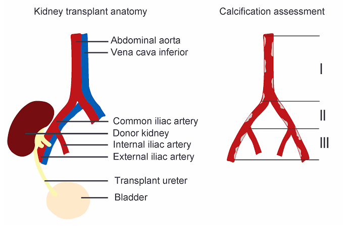
Procedures involving the isolation and vascular anastomosis of the il iliac vessels (common, external, and internal iliac arteries and veins) are fundamental skills in various surgical specialties, including transplant surgery (particularly kidney transplantation), vascular surgery, and trauma surgery. Mastery of these techniques is critical for successful outcomes and minimizing complications.
Essential Prerequisites
Before undertaking this procedure, the surgeon must possess a thorough understanding of retroperitoneal anatomy, including the precise location and relationships of the iliac arteries, veins, their branches, the ureter, genitofemoral nerve, and surrounding lymphatic structures. Familiarity with various surgical instruments, suture materials, and atraumatic clamping techniques is also prerequisite.
1. Patient Preparation and Surgical Approach
- Positioning: The patient is typically placed in a supine position on the operating table. Padding is used to protect pressure points. Depending on the specific procedure (e.g., unilateral vs. bilateral approach), slight table tilt may be beneficial.
- Anesthesia: General anesthesia is standard, ensuring muscle relaxation and hemodynamic stability. Proper monitoring lines are placed.
- Skin Preparation and Draping: The abdomen, flank, and potentially the upper thigh are prepped using an antiseptic solution (e.g., chlorhexidine or povidone-iodine). Sterile drapes are then applied to create a wide sterile field, allowing for potential extension of the incision or access to alternative sites.
- Incision: Access to the iliac vessels is commonly achieved via a lower midline laparotomy or an oblique incision (e.g., Gibson incision, particularly for unilateral access in kidney transplantation). The choice of incision depends on the specific procedure, required exposure, and surgeon preference. A midline incision provides excellent bilateral exposure of the common, external, and internal iliac vessels.
- Accessing the Retroperitoneum: Following fascial incision, the peritoneum is identified. The goal is to access the retroperitoneal space. For a midline approach, the peritoneum is carefully dissected away from the anterior abdominal wall and reflected medially. For an oblique approach, the peritoneum on the ipsilateral side is identified and gently swept medially and superiorly to expose the retroperitoneal structures, including the iliac vessels. The ureter, which descends along the medial aspect of the iliac vessels, must be identified and carefully preserved throughout the dissection.
2. Isolation of the Iliac Vessels
The objective is to expose and mobilize a sufficient length of healthy, non-diseased vessel to allow for safe clamping and anastomosis. This process requires meticulous, gentle dissection.
- Identification of Landmarks: The psoas muscle typically lies posterior to the iliac vessels. The ureter crosses the common iliac artery bifurcation or proximal external iliac artery anteriorly. The genitofemoral nerve runs along the psoas muscle, lateral to the iliac artery. Identifying these structures aids in orienting the dissection.
- Artery Isolation:
- Begin by identifying the common iliac artery as it bifurcates from the aorta or tracing the external iliac artery distally towards the inguinal ligament.
- Carefully incise the overlying posterior peritoneum or retroperitoneal fascia directly over the artery using fine scissors or a scalpel.
- Using blunt dissection (e.g., with a Kitner or peanut dissector, or a laparoscopic sponge stick) and gentle spreading with fine forceps, separate the adventitia of the artery from surrounding lymphatic tissue, fat, and connective tissue. Stay directly on the vessel wall to minimize the risk of injury to adjacent structures, particularly veins which are often adherent to the posterior aspect of the artery.
- Systematically dissect along the chosen segment of the external or common iliac artery. Identify and carefully ligate or clip any small branches encountered to prevent troublesome bleeding or avulsion during handling. Use fine ligatures (e.g., 3-0 or 4-0 silk or vicryl) or appropriately sized surgical clips.
- Once a sufficient length of vessel is isolated, carefully pass vessel loops or vascular slings (e.g., Silastic slings) around the artery at the planned sites for proximal and distal clamping. This provides convenient control and allows for gentle traction during the anastomosis phase. Ensure slings are placed around the fully dissected vessel.
- Vein Isolation:
- The iliac veins (common, external, internal) typically lie medial, posterior, and slightly deeper than their corresponding arteries. The external iliac vein runs medial to the external iliac artery.
- Vein dissection requires even greater care than artery dissection due to their thinner walls, friability, and common adherence to the posterior aspect of the artery.
- Using very gentle blunt dissection and fine forceps, carefully separate the vein from the artery and surrounding tissues. Avoid aggressive sweeping motions which can easily tear the vein wall or avulse tributaries.
- Identify and carefully ligate or clip tributaries, particularly the internal iliac vein (if dissecting common/external iliac veins) or branches draining into the external iliac vein. Venous tributaries are more prone to tearing than arterial branches; precise ligation is crucial.
- Pass vessel loops or slings around the vein at the chosen sites, similar to the artery. Handle the vein gently using the slings, avoiding direct grasping with forceps on the vessel wall whenever possible.
3. Performance of Vascular Anastomosis
Anastomosis is the surgical connection of two blood vessels. The technique varies slightly based on whether it is an end-to-end or end-to-side anastomosis. The principles of ensuring a patent, leak-free connection with appropriate suture material and technique are paramount.
- Choosing the Anastomotic Site: Select a segment of the recipient vessel (iliac artery/vein) that is healthy, free of significant atherosclerosis or scarring, and large enough to accommodate the inflow/outflow vessel.
- Clamping: Apply appropriate atraumatic vascular clamps (e.g., Fogarty, DeBakey, Bulldog clamps) to the recipient vessel. For an end-to-side anastomosis on the external iliac artery/vein:
- Place the proximal clamp first to control inflow.
- Place the distal clamp second to control outflow.
- A “backflow” clamp may be placed more distally if significant back bleeding is anticipated (less common on arteries, can be useful on veins).
- For an end-to-end anastomosis, simply apply a clamp to the proximal and distal ends of the vessel segment to be joined.
- Ensure clamps are applied perpendicular to the vessel and only engage the vessel wall, without trapping surrounding tissue.
- Vessel Preparation (Arteriotomy/Venotomy):
- Once clamped, an incision is made in the recipient vessel.
- For end-to-side anastomosis (common in kidney transplant, where the renal artery is sewn to the external iliac artery, and the renal vein to the external iliac vein), make a precise longitudinal incision (arteriotomy or venotomy) using a scalpel (e.g., #11 or #15 blade) or fine Pott’s scissors. The length of the incision should match the diameter of the vessel being anastomosed. Avoid creating a jagged edge.
- For end-to-end anastomosis, freshen the cut ends of both vessels to be joined, ensuring they are clean and bevelled appropriately if necessary.
- Flush the clamped segment of the recipient vessel with heparinized saline using a small cannula or syringe to clear any residual blood or debris. Repeat flushing of the donor vessel if applicable.
- Suture Material: Non-absorbable monofilament sutures are standard for vascular anastomoses. Polypropylene (Prolene) is commonly used, typically in sizes 5-0 or 6-0 for iliac vessels, sometimes 7-0 for smaller or more distal vessels. The choice depends on the vessel size and wall thickness. Needles should be cardiovascular needles (tapered or fine cutting).
- Suturing Technique (End-to-Side):
- Place initial stay sutures at the “heel” and “toe” of the planned anastomosis. These sutures are tied and used for gentle traction to align the vessels and tension the suture line.
- One side of the anastomosis is then typically closed using a continuous suture technique. Begin at one stay suture, place precise bites through the full thickness of both vessel walls (adventitia, media, intima), maintaining consistent spacing (e.g., 1-2 mm apart) and distance from the edge (e.g., 1-2 mm). Ensure that each bite incorporates visible intima from both sides to prevent intimal flaps.
- The suture line is run towards the other stay suture. Be careful not to overtighten the suture, which can cause purse-stringing and stenosis, or leave it too loose, which can lead to bleeding. Tension should be just enough to approximate the edges.
- Once one side is complete, pass the suture needle out and begin running the suture line back along the other side, again maintaining consistent bites and tension.
- Tie off the suture securely to the starting end or the remaining stay suture.
- Suturing Technique (End-to-End):
- Can be performed using interrupted sutures (more common in smaller vessels or pediatrics) or a continuous suture technique.
- For continuous technique, initial stay sutures can be placed in three corners (“triangulation”) to help align the vessels. Suture lines are then run between the stay sutures.
- Ensure precise alignment of the vessel ends.
- Completion of Anastomosis: Visually inspect the completed suture line to ensure regularity and absence of obvious gaps.
- Declamping and Hemostasis:
- Carefully release the vascular clamps in a controlled sequence. For an end-to-side anastomosis on the external iliac artery, typically release the distal clamp first (allowing backflow to gently expand the vessel and reveal any leaks), then the proximal clamp. For a vein, sequence may vary, sometimes releasing distal first then proximal to check for venous outflow.
- Observe the anastomosis for bleeding points. Minor leaks often self-seal with gentle pressure from a surgical sponge. More significant leaks require additional sutures placed precisely at the bleeding point. Avoid grasping the entire anastomosis with forceps.
- Confirm pulsatility (for arterial anastomoses) and adequate flow.
4. Strategies for Dealing with Complex Anatomy
Managing variations or pathologies in the iliac vessels requires adaptability, experience, and sometimes alternative techniques.
- Atherosclerosis and Calcification:
- Challenge: Rigid, brittle vessels prone to cracking or tearing during clamping or suturing; difficulty getting needles through calcified plaques; risk of intimal dissection or distal embolization.
- Strategies:
- Select the least diseased segment available for anastomosis. Obtain preoperative imaging (CT angiography) to map disease extent.
- Use gentle, appropriately sized vascular clamps; avoid crushing calcified segments.
- Perform the arteriorotomy/venotomy through a non-calcified area if possible.
- When placing sutures, aim to pass the needle through areas between calcified plaques if visible. Use a slightly more robust needle if necessary.
- Consider performing a limited endarterectomy at the anastomotic site if diffuse, obstructing plaque is present, followed by patch angioplasty (using a vein patch or synthetic material) to widen the lumen.
- Be prepared to change the anastomotic site if the initial choice proves too challenging.
- Previous Surgery and Scarring:
- Challenge: Distortion of normal anatomy, dense tissue planes, increased risk of injuring adjacent structures (ureter, nerves) embedded in scar tissue.
- Strategies:
- Thorough review of previous operative reports and imaging.
- Perform careful, sharp dissection layer by layer in scar tissue. Blunt dissection is less effective and carries higher risk of avulsion.
- Stay close to the expected anatomical course of the vessel, relying on knowledge of typical relationships.
- Early identification and protection of critical adjacent structures like the ureter and femoral/obturator nerves is paramount. Dissecting these structures free before tackling the heavily scarred vessel can be beneficial.
- Patience is key. This dissection takes significantly more time and precision.
- Friable or Thin-Walled Vessels:
- Challenge: Vessels that tear easily with minimal manipulation or suture tension (more common with veins, or in certain pathological states).
- Strategies:
- Use extremely gentle handling; minimize grasping the vessel wall directly. Rely on slings for traction.
- Use finer suture material (e.g., 6-0 or 7-0) and smaller needles.
- Space sutures closer together to distribute tension and provide better tissue approximation.
- Consider reinforcing the suture line with a patch angioplasty or using felt pledgets (though less common on iliacs than larger vessels like the aorta).
- Maintain very low tension on the suture line.
- Anatomical Variations:
- Challenge: Unexpected branching patterns, unusual vessel size or position.
- Strategies:
- Be aware of common variations.
- Perform thorough initial exposure and carefully trace vessels proximally and distally to understand the local anatomy before committing to dissection or clamping.
- Utilize preoperative imaging if variations were suspected.
- Be prepared to adapt the anastomotic site or technique based on the encountered anatomy.
- Proximity to Nerves and Ureter:
- Challenge: Risk of thermal, mechanical, or clip injury to the genitofemoral nerve (causing numbness in the thigh), obturator nerve (causing adductor weakness), or ureter (leading to fistula or obstruction).
- Strategies:
- Consciously identify and protect these structures early in the dissection. Trace the ureter’s path as it crosses the common iliac bifurcation. Identify the nerves on the posterior abdominal wall/psoas.
- Avoid placing retractors directly on these structures.
- Use care with electrocautery near nerves.
- Ensure ligatures and clips are placed securely on the vessel branch and do not inadvertently ensnare or impinge upon nearby nerves or the ureter. If in doubt, use ties instead of clips near critical structures.
Conclusion
The isolation of iliac vessels and performance of vascular anastomosis are intricate surgical procedures demanding meticulous technique, a solid understanding of anatomy, and the ability to adapt to individual patient variations and pathological conditions. Careful dissection, precise suture placement, and gentle handling of tissues are fundamental to achieving a successful, patent anastomosis and minimizing complications. Approaching challenging anatomy with caution, preparation, and a repertoire of specific strategies is essential for optimal outcomes in complex cases.


