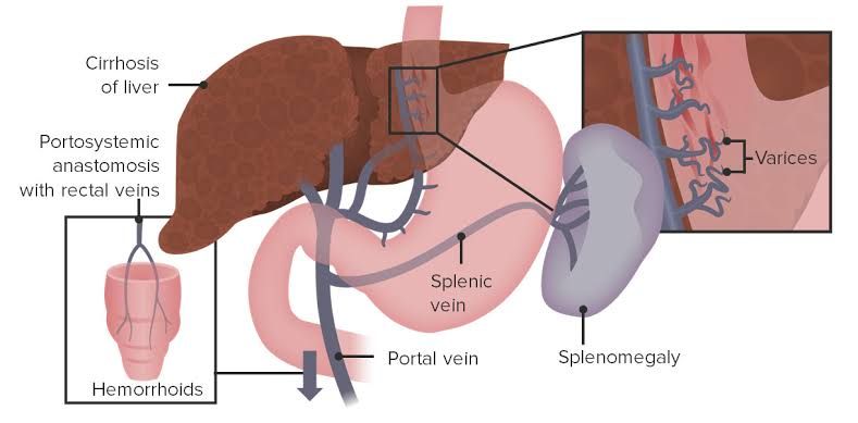
Portal hypertension is a significant clinical syndrome characterized by elevated pressure within the portal venous system. It is most commonly a consequence of advanced liver disease, particularly cirrhosis, but can result from various obstructions to blood flow through the portal vein, liver, or hepatic veins. Managing portal hypertension and its complications is crucial for improving patient outcomes.
Defining Portal Hypertension
The portal vein collects blood from the gastrointestinal tract, spleen, and pancreas, transporting it to the liver. Within the liver, this blood is processed before draining into the systemic circulation via the hepatic veins and inferior vena cava. Portal hypertension occurs when there is resistance to blood flow through the portal system, leading to an increase in pressure.
Clinically, portal hypertension is typically defined as a hepatic venous pressure gradient (HVPG) exceeding 5 mmHg. The HVPG is the difference between the wedged hepatic venous pressure (reflecting pressure in the portal vein system) and the free hepatic venous pressure (reflecting pressure in the inferior vena cava). While an HVPG > 5 mmHg constitutes portal hypertension, clinically significant consequences, such as the development of varices, usually occur when the HVPG exceeds 10 mmHg. Complications like variceal bleeding often arise when the HVPG surpasses 12 mmHg.
The elevated pressure triggers a cascade of events, including splanchnic vasodilation and hyperdynamic circulation, which further exacerbate the high portal pressure and contribute to the development of complications.
The Causes of Portal Hypertension
The causes of portal hypertension can be broadly categorized based on the location of the increased resistance to blood flow relative to the hepatic sinusoids:
2.1. Presinusoidal Causes: Increased resistance occurs before the hepatic sinusoids.
- Extrahepatic Portal Vein Obstruction (EHPVO): This is a common cause, particularly in children. It involves thrombosis or compression of the main portal vein or its branches. Causes include hypercoagulable states, abdominal infections (e.g., omphalitis in neonates leading to pylephlebitis), pancreatitis, malignancy, or surgical injury.
- Schistosomiasis: Infection with Schistosoma mansoni or S. japonicum leads to granulomatous inflammation and fibrosis in the portal venules before they enter the sinusoids, particularly in the periportal spaces. This is a major cause of portal hypertension in endemic regions.
- Sarcoidosis: Granulomas can form in the portal tracts, causing presinusoidal obstruction.
- Congenital Hepatic Fibrosis: Fibrosis is primarily periportal, affecting the portal venules.
- Arteriovenous Malformations or Fistulas: Abnormal connections between arteries and the portal vein can cause increased blood flow and pressure.
- Nontirrhotic Portal Fibrosis (Idiopathic Portal Hypertension): Characterized by fibrosis in the portal tracts and atrophy of associated hepatocytes, without true cirrhosis.
2.2. Sinusoidal Causes: Increased resistance occurs within the hepatic sinusoids.
- Cirrhosis: This is by far the most common cause globally. Fibrosis and nodule formation disrupt the normal liver architecture, compressing the sinusoids and increasing resistance to portal blood flow. Causes of cirrhosis include chronic viral hepatitis (Hepatitis B and C), alcoholic liver disease, non-alcoholic fatty liver disease (NAFLD)/non-alcoholic steatohepatitis (NASH), autoimmune hepatitis, primary biliary cholangitis (PBC), primary sclerosing cholangitis (PSC), hemochromatosis, Wilson’s disease, and alpha-1 antitrypsin deficiency.
- Acute Alcoholic Hepatitis: Severe inflammation and swelling can Transiently increase sinusoidal pressure.
- Veno-occlusive Disease (Sinusoidal Obstruction Syndrome – SOS): Damage to the sinusoidal endothelium and small hepatic venules leads to occlusion. This is often caused by chemotherapy (especially certain regimens used in hematopoietic stem cell transplantation) or pyrrolizidine alkaloids (found in some herbal remedies).
2.3. Post-sinusoidal Causes: Increased resistance occurs after the hepatic sinusoids, in the hepatic veins or beyond.
- Hepatic Vein Obstruction (Budd-Chiari Syndrome): See Step 5 for a detailed explanation. This involves thrombosis or compression of the hepatic veins or the suprahepatic vena cava.
- Cardiac Causes: Severe right-sided heart failure, constrictive pericarditis, or severe tricuspid regurgitation can lead to increased pressure in the inferior vena cava and hepatic veins, causing passive congestion and elevated portal pressure.
- Inferior Vena Cava Obstruction: Compression or thrombosis of the IVC superior to the hepatic vein drainage.
Clinical Presentation of Patients with Portal Hypertension
The clinical manifestations of portal hypertension result directly from the high pressure in the portal system and the ensuing hyperdynamic circulation. Patients may initially be asymptomatic, especially in the early stages. As portal pressure rises and complications develop, symptoms become apparent. Key clinical presentations include:
- Ascites: The accumulation of excess fluid in the peritoneal cavity. Increased hydrostatic pressure in the splanchnic capillaries, combined with impaired renal sodium and water excretion and decreased effective arterial blood volume, drives fluid into the abdomen. It is often the presenting symptom.
- Varices: Dilated veins that develop in areas where the portal and systemic circulations connect (portosystemic anastomoses). These occur because the high-pressure portal blood is forced into lower-pressure systemic veins. Most clinically significant varices occur in the esophagus and stomach (gastroesophageal varices), but they can also develop in the rectum (hemorrhoids), around the umbilicus (Caput Medusae), and retroperitoneally. Esophageal and gastric varices are prone to rupture and bleeding, a life-threatening complication.
- Hepatic Encephalopathy: A spectrum of neuropsychiatric abnormalities caused by the accumulation of toxins (primarily ammonia) in the bloodstream that are normally cleared by the liver. The shunting of portal blood directly into the systemic circulation (due to varices or shunts) bypasses the liver, allowing these toxins to reach the brain. Symptoms range from subtle cognitive changes, inverted sleep-wake cycles, and asterixis (flapping tremor) to confusion, stupor, and coma.
- Splenomegaly: Enlargement of the spleen due to congestion from increased pressure in the splenic vein (which drains into the portal vein). This can lead to hypersplenism, characterized by decreased peripheral blood cell counts (thrombocytopenia, leukopenia, anemia). Thrombocytopenia is common and contributes to the risk of bleeding.
- Coagulopathy: Impaired production of clotting factors by the diseased liver contributes to bleeding tendencies, exacerbated by thrombocytopenia from hypersplenism.
- Portopulmonary Hypertension: A rare but serious complication characterized by pulmonary arterial hypertension in the setting of portal hypertension.
- Hepatopulmonary Syndrome: A condition defined by liver disease, portal hypertension, intrapulmonary vascular dilations, and arterial hypoxemia.
Managing Acute Variceal Bleeding and Preventing Rebleeding
Acute variceal bleeding is a medical emergency with high mortality. Prompt and coordinated management is essential.
4.1. Management of Acute Bleeding:
- Resuscitation: Secure airway (if necessary, especially with encephalopathy or active vomiting/hematemesis), establish intravenous access, and initiate fluid resuscitation using crystalloids and blood products to maintain hemodynamic stability. Transfusion goals are typically lower than in other bleeding scenarios to avoid increasing portal pressure.
- Pharmacological Therapy: Administer vasoconstrictors that reduce splanchnic blood flow and portal pressure. Terlipressin is a vasopressin analogue often considered first-line where available. Somatostatin or its analogue, octreotide, are alternatives. These should be initiated as soon as bleeding is suspected and continued for 2-5 days.
- Antibiotic Prophylaxis: Patients with acute variceal bleeding are at high risk of bacterial infections, which can precipitate further bleeding or worsen encephalopathy. Broad-spectrum antibiotics (e.g., ceftriaxone) should be administered.
- Endoscopic Therapy: Upper endoscopy is performed once the patient is stabilized to confirm the source of bleeding and apply therapy. Endoscopic variceal ligation (banding) is generally preferred for esophageal varices. Sclerotherapy (injecting a sclerosant into or beside the varix) is an alternative, particularly when banding is technically difficult or for gastric varices of the cardiofundal type (although glue injection is often preferred for gastric varices like GVO type 2 or IGV type 1).
- Balloon Tamponade: If initial pharmacological and endoscopic therapies fail to control bleeding, a Sengstaken-Blakemore tube or Minnesota tube can be temporarily used to compress the varices. This is a bridge to more definitive therapy due to the risk of complications (esophageal rupture, aspiration).
- Transjugular Intrahepatic Portosystemic Shunt (TIPS): TIPS is a rescue therapy for bleeding that is refractory to pharmacological and endoscopic treatment. It involves creating a shunt within the liver to divert blood from the portal vein directly into the hepatic vein, significantly reducing portal pressure.
4.2. Prevention of Rebleeding (Secondary Prophylaxis): Once acute bleeding is controlled, preventing future episodes is critical, as rebleeding rates are high and associated with substantial mortality.
- Non-selective Beta-Blockers (NSBBs): Propranolol or nadolol are started to reduce heart rate and splanchnic vasoconstriction, thereby lowering portal pressure. Dosing is titrated to achieve a reduction in heart rate or systolic blood pressure while avoiding symptomatic hypotension. Carvedilol, a beta-blocker with alpha-1 blocking properties, can also be used.
- Endoscopic Variceal Ligation (EVL): For esophageal varices, repeated sessions of banding are performed every 1-4 weeks until the varices are eradicated. Surveillance endoscopy is then required. EVL is often used in combination with NSBBs, especially in patients with higher risk of rebleeding.
- TIPS: If bleeding recurs despite optimal combined medical and endoscopic therapy, TIPS is the preferred secondary prophylaxis method. TIPS is also considered in patients who cannot tolerate or have contraindications to NSBBs and EVL.
Addressing Budd-Chiari Syndrome
Budd-Chiari syndrome (BCS) is a post-sinusoidal cause of portal hypertension characterized by obstruction of hepatic venous outflow. This obstruction can occur anywhere from the small intrahepatic veins to the junction of the hepatic veins with the inferior vena cava (IVC), or in the IVC itself.
The underlying cause is frequently a hypercoagulable state (e.g., myeloproliferative neoplasms, inherited or acquired thrombophilias, oral contraceptive pill use, pregnancy, paroxysmal nocturnal hemoglobinuria). Other causes include compression by tumors or cysts, or webs within the veins.
Clinical presentation is variable, depending on the acuity and extent of the obstruction. It can range from subacute insidious onset with ascites and abdominal pain to fulminant hepatic failure. Chronic BCS often presents with ascites, hepatomegaly, abdominal pain, and signs of portal hypertension like varices.
Management of BCS aims to restore hepatic venous outflow, manage complications of portal hypertension, and treat the underlying prothrombotic condition. Treatment modalities include:
- Anticoagulation: Essential to prevent further thrombosis and potentially recanalize existing clots.
- Thrombolysis: May be attempted in acute presentations with fresh thrombus.
- Angioplasty and Stenting: Used to open stenosed veins or webs.
- TIPS: An important option to decompress the liver and manage portal hypertension if angioplasty fails or is not feasible.
- Liver Transplantation: The definitive treatment for patients with severe liver dysfunction or uncontrolled complications despite other therapies.
Comprehensive Management of Portal Hypertension and Portosystemic Shunts
Managing portal hypertension is multifaceted and depends on the underlying cause, the severity of liver disease, and the presence and severity of complications. The overall goal is to reduce portal pressure and prevent or treat complications.
Beyond the specific management of acute bleeding, broader management strategies include:
- Treating the Underlying Liver Disease: Addressing the root cause (e.g., antiviral therapy for hepatitis B/C, alcohol abstinence, managing metabolic syndrome, immunosuppression for autoimmune diseases) is crucial to slow disease progression and potentially reduce portal pressure.
- Management of Ascites: Dietary sodium restriction and diuretics (spironolactone often combined with furosemide) are the mainstays. Therapeutic paracentesis is used for large or refractory ascites.
- Management of Hepatic Encephalopathy: Identifying and treating precipitating factors (e.g., infection, bleeding, dehydration, certain medications) is key. Lactulose is used to reduce ammonia production and absorption. Rifaximin, a non-absorbable antibiotic, is also used.
- Primary Prophylaxis of Variceal Bleeding: In patients with medium to large esophageal varices who have not bled, prophylaxis is indicated to prevent the first bleed. This involves either non-selective beta-blockers (NSBBs) or endoscopic variceal ligation (EVL). The choice depends on factors like varice size, liver function, and patient tolerance/preference.
- Nutritional Support: Addressing malnutrition, common in advanced liver disease.
Portosystemic Shunts: These procedures create a connection between the portal venous system and the systemic venous circulation to decompress the portal system and reduce pressure.
- Transjugular Intrahepatic Portosystemic Shunt (TIPS): As mentioned earlier, TIPS is a minimally invasive procedure performed by interventional radiologists. A tract is created through the liver parenchyma connecting a branch of the portal vein to a hepatic vein, and a stent is placed to keep it open. TIPS is primarily used for refractory variceal bleeding or intractable ascites unresponsive to medical therapy. It is not without risks, notably potentially worsening hepatic encephalopathy due to increased shunting of toxins past the liver. Stent stenosis or occlusion requiring revision is also a potential complication.
- Surgical Portosystemic Shunts: These involve direct surgical creation of a connection between portal and systemic veins (e.g., portacaval shunt, distal splenorenal shunt). While effective at decompressing the portal system, they are more invasive, associated with higher procedural morbidity, and also carry a significant risk of hepatic encephalopathy. Their use has significantly decreased since the advent of TIPS, and they are now typically reserved for specific indications or situations where TIPS is not feasible.
Liver Transplantation: For patients with end-stage liver disease and complications of portal hypertension that are refractory to other treatments, liver transplantation is the definitive therapy. It addresses both the underlying liver disease and the portal hypertension.
In conclusion, portal hypertension is a serious consequence of impaired blood flow through the portal system, most commonly due to cirrhosis. Its management requires a thorough understanding of its causes and clinical manifestations, focusing on preventing and treating complications like variceal bleeding, ascites, and hepatic encephalopathy. Therapeutic strategies range from pharmacological interventions and endoscopic procedures to interventional radiological techniques like TIPS and, ultimately, liver transplantation. Effective management necessitates a multidisciplinary approach tailored to the individual patient’s condition.


