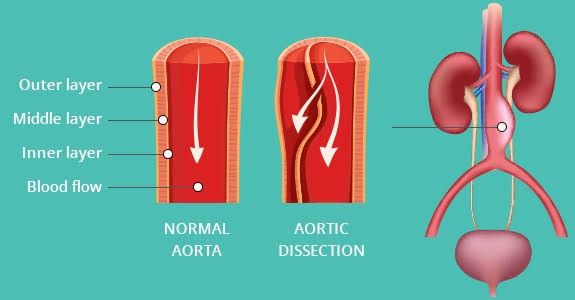
The aorta is the body’s largest artery, originating from the left ventricle of the heart and extending down through the chest and abdomen, branching off to supply oxygenated blood to all major organs and limbs. Given its critical role and the high pressure it carries, the aorta is susceptible to various serious conditions.
1. Aortic Dissection
Aortic dissection is an acute, life-threatening medical emergency involving a tear in the inner layer (intima) of the aorta. This tear allows blood to surge between the inner and middle layers (media) of the aortic wall, creating a false lumen (channel). As blood flows through this false lumen, it separates (dissects) the layers of the aortic wall, which can extend along the length of the aorta. This process weakens the wall, can impede blood flow to vital organs branching off the aorta, and carries a high risk of rupture.
- Definition: A tear in the arterial wall intima that allows blood to enter and separate the layers of the aorta, forming a false lumen alongside the true lumen.
- Risk Factors:
- Hypertension: Long-standing, uncontrolled high blood pressure is the most significant risk factor, placing excessive stress on the aortic wall.
- Pre-existing Aortic Aneurysm: A dilated aorta is structurally weaker and more prone to dissection.
- Connective Tissue Disorders: Conditions like Marfan syndrome, Ehlers-Danlos syndrome, and Loeys-Dietz syndrome weaken the connective tissue components of the aortic wall.
- Bicuspid Aortic Valve: A congenial heart defect where the aortic valve has two leaflets instead of the usual three, often associated with abnormalities of the ascending aorta.
- Atherosclerosis: While less direct a cause than hypertension, severe hardening and narrowing of the arteries can contribute to aortic wall damage.
- Aortitis: Inflammation of the aorta (e.g., Takayasu’s arteritis, Giant cell arteritis).
- Trauma: Severe chest trauma (e.g., deceleration injuries in motor vehicle accidents).
- Cocaine Use: Can cause a sudden, severe spike in blood pressure.
- Pregnancy: Particularly in the third trimester, due to hormonal changes and increased blood volume/pressure, especially in women with underlying connective tissue disorders or hypertension.
- Family History: A predisposition to aortic disease.
- Clinical Presentation:
- Severe Pain: Typically described as sudden onset, excruciating, sharp, tearing, or ripping pain. The location often correlates with the site of dissection:
- Anterior chest pain: Ascending aorta dissection (Type A).
- Back pain (interscapular): Descending aorta dissection (Type B).
- Pain may radiate downwards as the dissection extends.
- Neurological Deficits: Stroke symptoms (weakness, numbness, difficulty speaking), syncope (fainting), or paralysis may occur if the dissection extends into arteries supplying the brain or spinal cord.
- Pulse/Blood Pressure Discrepancies: Asymmetric pulses or significant blood pressure differences between the arms.
- Signs of Organ Ischemia: Symptoms related to reduced blood flow to organs, such as kidney failure (reduced urine output), mesenteric ischemia (abdominal pain), or limb ischemia (pain, pallor, pulselessness in a limb).
- Aortic Regurgitation: A new or worsening heart murmur if the dissection involves the aortic valve.
- Pericardial Tamponade: Accumulation of blood in the sac around the heart if the dissection ruptures outwards, leading to dangerously low blood pressure.
- Severe Pain: Typically described as sudden onset, excruciating, sharp, tearing, or ripping pain. The location often correlates with the site of dissection:
- Diagnosis:
- Clinical Suspicion: Based on the characteristic severe, sudden pain, particularly in patients with risk factors.
- Imaging Studies:
- CT Angiography (CTA): The most commonly used and fastest diagnostic test. Provides detailed images of the aorta, clearly showing the intimal flap, true and false lumens, and extent of dissection.
- Transesophageal Echocardiography (TEE): Useful in unstable patients or when CTA is contraindicated. Provides good visualization of the ascending aorta and aortic valve.
- Magnetic Resonance Angiography (MRA): Can provide excellent images but is more time-consuming and less available in emergency settings.
- Chest X-ray: May show a widened mediastinum (silhouette of the aorta), but is not definitive for diagnosis.
- Treatment:
- Immediate Medical Stabilization: Aggressive control of blood pressure and heart rate to reduce stress on the aortic wall and prevent extension of the dissection. Intravenous medications (e.g., beta-blockers, nitroprusside) are used. Pain management is also crucial.
- Type A Dissection (Involving the Ascending Aorta): This is a surgical emergency. Requires immediate open surgical repair to replace the damaged section of the ascending aorta and restore the integrity of the aortic wall, preventing rupture or extension into the aortic arch or coronary arteries.
- Type B Dissection (Involving only the Descending Aorta): Initial management is typically medical, focusing on blood pressure and heart rate control. Surgery or endovascular repair (Thoracic Endovascular Aortic Repair – TEVAR) is indicated if complications arise, such as rupture, malperfusion of organs/limbs (blood flow blockage), uncontrolled pain, or rapid expansion of the aorta.
- Long-term Management: Lifelong blood pressure control, serial imaging surveillance, and management of underlying risk factors are essential for all patients after dissection, regardless of initial treatment type.
2. Thoracic Aortic Aneurysms (TAA)
A thoracic aortic aneurysm (TAA) is a localized or diffuse dilation (bulging) of the aorta within the chest cavity. These aneurysms can occur in the ascending aorta (most common), the aortic arch, or the descending aorta. TAAs weaken the aortic wall and are primarily dangerous because of their potential to rupture, causing massive, often fatal, internal bleeding, or to lead to aortic dissection.
- Definition: A permanent, localized widening or bulging of the thoracic aorta to more than 1.5 times its normal diameter.
- Risk Factors:
- Atherosclerosis: The most common cause of descending and thoracoabdominal aortic aneurysms.
- Hypertension: Contributes to the development and growth of aneurysms by increasing stress on the aortic wall.
- Connective Tissue Disorders: Marfan syndrome, Ehlers-Danlos syndrome, Loeys-Dietz syndrome are strongly associated with ascending aortic and arch aneurysms and are often identified at younger ages.
- Bicuspid Aortic Valve: A common risk factor for ascending TAA.
- Family History: A genetic predisposition exists for thoracic aneurysms.
- Smoking: Damages the arterial walls and is a major risk factor for atherosclerotic aneurysms.
- Advanced Age: Risk generally increases with age.
- Aortitis: Inflammatory conditions affecting the aorta.
- Trauma: Less common, but blunt chest trauma can potentially lead to aneurysm formation.
- Clinical Presentation:
- Often Asymptomatic: Many TAAs are discovered incidentally on imaging performed for other reasons.
- Pain: If symptoms occur, it is often a deep, aching pain in the chest, upper back, or neck. Pain may indicate rapid expansion or impending rupture.
- Symptoms due to Compression of Nearby Structures: If the aneurysm grows large enough to press on adjacent structures:
- Cough or shortness of breath (compressing trachea or bronchi).
- Hoarseness (compressing the recurrent laryngeal nerve).
- Difficulty swallowing (compressing the esophagus).
- Swelling of the face and arms (compressing the superior vena cava – Superior Vena Cava Syndrome).
- Rupture Symptoms: Sudden onset of severe, tearing chest or back pain, often radiating outwards, accompanied by signs of shock (sudden weakness, low blood pressure, rapid heart rate). This is a catastrophic event.
- Diagnosis:
- Incidental Findings: Often first suspected on a routine chest X-ray (showing mediastinal widening).
- Imaging Confirmation and Sizing:
- CT Angiography (CTA): Gold standard for diagnosing, sizing, and planning treatment for TAAs. Provides detailed 3D reconstruction.
- Magnetic Resonance Angiography (MRA): Useful alternative, especially for patients with kidney problems or contrast allergies, but slower than CTA.
- Echocardiography: Can visualize the ascending aorta and aortic root well, particularly useful in assessing patients with bicuspid aortic valve or known connective tissue disorders. Limited visualization of the descending aorta.
- Transesophageal Echocardiography (TEE): Provides better views of the thoracic aorta than transthoracic echo.
- Treatment:
- Medical Management: For smaller, stable aneurysms and to manage risk factors:
- Strict blood pressure control (often with beta-blockers, especially for aneurysms related to connective tissue disorders).
- Statin therapy to manage atherosclerosis.
- Smoking cessation counseling and support.
- Surveillance: Regular imaging (usually CTA or MRA) to monitor aneurysm size and growth rate. Frequency depends on the size and risk factors.
- Intervention (Surgical or Endovascular Repair): Indicated for larger aneurysms (typically >5-6 cm, though size criteria vary based on location, growth rate, etiology like Marfan syndrome, and patient factors), symptomatic aneurysms, or rapidly growing aneurysms.
- Open Surgical Repair: Involves replacing the weakened segment of the aorta with a synthetic graft through a large incision. Traditional approach, often used for complex or ascending/arch aneurysms.
- Endovascular Aneurysm Repair (TEVAR): Less invasive approach involving inserting a stent graft through blood vessels (usually in the groin) and deploying it inside the aneurysm to exclude it from blood flow. Preferred for descending TAAs when feasible.
- Genetic Counseling: Recommended for patients with TAAs suspected to be hereditary.
- Medical Management: For smaller, stable aneurysms and to manage risk factors:
3. Abdominal Aortic Aneurysms (AAA)
An abdominal aortic aneurysm (AAA) is a localized or diffuse dilation (bulging) of the abdominal segment of the aorta, most commonly occurring below the level where the renal arteries branch off. Like TAAs, AAAs are dangerous primarily due to the risk of rupture, which is a highly lethal event.
- Definition: A permanent, localized widening or bulging of the abdominal aorta to more than 1.5 times its normal diameter, typically defined as >3 cm.
- Risk Factors:
- Atherosclerosis: The dominant underlying cause.
- Smoking: The single strongest risk factor for AAA development and growth.
- Age: Risk increases significantly after the age of 65.
- Male Sex: Males are significantly more likely to develop AAAs than females.
- Family History: A strong family history of AAA increases risk.
- Hypertension: Contributes to aneurysm formation and growth.
- Hyperlipidemia (High Cholesterol): A key component of atherosclerosis.
- Caucasian Race: More prevalent in this group than other racial groups.
- Prior Aneurysm in Another Location: Presence of an aneurysm elsewhere (e.g., popliteal artery aneurysm) increases the risk of AAA.
- Clinical Presentation:
- Often Asymptomatic: The vast majority of AAAs cause no symptoms and are found incidentally during examinations or imaging for other conditions.
- Palpable Abdominal Mass: In some individuals, a large AAA may be felt as a pulsating mass in the abdomen during a physical examination.
- Pain: If symptoms occur, it may be a deep, persistent ache in the lower back, flank, or abdomen. Pain can indicate rapid expansion or leakage.
- AAA Rupture: A catastrophic event presenting with:
- Sudden, severe, tearing abdominal or back pain.
- Hypotension (low blood pressure) and shock.
- A pulsatile abdominal mass (if palpable).
- Loss of consciousness. Rupture is often fatal.
- Symptoms from Embolization: Clots from within the aneurysm sac can break off and travel downstream, causing sudden pain, pallor, pulselessness, paresthesia, and paralysis in the legs or feet (“trash foot”).
- Diagnosis:
- Screening Ultrasound: Non-invasive, cost-effective, and widely used for screening populations at high risk (e.g., men aged 65-75 who have ever smoked).
- CT Angiography (CTA): Used to confirm the diagnosis, accurately measure the size, determine the anatomical extent, and plan potential surgical or endovascular repair. Essential for assessing ruptured or symptomatic AAAs.
- Magnetic Resonance Angiography (MRA): Alternative imaging modality, useful in certain cases.
- Physical Examination: Palpating a pulsatile abdominal mass may suggest an AAA, but palpation alone is unreliable for diagnosis or sizing, especially in obese patients, and should not be attempted vigorously if rupture is suspected.
- Treatment:
- Medical Management and Surveillance: For smaller AAAs (<5.5 cm in men, smaller thresholds often considered for women or those with specific risk factors/anatomy) that are not causing symptoms:
- Aggressive management of risk factors (smoking cessation is paramount, blood pressure control, statins for cholesterol).
- Regular ultrasound or CT surveillance to monitor size and growth rate.
- Intervention (Surgical or Endovascular Repair): Indicated for AAAs that are large (typically ≥5.5 cm), growing rapidly (>0.5 cm in 6 months or >1 cm per year), or causing symptoms (pain, leakage).
- Endovascular Aneurysm Repair (EVAR): The preferred method for many patients. It is less invasive than open surgery, involving insertion of a stent graft via catheters through the femoral arteries in the groin to exclude the aneurysm from blood flow.
- Open Surgical Repair: Involves replacing the aneurysmal segment of the aorta with a synthetic graft through a large abdominal incision. A more invasive procedure, but necessary in some complex cases or when EVAR is not anatomically suitable.
- Emergency Repair: Ruptured AAAs require immediate emergency surgical or endovascular repair, although mortality rates remain high.
- Medical Management and Surveillance: For smaller AAAs (<5.5 cm in men, smaller thresholds often considered for women or those with specific risk factors/anatomy) that are not causing symptoms:
4. Aorto-iliac Occlusion
Aorto-iliac occlusion refers to the narrowing (stenosis) or complete blockage (occlusion) of the terminal segment of the abdominal aorta and/or the iliac arteries, which are major blood vessels supplying blood to the pelvis and lower extremities. This condition is a form of Peripheral Artery Disease (PAD) and is most commonly caused by atherosclerosis. Adequate blood flow through this segment is crucial for lower limb function and viability.
Risk Factors
Development of aorto-iliac occlusion is strongly associated with established risk factors for atherosclerosis. These include:
- Smoking: The most significant modifiable risk factor, severely accelerating atherosclerotic progression.
- Diabetes Mellitus: Contributes to widespread micro- and macrovascular disease.
- Hypertension (High Blood Pressure): Increases arterial wall stress and promotes atherosclerosis.
- Hyperlipidemia (High Cholesterol): Particularly elevated LDL cholesterol, promoting plaque formation.
- Advancing Age: Atherosclerosis is more common with age.
- Family History: Genetic predisposition to atherosclerosis or PAD.
- Obesity
- Sedentary Lifestyle
Clinical Presentation
Symptoms result from insufficient blood flow to the muscles and tissues of the buttocks, thighs, and calves. The severity of symptoms depends on the degree and location of the blockage and the adequacy of collateral circulation. Common presentations include:
- Claudication: Pain, cramping, or fatigue in the muscles (buttocks, thighs, calves) that is triggered by walking or exercise and relieved by rest. Buttock and thigh claudication are particularly suggestive of aorto-iliac disease.
- Rest Pain: Severe pain, typically in the feet or toes, that occurs while at rest, often at night. This indicates critical limb ischemia, a severe form of the disease.
- Non-healing Wounds, Ulcers, or Gangrene: Extremely poor perfusion can lead to tissue breakdown, delayed wound healing, and potentially tissue death, especially in the feet and toes.
- Erectile Dysfunction: In males, occlusion of the internal iliac arteries (which supply pelvic organs) can cause impotence. This, combined with bilateral buttock/thigh claudication and absent femoral pulses, forms the classic triad known as Leriche Syndrome.
- Physical Examination Findings: Diminished or absent femoral, popliteal, and pedal pulses; pallor, coolness, or trophic changes (hair loss, nail changes, skin thinning) in the lower extremities; muscular atrophy; presence of arterial bruits over the iliac or femoral arteries.
Diagnosis
Diagnosis typically involves a combination of clinical evaluation and objective vascular tests:
- Clinical History and Physical Examination: Detailed assessment of symptoms, risk factors, and physical findings (especially pulse palpation).
- Ankle-Brachial Index (ABI): A simple, non-invasive test comparing blood pressure in the ankle to the arm. A low ABI (< 0.90) is indicative of PAD. Exercise ABI may uncover disease not apparent at rest.
- Duplex Ultrasound: Provides non-invasive imaging to visualize the arteries, assess blood flow velocity, and identify stenoses or occlusions.
- CT Angiography (CTA): Offers detailed cross-sectional images of the arterial anatomy, useful for planning revascularization procedures. Requires intravenous contrast.
- MR Angiography (MRA): Similar to CTA but uses magnetic fields. Useful, particularly in patients with renal insufficiency or contrast allergies (though gadolinium contrast used in MRA is not without risks).
- Digital Subtraction Angiography (DSA): An invasive procedure involving contrast injection directly into the arteries under X-ray guidance. Provides the most detailed arterial mapping and is often performed immediately before or during endovascular intervention.
Treatment
Management strategies aim to alleviate symptoms, prevent limb loss, and reduce cardiovascular morbidity and mortality. Treatment approaches include:
- Conservative Management:
- Risk Factor Modification: Aggressive control of blood pressure, cholesterol, and diabetes. Smoking cessation is critical and the single most impactful intervention.
- Supervised Exercise Program: Can significantly improve walking distance and claudication symptoms.
- Pharmacotherapy:
- Antiplatelet agents (e.g., aspirin, clopidogrel) to reduce the risk of cardiovascular events (heart attack, stroke).
- Statins for lipid management.
- Medications like Cilostazol may improve claudication but are less effective for severe proximal disease.
- Revascularization (for significant symptoms or critical limb ischemia): Procedures to restore blood flow.
- Endovascular Therapy: Less invasive techniques suitable for many aorto-iliac lesions. Includes balloon angioplasty ± stenting to open blockages. Offers quicker recovery but may have lower long-term patency rates compared to surgery for extensive disease.
- Surgical Bypass: More invasive options used for extensive or complex lesions. Common procedures include aortobifemoral bypass (creating a new path from the aorta to the femoral arteries using a graft) or femorofemoral bypass (graft from one femoral artery to the other). Provides durable long-term results but involves a larger operation.
- Hybrid Procedures: Combining endovascular and open surgical techniques in a single setting.
The choice of treatment depends on the patient’s overall health, the severity of symptoms, the location and extent of the blockage, and anatomical considerations. Timely diagnosis and management are essential to prevent progression to critical limb ischemia and improve patient outcomes.


