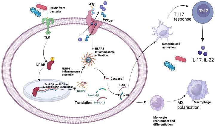
Allogeneic organ transplantation is a life-saving treatment for end-stage organ failure. However, the success of transplantation is constantly challenged by the recipient’s immune system recognizing the donor organ as foreign, leading to immune-mediated rejection. While the adaptive immune response, particularly T cells and antibodies directed against donor-specific antigens (DSAs), is the primary driver of chronic and acute rejection, innate immunity and inflammation play a crucial, often insidious, role in exacerbating this process. Inflammation, triggered by various factors ranging from surgical trauma and ischemia-reperfusion injury (IRI) to infections and rejection itself, creates a permissive or even enhancing environment that amplifies adaptive immune responses, leading to increased allograft damage and ultimately, graft loss.
Mechanisms by Which Inflammatory Reactions Can Exacerbate Donor-Specific Immune Mediated Allograft Damage
Inflammation is a complex biological response involving a cascade of cellular and molecular events. Within the context of transplantation, this response significantly influences how the recipient’s adaptive immune system encounters and reacts to the donated organ. The inflammatory milieu within the graft and surrounding recipient tissues provides critical signals and conditions that boost the potency and duration of the donor-specific adaptive immune attack.
Here are the principal mechanisms:
- Enhanced Antigen Presentation:
- Inflammation is a potent inducer of antigen-presenting cell (APC) activation and maturation. In the context of transplantation, resident APCs within the graft (e.g., dendritic cells, macrophages) and recipient APCs infiltrating the graft are centrally involved in presenting donor antigens (primarily MHC molecules, but also minor histocompatibility antigens) to recipient T cells.
- Inflammatory mediators, such as cytokines (e.g., TNF-α, IL-1β, IL-6, Type I IFNs) and damage-associated molecular patterns (DAMPs) released from injured graft cells, strongly upregulate the expression of MHC molecules (Class I and Class II) and co-stimulatory molecules (e.g., CD80, CD86, CD40) on these APCs.
- This enhanced expression increases the efficiency with which donor peptides are presented in the context of MHC to recipient T cells and provides the necessary second signals required for full T cell activation and proliferation.
- Both direct pathway presentation (where recipient T cells recognize intact donor MHC molecules on donor APCs migrating from the graft to recipient lymphoid organs) and indirect pathway presentation (where recipient APCs process donor antigens and present donor peptides on recipient MHC molecules) are significantly amplified by inflammation-induced APC maturation. Inflammation promotes the migration of donor APCs and enhances the uptake and processing of donor material by recipient APCs.
- Recruitment and Activation of Immune Cells:
- A hallmark of inflammation is the increased permeability of blood vessels and the production of chemokines (chemoattractant cytokines). These molecules create gradients that guide circulating recipient leukocytes into the inflamed graft tissue.
- Inflammation leads to the activation of graft endothelial cells, causing them to express higher levels of adhesion molecules (e.g., E-selectin, P-selectin, ICAM-1, VCAM-1). These molecules facilitate the rolling, adhesion, and transendothelial migration of various immune cells, including neutrophils, monocytes/macrophages, lymphocytes (T and B cells), and natural killer (NK) cells, into the graft interstitium.
- Once inside the graft, these recruited innate immune cells (macrophages, neutrophils) contribute to tissue injury through the release of reactive oxygen species (ROS), proteases, and pro-inflammatory cytokines, further perpetuating the inflammatory cycle.
- More critically for exacerbating adaptive immunity, the increased influx of recipient lymphocytes, particularly T and B cells, brings the effectors of adaptive immunity into direct contact with donor antigens presented by the activated APCs within the graft or draining lymphoid tissues. The inflammatory environment within the graft also provides survival signals for these infiltrating lymphocytes.
- Activation of Endothelial Cells and Vascular Damage:
- Graft endothelial cells are highly sensitive to inflammatory stimuli. Activation of endothelial cells by cytokines (TNF-α, IL-1β), DAMPs, or complement components leads to endothelial dysfunction.
- This activation results in increased vascular permeability, facilitating immune cell infiltration, and enhanced expression of pro-coagulant factors, potentially contributing to microvascular thrombosis.
- Chemokine production by activated endothelial cells further drives the recruitment of inflammatory and immune cells.
- Endothelial cell activation can also directly damage the microvasculature of the graft. This vascular injury contributes to ischemia and perpetuates the release of DAMPs, creating a vicious cycle of inflammation and damage. Antibody-mediated rejection (AMR), a key form of DSA-driven damage, heavily targets the endothelium, and pre-existing inflammation in the graft can prime the endothelium for greater susceptibility to antibody and complement attack.
- Shaping the Cytokine and Chemokine Milieu:
- The inflammatory environment within and around the graft generates a specific profile of cytokines and chemokines that profoundly influences the differentiation and function of infiltrating lymphocytes.
- Pro-inflammatory cytokines (e.g., IL-1β, IL-6, IL-12, IL-18) produced by activated innate cells (macrophages, dendritic cells) and damaged graft cells steer the differentiation of naive T cells towards specific effector subsets known for mediating rejection. For example, IL-12 and IL-18 promote the differentiation of cytotoxic CD8+ T cells and T helper 1 (Th1) cells, which produce IFN-γ and are critical for cell-mediated rejection. IL-1β and IL-6 promote the differentiation of Th17 cells, which produce IL-17 and contribute to neutrophil recruitment and tissue pathology.
- Chemokines (e.g., CXCL9, CXCL10, CXCL11 which attract CXCR3+ T cells; CCL2, CCL5 which attract macrophages and T cells) produced during inflammation guide the specific localization of these effector cells within the graft, directing them to sites of potential damage.
- Release of Damage-Associated Molecular Patterns (DAMPs):
- Tissue injury inherent in transplantation, such as surgical trauma, organ preservation, and particularly ischemia-reperfusion injury (IRI), leads to the release of intracellular components into the extracellular space. These are known as DAMPs or “alarmins.” Examples include High Mobility Group Box 1 (HMGB1), heat shock proteins (HSPs), uric acid, S100 proteins, and extracellular ATP.
- DAMPs act as endogenous ligands for pattern recognition receptors (PRRs), such as Toll-like receptors (TLRs) and NOD-like receptors (NLRs), expressed on both innate immune cells and cells within the graft parenchyma and endothelium.
- Binding of DAMPs to PRRs triggers intracellular signaling pathways (e.g., NF-κB, inflammasome activation) that result in the production of pro-inflammatory cytokines (IL-1β, TNF-α, IL-6) and chemokines.
- This DAMP-induced innate immune activation initiates and amplifies the local inflammatory response, effectively ‘sounding an alarm’ in the transplant setting, which then fuels the recruitment, activation, and differentiation of adaptive immune cells against donor antigens. It bridges the innate and adaptive immune responses.
- Promoting Humoral Immunity:
- While T cells are central, inflammation also contributes significantly to the development and efficacy of donor-specific antibodies (DSAs), a key component of antibody-mediated rejection (AMR).
- Inflammatory signals, particularly from activated T follicular helper (Tfh) cells (whose differentiation can be influenced by the inflammatory cytokine milieu, such as IL-6), help B cells proliferate, undergo somatic hypermutation, and differentiate into antibody-producing plasma cells.
- Complement system activation is often a consequence of inflammation and is a major effector pathway in AMR. Inflammatory mediators can prime the tissue for complement deposition and enhance the damage caused by complement cascade activation initiated by DSAs binding to the graft endothelium.
- Macrophages and neutrophils recruited during inflammation can also contribute to antibody-dependent cellular cytotoxicity (ADCC) mechanisms against DSA-coated graft cells.
- Interfering with Regulatory Mechanisms:
- The immune system has intrinsic regulatory mechanisms, such as regulatory T cells (Tregs) and myeloid-derived suppressor cells (MDSCs), designed to dampen immune responses and maintain tolerance.
- Acute and chronic inflammation can impair the function, stability, and suppressive capacity of these regulatory cell populations. Pro-inflammatory cytokines like IL-6, IL-1β, and TNF-α have been shown to inhibit Treg differentiation or function.
- By compromising these natural brakes on the immune system, inflammation allows effector T and B cell responses against the allograft to proceed more vigorously and for a longer duration, contributing to more severe and persistent damage.
- Contribution to Fibrosis and Chronic Allograft Dysfunction:
- While not solely an initial trigger of adaptive immunity, chronic inflammation within the graft promotes tissue remodeling and fibrosis. This process involves the activation of fibroblasts and deposition of extracellular matrix proteins, leading to scarring and loss of organ function.
- The persistence of inflammatory cells and mediators within the graft contributes to chronic tissue injury and further attracts immune cells, perpetuating a cycle that culminates in chronic allograft dysfunction (CAD). Inflammation therefore underpins both the initial immune attack and the long-term consequences that lead to graft failure.
Inflammation-Associated Cytokines that Enhance the Initial Phases of Adaptive Immune Responses
The early inflammatory response following transplantation releases a battery of cytokines that are crucial in shaping and enhancing the recipient’s nascent adaptive immune response against the donor organ. These cytokines often act on APCs and naive lymphocytes to promote activation, proliferation, and differentiation.
Here are some key inflammation-associated cytokines involved in enhancing the initial phases of adaptive immune responses:
- Interleukin-1 (IL-1α and IL-1β): Produced primarily by activated macrophages, dendritic cells, and damaged tissues, IL-1 is a potent pro-inflammatory cytokine. It enhances the expression of MHC molecules and co-stimulatory molecules on APCs, making them more effective at presenting antigens. IL-1β, often produced following inflammasome activation by DAMPs, also directly stimulates the proliferation of T cells and drives the differentiation of naive T cells into Th17 cells.
- Tumor Necrosis Factor-alpha (TNF-α): Secreted by macrophages, dendritic cells, and T cells, TNF-α is a central mediator of inflammation. It promotes the activation and maturation of APCs and endothelial cells, increasing adhesion molecule expression and facilitating immune cell recruitment. TNF-α also has direct cytotoxic effects on graft cells and can enhance the production of other pro-inflammatory cytokines and chemokines, creating a highly inflammatory environment conducive to adaptive immunity.
- Interleukin-6 (IL-6): Produced by various cells including macrophages, dendritic cells, endothelial cells, and fibroblasts, IL-6 has pleiotropic effects. It is a key cytokine for driving Th17 differentiation in concert with IL-1β. IL-6 also promotes the proliferation and differentiation of T cells and is essential for optimal B cell activation and differentiation into antibody-producing plasma cells, particularly contributing to the germinal center reaction and Tfh cell function that supports B cell help.
- Interleukin-12 (IL-12): Primarily produced by dendritic cells and macrophages upon activation (e.g., by DAMPs or microbial products), IL-12 is the critical cytokine that drives the differentiation of naive CD4+ T cells into IFN-γ-producing Th1 cells. Th1 cells are major players in cell-mediated rejection. IL-12 also enhances the activity of cytotoxic CD8+ T cells and NK cells.
- Interleukin-18 (IL-18): Also produced by macrophages and dendritic cells, often after inflammasome activation, IL-18 works synergistically with IL-12 to powerfully promote the differentiation of naive T cells into Th1 cells and enhance IFN-γ production. It significantly contributes to cell-mediated inflammatory responses.
- Granulocyte-Macrophage Colony-Stimulating Factor (GM-CSF): Produced by various cells including activated T cells, macrophages, and stromal cells, GM-CSF promotes the differentiation, proliferation, and maturation of myeloid lineage cells, including monocytes, macrophages, and crucially, dendritic cells. By enhancing the numbers and activation state of dendritic cells, GM-CSF augments their capacity to capture, process, and present donor antigens, thereby boosting the initiation of the adaptive immune response.
- Type I Interferons (IFN-α/β): While classically associated with anti-viral responses, Type I IFNs, produced by plasmacytoid dendritic cells and other cells in response to viral sensing or certain DAMPs, are potent activators of conventional dendritic cells. They enhance MHC class I expression and co-stimulatory molecule expression on APCs and directly promote CD8+ T cell activation and cytotoxic function, contributing to cell-mediated immunity against the graft.
In summary, inflammation is not merely a consequence of allograft rejection but a powerful accomplice that significantly exacerbates donor-specific immune-mediated damage. By enhancing antigen presentation, recruiting effector cells, damaging the endothelium, shaping the cytokine environment, triggering DAMP signaling, supporting humoral immunity, and impairing regulatory mechanisms, the inflammatory cascade amplifies the adaptive immune response against the allograft, driving both acute and chronic rejection processes. Targeting key inflammatory pathways and mediators, including the cytokines listed, represents a critical strategy for improving transplant outcomes and promoting long-term graft survival.


